
We use cookies to ensure our website works properly and to personalise your experience. Cookies policy
Department of Pharmacology, AISSMS College of Pharmacy (Affiliated with SSPU), RB Kennedy Road, near RTO Pune, Sangamvadi, Pune, Maharashtra, 411001
The goal of this work was to assess the anti-inflammatory efficacy of Phyllanthus emblica and Camellia sinensis aqueous extracts using an in vitro protein denaturation assay. The study sought to assess the efficacy of plant extracts to reduce protein denaturation, which is linked to inflammatory diseases. It also sought to determine the impact of phytochemicals such as polyphenols and flavonoids in lowering inflammation, as well as to investigate the possibility of these plants as safer natural alternatives to pharmaceutical anti-inflammatory medications. The in vitro anti-inflammatory efficacy of aqueous extracts of Phyllanthus emblica (Amla), Camellia sinensis (Green Tea), and their combination was assessed using a protein denaturation test. The extracts exhibited concentration-dependent anti-inflammatory activity, with the combination of amla and green tea showing greater percentage inhibition of protein denaturation compared to the individual extracts and activity comparable to diclofenac sodium at higher concentrations. The findings suggest that the combination of amla and green tea possesses enhanced in vitro anti-inflammatory potential, possibly due to synergistic interactions between their phytoconstituents
Inflammation is the body's natural way of protecting itself against harmful factors like pathogens, damaged tissues, allergens, toxins, or irritants and it assists in eliminating harmful agents, repairing damaged tissues, and restoring normal body function [1]. While inflammation is essential for survival, excessive or uncontrolled inflammation can cause tissue damage and lead to chronic diseases [1].Inflammation is a complex physiological response triggered by harmful stimuli including pathogens, tissue injury, or irritants. Although acute inflammation serves as a protective mechanism, chronic inflammation contributes to the progression of numerous diseases such as arthritis, cardiovascular disorders, and metabolic syndromes. The inflammatory process involves the production of reactive oxygen species (ROS), cytokines, and various signaling mediators, making it an important target for therapeutic intervention (2).
Inflammation activates immune cells, blood vessels, and various chemical messengers. During this response, the body attempts to isolate harmful substances and begin healing. Acute inflammation is typically beneficial and short-term, while chronic inflammation may persist for long periods and contribute to diseases such as arthritis, asthma, cardiovascular diseases, and autoimmune disorders [1].
Concept and mechanism of protein denaturation:
The concept of protein denaturation is characterized by loss of natural three-dimensional shape of a secondary, tertiary or quaternary structure of proteins [3]. Protein denaturation maintains primary structure but destroys non-covalent bonds like hydrogen bonds, hydrophobic forces, ion pairs, and van der Waals interactions, causing changes in secondary and higher structures [3]. Denaturation can be brought about by agents such as heating, highly acidic/alkaline conditions, exposure to organic solvents, detergents, chaotropic agents, or oxidation, causing loss of stability in native conformations [3]. On denaturation, proteins tend to become insoluble and prone to aggregate and/or degradation, thus becoming non-functional [3]. Protein denaturation within the body causes malfunctioning and cell death by altering the structure of structural proteins, enzymes, and receptors, thus affecting cellular metabolism and architecture [4]. In inflammation, denatured extracellular or plasma proteins can create neoepitopes that serve as DAMPs [4].
Protein denaturation caused by oxidative stress:
Protein denaturation and cross linking may result from free radicals and other oxidants produced during oxidative stress [5]. In addition, oxidative damage to critical residues in the chaperones and proteasomes may disrupt their functions, resulting in the accumulation of denatured proteins [5]. Denatured proteins usually have high amounts of carbonyls, altered electrophoretic mobility, and decreased enzymatic activities, which are used as markers for oxidative protein damage during experimental procedures [5].
These oxidized proteins can either be degraded by the ubiquitin-proteasome pathway or autophagy, although excess oxidative stress may surpass the capacity of these degradation mechanisms and result in aggregates [4].
Anti-inflammatory assays based on the mechanism of protein denaturation:
There are numerous in vitro studies in which inhibition of protein denaturation was used as an indicator of potential anti-inflammatory activity [6]. In these tests, thermal or chemical denaturation of serum or egg albumin is carried out, and then stabilization of proteins, leading to decreased turbidity or precipitation, is monitored [6]. Inhibition of the denaturation and aggregation of proteins under stressful conditions might lead to the prevention of such processes in vivo, resulting in prevention of generation of denatured or aggregated DAMPs, which trigger the inflammatory response [6]. Moreover, antioxidants, by virtue of their antioxidant activity, prevent protein oxidation, thus contributing to stabilization [6].
Role of herbal medicine in Inflammation:
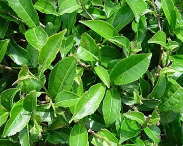
Fig no. 1: Camellia sinensis
Botanical Information:
• Scientific Name: Camellia sinensis
• Family: Theaceae
Major Phytochemical Constituents:
• Catechins
• Epigallocatechin gallate (EGCG)
• Polyphenols
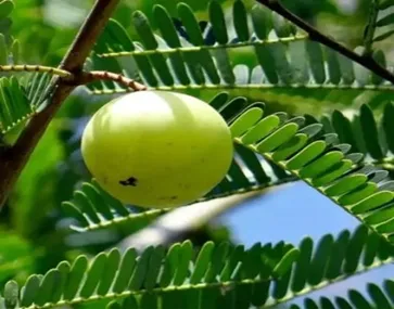
Fig no 2: Phyllanthus emblica
Botanical information:
• Scientific Name: Phyllanthus emblica
• Family: Phyllanthaceae
Major Phytochemical Constituents:
• Vitamin C
• Polyphenols
• Tannins (Emblicanin A and B)
• Flavonoids
Plant-based bioactive compounds have gained significant importance in the development of therapeutic agents, especially for the treatment and management of inflammatory disorders. Compared with synthetic medications, natural compounds are generally considered to possess better safety profiles and fewer side effects, which has encouraged extensive research into their pharmacological activities. Among the medicinal plants investigated for such properties, Phyllanthus emblica (Amla) and Camellia sinensis (green tea) are widely recognized for their potent antioxidant and anti-inflammatory effects [7].
Phyllanthus emblica is an important medicinal plant used in traditional systems of medicine such as Ayurveda. It possesses a rich phytochemical composition containing ascorbic acid, tannins, flavonoids, and polyphenols. Various experimental investigations have demonstrated that extracts of P. emblica can reduce oxidative stress and regulate inflammatory responses by inhibiting enzymes such as lipoxygenase and suppressing the formation of inflammatory mediators [8].
Similarly, green tea derived from Camellia sinensis has been extensively studied for its therapeutic benefits. The biological activity of green tea is mainly attributed to catechins, particularly epigallocatechin-3-gallate (EGCG). These phytoconstituents are known to modulate several inflammatory pathways through inhibition of pro-inflammatory cytokines such as tumor necrosis factor-alpha (TNF-α) and interleukins, along with regulation of transcription factors associated with inflammatory signaling pathways [2].Among the available in vitro techniques for evaluating anti-inflammatory activity, the protein denaturation assay is commonly employed. Protein denaturation refers to structural modifications in proteins caused by heat, chemicals, or other stress conditions, leading to loss of their biological function. During inflammation, such alterations may aggravate tissue damage. Therefore, agents capable of preventing protein denaturation are considered to exhibit anti-inflammatory activity by stabilizing protein structure and minimizing inflammatory progression [9].The anti-inflammatory potential of Phyllanthus emblica has been linked to its ability to inhibit enzymes such as 15-lipoxygenase, reduce oxidative stress, and maintain cellular integrity [10]. Likewise, catechins present in green tea, especially EGCG, have been reported to suppress inflammatory signaling pathways such as nuclear factor-kappa B (NF-κB), thereby decreasing the expression of inflammatory mediators and genes [11].The therapeutic activities of these plants are primarily associated with their phytochemical constituents. In amla, compounds including gallic acid, ellagic acid, emblicanin A, emblicanin B, and several flavonoids contribute significantly to its antioxidant and anti-inflammatory properties [12]. In green tea, catechins such as EGCG, epicatechin (EC), epigallocatechin (EGC), and epicatechin gallate (ECG) are mainly responsible for its pharmacological effects (2).Considering their individual therapeutic benefits, a combination of amla and green tea may provide enhanced anti-inflammatory activity. The diverse range of polyphenolic compounds present in both plants may act synergistically to modulate multiple inflammatory pathways simultaneously. Such a combinational strategy may improve therapeutic efficacy by enhancing antioxidant protection and reducing inflammatory responses more effectively than individual treatments alone [13].
Principle of In vitro egg albumin method:
The principal objective behind the egg albumin denaturation assay is to determine whether agents or compounds can stop or hinder egg albumin from becoming denatured under particular circumstances. Denaturation is the term used to describe how a protein changes in structure and loses its biological activity [14]. Egg albumin is employed as a model protein in the experiment, and denaturation is brought about by exposing it to extremes of heat, pH, or other denaturing agents. Egg albumin’s original conformation is disrupted during denaturation, changing its physical characteristics, and causing it to lose its functional activity. The egg albumin denaturation assay measures a drug or compound’s capacity to prevent or lessen egg albumin denaturation to evaluate its anti-inflammatory effects [15]. The egg albumin denaturation assay is based on the idea that substances with anti-inflammatory qualities may be able to stabilize protein structures and prevent denaturation, which is frequently linked to inflammation and tissue damage. As a result, agents or chemicals that significantly decrease the denaturation of egg albumin in this assay may have potential anti-inflammatory properties [16]. One of the causes of inflammation is assumed to be protein denaturation. NSAIDs prevent protein denaturation and inhibit the COX enzyme at the same time [17]. The different concentrations of the test sample can be incubated with egg albumin solution in controlled experimental conditions and let the reactions happen and then the determination of absorbance for determination of percent inhibition.
MATERIALS AND METHODS [18]
Materials:
Chemical and Reagents
• Phosphate buffered saline
• Distilled water/DMSO
• Egg albumin solution
Equipment
Clean pipettes and puppet tips
• Khan tubes or test tubes
• Incubator
• Spectrophotometer
Procedure
Egg Albumin Denaturation Assay Methodology [18]:
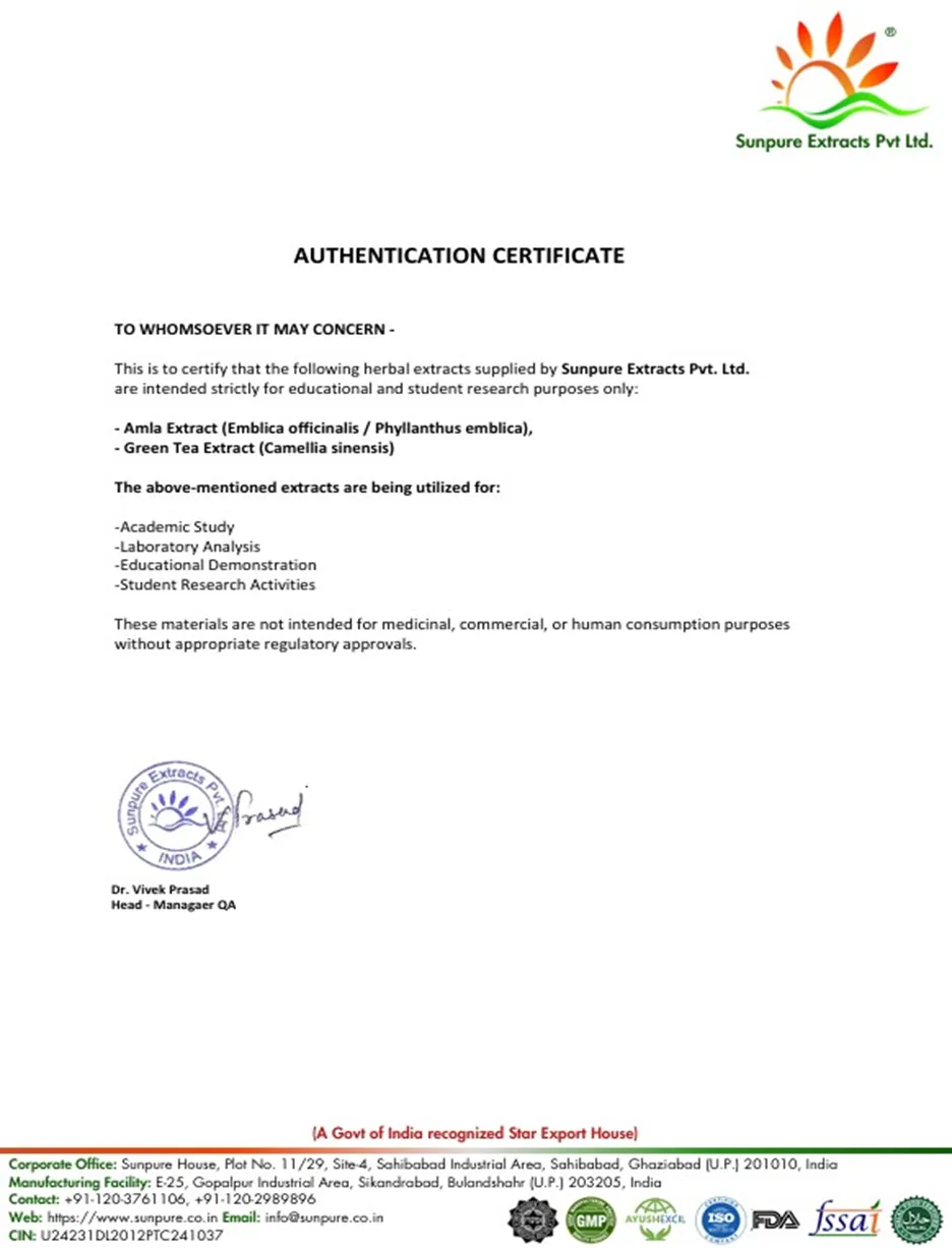
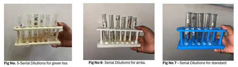
Preparation of plant extract: To initiate the dilution series, the extracts of Amla (Phyllanthus emblica) and green tea (Camellia sinensis) plants were used. The plant extracts were procured from Sunpure Extracts Private Limited, and the corresponding Certificate of Analysis (CoA) for the extracts was provided by the company. Due to variations in the concentration of tannins and polyphenols present in the amla and green tea extracts respectively, normalization of the extracts was carried out prior to the experimental analysis to ensure uniformity and comparability of the samples. Initially, stock solutions of amla and green tea extracts were prepared at a concentration of 2 mg/mL. Further dilution was carried out by taking 1 mL of the stock solution and making up the volume to 10 mL using distilled water as the solvent, resulting in a working solution of 200 µg/mL. Similarly, stock solution for the standard (Diclofenac sodium) was created. Then, 2ml of the prepared solution was taken into the respective tubes and the assay was conducted.
Preparation of 1% of egg albumin solution [18]:
Fresh hen’s eggs or egg albumin powder that is readily accessible in stores can be used to make a 1% egg albumin solution. Making egg-albumin solution using a fresh hen’s egg properly involves carefully cracking an egg, transferring 1 mL of the translucent portion to 100 mL of w/V distilled water, and stirring thoroughly. The clear component of the egg is called egg albumin. The water should be cold when making the solution. Water will coagulate if it is heated to a boil.
PERCENTAGE INHIBITION = Absorbance control - Absorbance of test × 100
Absorbance of control
Egg Albumin Assay [18]:
• 0.2 mL of 1-2% egg albumin solution (from fresh hen’s egg/ or commercially available egg albumin powder), 2mL of sample extract or standard (Diclofenac sodium) at varying concentrations, and 2.8mL of phosphate buffered saline (pH 7.4) were mixed to form a reaction mixture of a total volume of 5 mL.
• A total volume of 5 mL of the control was created by combining 2 mL of triple-distilled water, 0.2 mL of 1-2% egg albumin solution, and 2.8 mL of phosphate-buffered saline.
• The reaction mixtures were then incubated at 37±2°C for 30 min and will be heated in a water bath at 70±2°C for 15 min.
• After cooling, the absorbance was measured at 280 nm by a suitable UV/Vis spectrophotometer using distilled water as the blank. % inhibition of protein denaturation was calculated using the formula mentioned.
OBSERVATIONS AND RESULT:
The anti-inflammatory activity of amla extract, green tea extract, and their combination was evaluated using the egg albumin denaturation assay. Diclofenac sodium was used as the standard drug. The absorbance was measured spectrophotometrically, and percentage inhibition of protein denaturation was calculated.
Table no 1: Absorbance Values of Diclofenac Sodium, Amla Extract, Green Tea Extract, and Their Combination in Egg Albumin Denaturation Assay
|
Concentration (µg/mL) |
Standard |
Amla |
Green Tea |
Combination |
|
10 |
0.67 |
0.75 |
0.69 |
0.69 |
|
20 |
0.55 |
0.68 |
0.58 |
0.58 |
|
30 |
0.43 |
0.57 |
0.49 |
0.49 |
|
40 |
0.31 |
0.48 |
0.38 |
0.36 |
|
50 |
0.20 |
0.39 |
0.31 |
0.28 |
Table no 2: Comparative Percentage Inhibition of Protein Denaturation by Diclofenac Sodium, Amla Extract, Green Tea Extract, and Their Combination
|
Concentration (µg/mL) |
Standard |
Amla |
Green Tea |
Combination |
|
10 |
0.67 |
0.75 |
0.69 |
0.69 |
|
20 |
0.55 |
0.68 |
.0.58 |
0.58 |
|
30 |
0.43 |
0.57 |
0.49 |
0.49 |
|
40 |
0.31 |
0.48 |
0.38 |
0.36 |
|
50 |
0.20 |
0.39 |
0.31 |
0.28 |
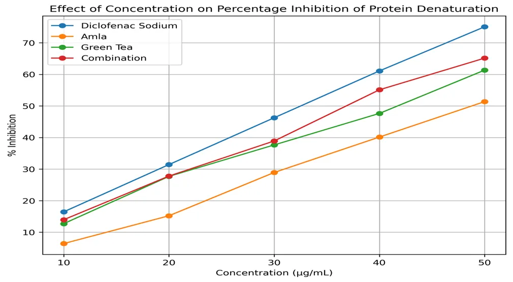
Graph: Effect of concentrations of amla, green tea and their combination on their percent inhibition
CONCLUSION
The present study evaluated the in vitro anti-inflammatory activity of amla extract, green tea extract, and their combination using the egg albumin denaturation assay. All the tested samples exhibited concentration-dependent inhibition of protein denaturation, indicating the presence of anti- inflammatory potential. Among the individual extracts, green tea demonstrated comparatively higher activity than amla at the tested concentrations. The combination of amla and green tea showed greater percentage inhibition than the individual extracts, suggesting a possible synergistic interaction between the phytoconstituents present in both extracts. Although the activity observed was lower than the standard drug diclofenac sodium at certain concentrations, the results indicate that the herbal extracts possess appreciable protein-stabilizing properties. The observed effects may be associated with the presence of polyphenols, flavonoids, tannins, catechins, and antioxidant compounds in the extracts. The findings of the study suggest that the combination of amla and green tea may have potential as a natural anti-inflammatory formulation.
REFERENCES


K. Shinde, S. Ambilwade, P. Gaulkar, S. Dhotrikar, S. Kudale, G. Badgujar, Comparative Study of The Anti-Inflammatory and Synergistic Activity of Phyllanthus Emblica and Camellia Sinensis Extracts Using The In Vitro Egg Albumin Denaturation Method, Int. J. of Pharm. Sci., 2026, Vol 4, Issue 6, 695-703, https://doi.org/10.5281/zenodo.20526042
 10.5281/zenodo.20526042