
We use cookies to ensure our website works properly and to personalise your experience. Cookies policy
The Oxford College of Pharmacy, Hongasandra, Bommanahalli, Bengaluru, Karnataka, India.
Aim: The aim of the present study was to develop and evaluate a controlled-release oral in-situ gel containing curcumin (Curcuma longa) and Punica granatum peel extract for the effective management of oral candidiasis. Materials and Methods: Oral in-situ gels were prepared using Carbopol Ultrez 10 as the gelling agent and HPMC K100 as the viscosity-modifying polymer. Five formulations (F1–F5) were developed and evaluated for physicochemical properties such as pH, viscosity, gelation temperature, gelation time, spreadability, and mucoadhesive strength. Antifungal activity was assessed against Candida albicans, and FTIR analysis was performed to study drug–polymer compatibility. Results and Discussion: All formulations showed acceptable physicochemical characteristics suitable for oral application. Among them, formulation F3 exhibited optimal viscosity, rapid gelation at physiological temperature (37 °C), good spreadability (12.5 cm), and effective mucoadhesive strength. The antifungal study revealed a zone of inhibition of 1.2 cm for F3, which was comparable to the standard drug fluconazole. FTIR results confirmed the absence of chemical interaction between the drug and polymers, indicating good stability of the formulation. Conclusion: The study demonstrated that an oral in-situ gel containing curcumin and Punica granatum peel extract provides sustained drug release, enhanced mucosal retention, and effective antifungal activity. This herbal-based formulation shows promise as a safe, natural, and patient-friendly alternative for the treatment of oral candidiasis.
The oral mucosa plays a vital role in protecting the oral cavity and maintaining overall oral health. Under normal conditions, the mouth contains a balanced microbial flora; however, disruption of this balance can lead to infections1. Oral candidiasis, commonly known as oral thrush, is one of the most frequently encountered opportunistic infections of the oral cavity. It is mainly caused by Candida albicans, a commensal yeast that becomes pathogenic under favourable conditions2,3. The infection is commonly observed in infants, elderly individuals, denture wearers, and immunocompromised patients3. Clinically, it presents as white or erythematous lesions associated with pain, burning sensation, and difficulty in swallowing, which can significantly affect quality of life2.
The prevalence of oral candidiasis has increased due to factors such as immunosuppressive therapy, HIV infection, diabetes, reduced salivary flow, and prolonged drug use2,3. Although C. albicans is the primary causative organism, other species including C. glabrata and C. tropicalis may also be involved3,4. In severe cases, particularly in immunocompromised individuals, the infection may spread beyond the oral cavity and lead to serious complications.5
Topical antifungal agents are commonly used for the treatment of mild oral candidiasis because they act locally and produce fewer systemic side effects6. However, conventional oral formulations such as mouth rinses, gels, and suspensions suffer from poor retention in the oral cavity due to salivary washout, resulting in reduced therapeutic efficacy and frequent dosing.3,4
In situ gelling systems have emerged as an effective approach to overcome these limitations7. These formulations are administered as liquids and undergo gelation upon exposure to physiological conditions in the oral cavity, forming a mucoadhesive gel that prolongs drug residence time8,9. pH-sensitive in situ gels, in particular, utilize changes in oral pH to trigger gel formation, enabling sustained drug release and improved therapeutic outcomes7,10. Thus, in situ gel-based delivery systems offer a promising strategy for the effective management of oral candidiasis.11
DRUG PROFILE
1. POMEGRANATE (Punica granatum)
Taxonomy:
Biological source: Peel of Punica granatum
Family: Lythraceae
Kingdom: Plantae
Genus: Punica
Species: P. granatum
Plant Description:
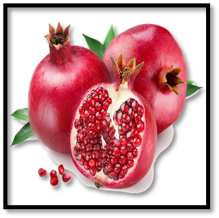
Fig. No. 1: Pomegranate peel
Pomegranate is a small tree or shrub grown in many parts of the world. The fruit has a hard outer peel and contains many seeds surrounded by juicy arils. The peel is thick, leathery, and not edible, but it is rich in medicinally important compounds.
Chemical Composition:
The peel mainly contains polyphenols such as gallic acid and ellagic acid, tannins like punicalagin, and flavonoids. It also has small amounts of alkaloids, saponins, terpenoids, vitamins, minerals, and dietary fibre.
Uses:
Pomegranate peel is known for its antioxidant, antimicrobial, anti-inflammatory, wound-healing, cardioprotective, antidiabetic, and anticancer properties.12-16
2. CURCUMIN (Curcuma longa)
Taxonomy:
Biological source: Rhizomes of Curcuma longa
Family: Zingiberaceae
Kingdom: Plantae
Genus: Curcuma
Species: C. longa
Plant Description:
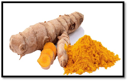
Fig. No. 2: Curcumin
Curcuma longa is a perennial herb commonly grown in tropical regions. The plant has underground rhizomes that are yellow-orange in colour and aromatic in nature. These rhizomes are the source of turmeric and its active compound, curcumin.
Chemical Composition:
Curcumin is the major bioactive constituent of turmeric. Other curcuminoids and essential oils are also present, along with carbohydrates, proteins, starch, fats, dietary fibre, and trace minerals.
Uses:
Curcumin shows anti-inflammatory, antioxidant, antimicrobial, anticancer, and wound-healing activities. It is widely used in traditional medicine and in cosmetic preparations.17-23
MATERIALS AND METHODS:
2.1. DRUGS:
Pomegranate peel: Dried and powdered extract obtained from the pomegranate peel, used as an active therapeutic agent.
Curcumin: A bioactive compound derived from turmeric, used as an active therapeutic agent.
2.2. EXCIPIENTS AND OTHER MATERIALS:
2.3. Collection and Preparation of Plant Materials
2.3.1. Pomegranate Peel
Fresh fruits of Punica granatum were obtained from a local market and washed thoroughly with distilled water to remove adhering impurities. The peels were separated from the pulp, cut into small pieces, and authenticated by a taxonomist at the Central Ayurveda Research Institute, Bengaluru. The collected peels were shade-dried at room temperature (25–30 °C) for about one week, powdered using a grinder, sieved, and stored in an airtight container for further studies.
2.3.2. Curcumin
Fresh rhizomes of Curcuma longa were collected locally in Bengaluru, Karnataka. The rhizomes were cleaned, peeled, sliced, and shade-dried at room temperature (25–30 °C) to preserve the active constituents. After drying, the material was powdered, passed through a sieve, and stored in airtight containers. The plant material was authenticated by a taxonomist at the Central Ayurveda Research Institute, Bengaluru.
2.4. EXTRACTION PROCEDURE
2.4.1. Punica granatum Peel
The dried pomegranate peels were finely powdered and stored in a dry place at room temperature. About 1000 g of the powder was subjected to Soxhlet extraction using ethanol as the solvent. The powdered material was placed in a thimble and extracted continuously to ensure maximum recovery of phytoconstituents. During extraction, fresh solvent repeatedly passed through the material. After completion of extraction, the solvent containing the extract was collected. The solvent was removed under reduced pressure using a rotary evaporator to obtain a concentrated peel extract24.
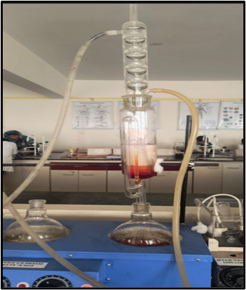
Fig. No. 3: Extraction of Punica granatum
2.4.2. Curcuma longa
The dried turmeric rhizomes were ground into a fine powder and used for extraction. The powder was soaked in alcohol using the maceration method in a closed container. The mixture was kept undisturbed for several days with occasional shaking to enhance extraction. After the maceration period, the liquid extract was separated by filtration. The filtrate was further clarified to remove fine particles. The solvent was evaporated to obtain the concentrated turmeric extract25.
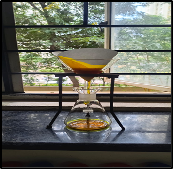
Fig. No. 4: Extraction of Curcuma longa
2.5. FTIR Analysis:
Fourier Transform Infrared (FTIR) spectroscopy was carried out to identify the characteristic functional groups of the extracts and polymers used. The study was also performed to assess possible interactions between the drug and excipients in the formulation.
2.6. Formulation of Insitu gel:
A. Preparation of Simulated Salivary Fluid (SSF)
Composition:
Sodium chloride (NaCl), Potassium chloride (KCl), Calcium chloride dihydrate (CaCl?·2H?O), Disodium hydrogen phosphate (Na?HPO?), Sodium dihydrogen phosphate (NaH?PO?).
Procedure:
B. Preparation of Gel Base (Carbopol Ultrez 10 and HPMC K100M)
C. Preparation of Curcumin and Pomegranate Peel Extract-Loaded Gel
D. In-situ Gelation Test
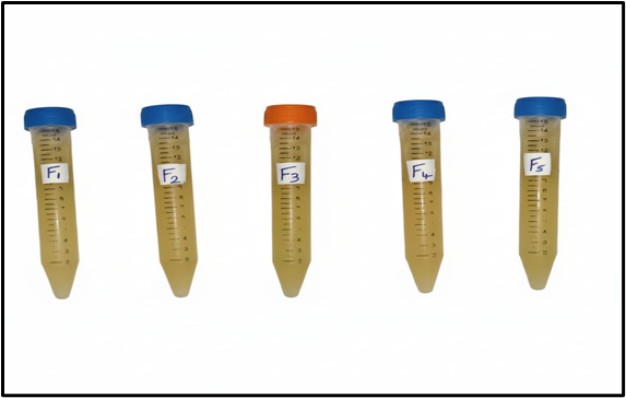
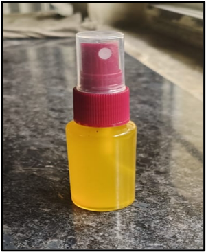
Fig. No. 5: Formulation of In-situ Gel
Table 1: Composition of Insitu gel (30 ml)
|
INGREDIENTS |
F1 |
F2 |
F3 |
F4 |
F5 |
|
Curcumin extract |
1.0 ml |
1.0 ml |
1.5 ml |
1.0 ml |
1.0 ml |
|
Pomegranate peel extract |
100 mg |
100 mg |
100 mg |
150 mg |
150 mg |
|
Carbopol Ultrez 10 |
0.21 g |
0.30 g |
0.21 g |
0.35 g |
0.21 g |
|
HPMC K 100M |
0.09 g |
- |
0.09 g |
- |
0.09 g |
|
Purified water |
q.s |
q.s |
q.s |
q.s |
q.s |
2.7. Evaluation parameters of Insitu gel:
2.7.1. Physical Appearance
The gels are ideally expected to be transparent. Each formulation was visually examined to observe characteristics such as colour, odour, texture, and the presence of any suspended particles. Additionally, the movement of these particles was checked by gently tilting the container26.
2.7.2. pH
The pH of the formulations was determined by placing the gel in a beaker and gradually adding 1 ml of NaOH while continuously stirring. The final pH was measured using a calibrated pH meter27,28.
2.7.3. Viscosity and Rheological Studies
Viscosity plays a crucial role in the performance and ease of application of in-situ gels. The flow behaviour and consistency of the gels were assessed using instruments such as a Brookfield viscometer or an Ostwald viscometer. The formulations were designed to have a viscosity between 5 and 100 mPas, allowing smooth application in the oral cavity and ensuring patient comfort9.
2.7.4. Gel Strength
Gel strength was measured using a rheometer and depends on the gelation properties of the chosen gelling agent. A sample of gel was prepared, and the probe of the instrument was gradually lowered into the gel. The resistance encountered by the probe was recorded as it penetrated the gel, providing a measure of the gel’s mechanical strength26.
2.7.5. Gelling Capacity
The ability of the gel to form and maintain a gel structure in the oral environment was tested using simulated salivary fluid (SSF). A small volume of the formulation was mixed with SSF and maintained at 37 ± 0.5°C. The time taken for gel formation and the duration for which it retained its structure were visually observed. An effective in-situ gel should form rapidly and maintain integrity to allow sustained drug release in the oral cavity29.
2.7.6. Sol-Gel Transition Temperature and Gelling Time
For temperature-sensitive in-situ gels, the sol-gel transition temperature is the point at which the liquid begins to gel. This was determined by gradually heating the sample in a test tube and observing the point where the meniscus stopped moving upon tilting. The time taken for the sol to convert into gel was also recorded8,33.
2.7.7. Clarity
The clarity of the formulations was assessed by observing them under light against both black and white backgrounds. This helps identify any cloudiness or suspended particles in the solution27,28.
2.7.8. Spreadability
Spreadability reflects how easily the gel can be applied. A measured amount of gel was placed between two glass slides and pressed to a uniform thickness using a 1000 g weight for 5 minutes. A 50 g weight was then attached to the upper slide, and the time taken for the slides to separate was recorded. The spreadability (S) was calculated using the formula:31
???? = ???? × ????/T
Where:
M = weight attached to the upper slide
L = length of the glass slides
T = time taken for the slides to separate
2.7.9. Sterility Testing
Sterility tests were conducted according to IP 1996. The formulations were incubated for a minimum of 40 days. Bacterial contamination was checked using fluid thioglycolate medium at 30–35°C, while fungal contamination was assessed in soya casein digest medium at 20–25°C26.
2.7.10. Mucoadhesion Study
Mucoadhesive strength was measured as the force required to detach the gel from the oral mucosa. Chicken buccal mucosa was mounted between two vials and maintained at 32–34°C. A known amount of gel was placed between the mucosal tissues, and the detachment force was recorded using a modified balance method. This test indicates the gel’s ability to adhere to oral tissues and maintain contact for effective drug delivery4.
2.7.11. ANTI-MICROBIAL ACTIVITY BY WELL DIFFUSION METHOD
1.1. Materials & Methods:
|
Sr. no |
Particulars |
Source |
Catalogue No. |
|
1 |
Antibiotic Assay Medium A |
Hi-media |
ME003 |
|
2 |
Petri plates |
Tarson |
460096 |
|
3 |
1000 µl tips |
Tarson |
521020 |
|
4 |
200 µl tips |
Tarson |
521014 |
1.2 Test organisms:
Antifungal (Candida albicans)
1.3. Test compound:
In-situ gel.
1.4 Inoculum:
Candida albicans cell suspension was prepared and grown on media, and cultures were incubated for 48hrs at 35°C. The cell suspensions of the cultures were adjusted to 2x 10⁶cells/ml.
1.5 Test compound:
1.6. Procedure:
a) Determination of Antimicrobial Activity
Candida albicans were inoculated on media (90 mm), and wells were created.
Test compounds: Sample: - In-situ gel (30µl).
The treated plates with Candida albicans were incubated in an aerobic chamber at 37°C for 24hrs. The treated plates were observed for zones of inhibition around the wells.
RESULTS AND DISCUSSION:
All the evaluation tests were done and the results were recorded and reported.
3.1. FTIR Studies:
FTIR studies were carried out to check the compatibility between curcumin, Punica granatum peel extract, and the excipients used in the formulation. The recorded spectra showed that the characteristic functional groups of the actives were retained. No major changes or peak shifts were observed in the formulation spectrum. This indicates that there was no interaction between the components and they are compatible with each other.
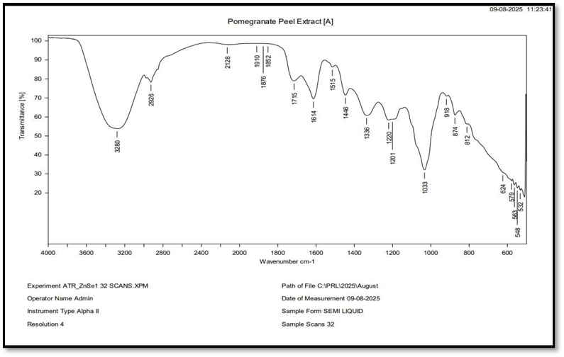
Fig. No. 6: FTIR report for extraction of Punica granatum
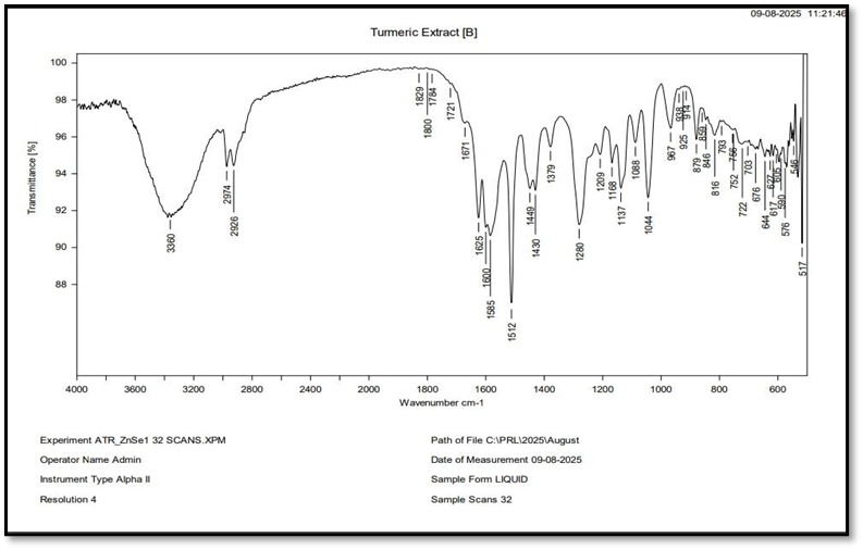
Fig. No. 7: FTIR report for extraction of Curcuma longa
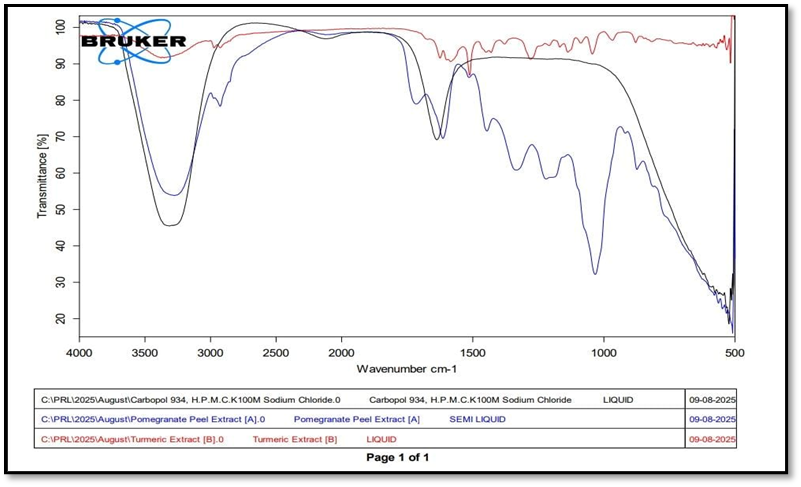
Fig. No. 8: FTIR report for comparison
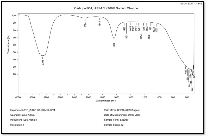
Fig. No. 9: FTIR report for Gel Formulation
3.2. Physical Appearance
All the formulations (F1–F5) appeared as homogeneous gels with a yellowish-brown colour and a characteristic herbal odour. The smooth texture observed in all batches indicates uniform composition and good physical consistency of the in-situ gel formulations.
Table 2: Organoleptic properties
|
Formulation (F) |
Appearance |
Colour |
Odour |
Texture |
|
F1 |
Homogeneous gel |
Yellowish-brown |
Characteristic herbal |
Smooth |
|
F2 |
Homogeneous gel |
Yellowish-brown |
Characteristic herbal |
Smooth |
|
F3 |
Homogeneous gel |
Yellowish-brown |
Characteristic herbal |
Smooth |
|
F4 |
Homogeneous gel |
Yellowish-brown |
Characteristic herbal |
Smooth |
|
F5 |
Homogeneous gel |
Yellowish-brown |
Characteristic herbal |
Smooth |
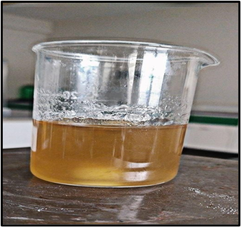
Fig. No.10: Physical Appearance
3.3. pH
The pH of the in-situ gel formulations was measured using a calibrated pH meter after gradual addition of NaOH with continuous stirring. All formulations showed pH values between 6.11 and 6.61, indicating that the gels are within a suitable range for oral use and are unlikely to cause irritation.
Table 3:pH of Insitu gel
|
FORMULATION |
pH of the gel |
|
F1 |
6.11 |
|
F2 |
6.3 |
|
F3 |
6.61 |
|
F4 |
6.4 |
|
F5 |
6.49 |
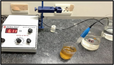
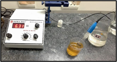
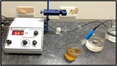
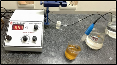
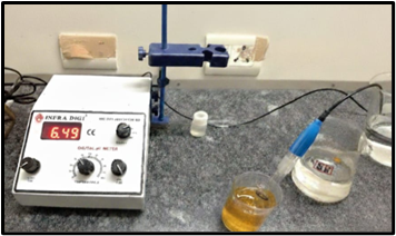
Fig. no.11: pH of Insitu gel formulation
3.4. Viscosity and rheological studies
The viscosity of the in-situ gel formulations was evaluated before and after gelation using a Brookfield viscometer. The pre-gelling viscosity ranged from 610 to 1400 mPas, while the viscosity increased to 1280–2600 mPas after exposure to simulated salivary fluid, indicating good flow before administration and sufficient gel strength after gelation for prolonged retention in the oral cavity.
Table 5:Viscosity of Insitu gel
|
FORMULATION |
PREGELLING VISCOSITY |
GELLING VISCOSITY WITH SFF FLUID |
|
F1 |
780 |
1600 |
|
F2 |
850 |
1850 |
|
F3 |
610 |
1280 |
|
F4 |
1100 |
2200 |
|
F5 |
1400 |
2600 |
3.5. Sol-gel transition temperature and gelling time
The sol-gel transition of the temperature-sensitive in-situ gels was determined by gradually heating the formulations and observing the point where the liquid began to gel. The gels remained fluid at 25–2?°C and solidified at 37–38?°C, ensuring easy administration and stable gel formation under oral conditions. This confirms their suitability for prolonged retention in the oral cavity.
Table 6: Sol-gel transition temperature of Insitu gel
|
FORMULATION |
PREGELLING TEMPERATURE |
GELLING VISCOSITY WITH SSF FLUID |
|
F1 |
25.3 |
37.0 |
|
F2 |
25.8 |
37.9 |
|
F3 |
25.2 |
37.6 |
|
F4 |
25.6 |
37.0 |
|
F5 |
25.5 |
37.2 |
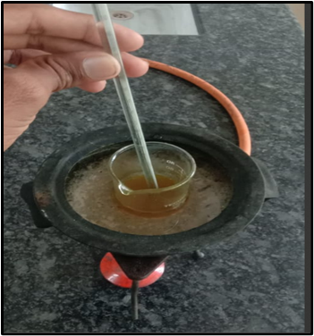
Fig. no.12: Sol-gel transition temperature of Insitu gel formulation
3.6. Spreadability
The in-situ gel formulations showed variation in spreadability, ranging from 4.5 to 12.?g.cm/s. Among them, F3 spread most easily, while F5 was the least spreadable, reflecting differences in how smoothly each formulation can be applied.
Table 7: Spreadability of Insitu gel
|
FORMULATION |
SPREADABILITY |
|
F1 |
9.0 |
|
F2 |
10.5 |
|
F3 |
12.5 |
|
F4 |
6.0 |
|
F5 |
4.5 |
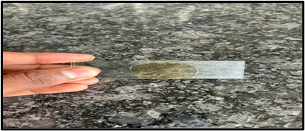
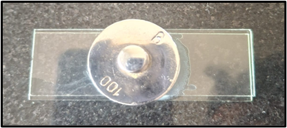
Fig. No. 13: Spreadability of Insitu gel formulation
3.7. ANTIMICROBIAL ACTIVITY:
The antifungal activity of the in-situ gel was evaluated against Candida albicans and compared with standard Fluconazole (2?µg). The in-situ gel produced a clear zone of inhibition measuring 1.2?cm, while the standard showed a slightly larger zone of 1.5?cm, indicating that the gel possesses significant antifungal activity.
Table 8: Inhibitory activity of test compounds against test organisms
|
Test Organisms |
Test Compounds |
Conc. per well |
Zone of inhibition (cm) |
|
Candida albicans |
Standard Fluconazole (25mcg) |
25 mcg disc |
1.5 |
|
In-situ gel |
30 µl |
1.2 |
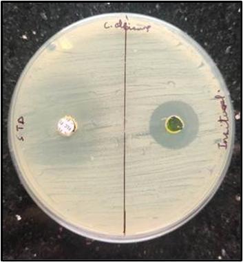
Fig. no. 14: Inhibitory activity of test sample against Candida albicans
CONCLUSION:
The study successfully developed a controlled-release oral in-situ gel incorporating curcumin and Punica granatum peel extract aimed at managing oral fungal infections. Among the five formulations, F3 proved to be the most effective, displaying ideal gelation at body temperature, suitable viscosity, good spreadability, strong mucoadhesive properties, and significant antifungal activity comparable to standard fluconazole.
The synergistic combination of curcumin and pomegranate peel extract provides multiple therapeutic benefits, including anti-inflammatory, antioxidant, antimicrobial, and wound-healing effects, which not only help control the infection but also promote oral mucosal health and patient comfort.
These results suggest that the F3 formulation is an optimized, safe, and patient-friendly herbal in-situ gel with sustained drug release, minimal side effects, and potential for improved compliance. This novel herbal-based system offers a promising, natural alternative to conventional antifungal therapies and could be further explored for clinical use and large-scale pharmaceutical applications in oral healthcare.
REFERENCES


R. Uma Prabha, Vishal A, Akshitha Y, Amrin M, Chandrakanth G, Controlled Release Oral In-Situ Gel of Curcumin and Punica Granatum Peel Extract: An Innovative Approach for Oral Thrush Management, Int. J. of Pharm. Sci., 2026, Vol 4, Issue 1, 2605-2618. https://doi.org/10.5281/zenodo.18351014
 10.5281/zenodo.18351014