View Article

Abstract

Superparamagnetic iron oxide nanoparticles (SPIONs) have emerged as promising nanocarriers for targeted cancer therapy owing to their magnetic responsiveness, biocompatibility, and large surface area. In the present study, a novel carboxymethyl Assam Bora rice starch (CM-ABRS) was synthesized and employed as a natural stabilizing and capping agent for SPIONs prepared via the co-precipitation method. The carboxymethylation process was optimized, yielding a maximum degree of substitution (DS) of 1.23 ± 0.01 under optimized reaction conditions. Successful carboxymethylation of ABRS was confirmed by FTIR and NMR analyses. Structural characterization by XRD verified the crystalline nature of both CM-ABRS and magnetite nanoparticles, while SEM analysis revealed the irregular granular morphology of CM-ABRS and uniform nanoscale dispersion of CM-ABRS-coated SPIONs. The optimized formulation exhibited an average particle size of 203.7 ± 1.09 nm, a polydispersity index (PDI) of 0.027 ± 0.005, and a favorable zeta potential, indicating excellent colloidal stability. Vibrating sample magnetometry (VSM) confirmed the superparamagnetic behavior of the nanoparticles, and in vitro magnetic localization studies demonstrated their efficient responsiveness to an external magnetic field. Overall, the findings demonstrate that CM-ABRS is an effective natural polymeric stabilizer for SPIONs and highlight the potential of the developed magnetic nanosystem as a promising platform for targeted anticancer drug delivery.

Keywords

SPIONs, CM?ABRS, magnetic targeting, natural polymer, nanoparticle stabilization, anticancer drug delivery.

Introduction

× Popup Image

Conventional chemotherapy suffers from poor tumor selectivity and systemic toxicity. Superparamagnetic iron oxide nanoparticles (SPIONs) can be magnetically guided to tumor sites and provide controlled drug delivery. Natural polymers are attractive stabilizers because of their biodegradability and biocompatibility. Assam Bora rice starch (ABRS) possesses mucoadhesive and release‑retarding properties, but its native form has limited applicability as a nanoparticle stabilizer due to poor solubility and excessive swelling. Therefore, ABRS was chemically modified by carboxymethylation and evaluated as a capping agent for SPIONs. The aim of the study was to develop CM‑ABRS stabilized SPIONs and investigate their physicochemical and magnetic properties for targeted drug delivery.

  1. MATERIALS AND METHODS

Polymer synthesis: ABRS was extracted and carboxymethylated using chloroacetic acid in isopropyl alcohol under alkaline conditions.

SPION synthesis: Magnetite nanoparticles were prepared by co‑precipitation of FeCl₃ and FeSO₄ using NH₄OH (pH 11–14) at 60–65°C under nitrogen atmosphere.

Characterization: FTIR, ¹H/¹³C NMR, DSC, XRD, SEM, TEM, DLS, zeta potential, and VSM analyses were performed.

Magnetic localization: FITC‑labeled red blood cells were used to evaluate nanoparticle localization in a microcapillary under an external magnetic field.

2.1 PREPARATION OF CARBOXYMETHYL ASSAM BORA-RICE STARCH (CM-ABRS):
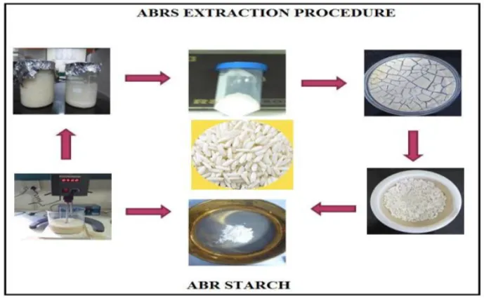
Figure 1. Extraction procedure of ABRS from Assam bora rice
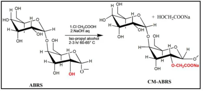
Figure 2. Reaction scheme for the synthesis of CM-ABRS

Table 1. Physicochemical Properties of CM-ABRS Compared with Native ABRS

Parameter

ABRS

CM-ABRS

Bulk density (g/cm³)

0.387

0.476

Tapped density (g/cm³)

0.525

0.534

Hausner ratio

1.358

1.122

Carr's Index (%)

26.34

10.11

Angle of repose (°)

34.64

18.34

Degree of substitution

1.23

The carboxymethylation process improved powder flow characteristics and produced CM-ABRS with a degree of substitution of 1.23.

2.2 Formulation of pristine magnetite (SPIONs)

SPIONs were synthesized by the co-precipitation method using a 2:1 molar ratio of FeCl₃ and FeSO₄·7H₂O. Ammonium hydroxide (0.1 M, pH 11–14) was added dropwise under continuous stirring (1500 rpm) at 60–65°C. The reaction mixture was aged for 30 min under a nitrogen atmosphere until black magnetite formed. The nanoparticles were separated by magnetic decantation, washed three times with deoxygenated double-distilled water, and dried for further characterization.

2.3 PHYSICOCHEMICAL CHARACTERIZATION

Dynamic Light Scattering (DLS)

The particle size, polydispersity index (PDI), and zeta potential of CM-ABRS SPIONs were measured using a Malvern Zetasizer Nano ZS. Samples were ultrasonicated, diluted with nitrogen-purged double-distilled water, and analyzed at 25°C.

Transmission Electron Microscopy (TEM)

The morphology of CM-ABRS SPIONs was examined using HR-TEM (TECNAI G2, 200 kV). Ultrasonically dispersed samples were placed on copper grids and imaged under high vacuum.

Fourier Transform Infrared Spectroscopy (FTIR)

The interaction between CM-ABRS and magnetite was analyzed using FTIR (Shimadzu IR Affinity-1) over the 4000–400 cm⁻¹ range. Samples were prepared as KBr pellets.

X-Ray Diffraction (XRD)

The crystalline structure of pure magnetite and CM-ABRS SPIONs was characterized using a Rigaku X-ray diffractometer with Cu Kα radiation, scanning from 10°–80° (2θ).

Vibrating Sample Magnetometry (VSM)

The magnetic properties of pure magnetite and CM-ABRS SPIONs were evaluated using a VSM (LDJ9600-1) over a magnetic field range of 0–18 kOe

2.4 In vitro Magnetic Targeting of CM-ABRS SPIONs

Blood Cell Labeling: Red blood cells (RBCs) collected from healthy mice were isolated by centrifugation, washed with glucose-saline buffer, and labeled with fluorescein isothiocyanate (FITC) following the method of Hudetz et al. The labeled RBCs were adjusted to a hematocrit of 45% with normal saline and stored in citrate-phosphate-dextrose buffer until use. All animal procedures were approved by the Institutional Animal Ethics Committee, Jamia Hamdard (Approval No. 1119).

In vitro Magnetic Localization: Lyophilized CM-ABRS SPIONs were dispersed in PBS (pH 7.4) to prepare a 5% (w/v) suspension and mixed with FITC-labeled RBCs in a 1:1 (v/v) ratio. The suspension (0.8% w/v) was passed through a 500 × 500 μm square glass capillary at a flow rate of 5 μL/min using a syringe pump. A NdFeB permanent magnet (150 mT) was positioned adjacent to the capillary to generate an external magnetic field. The localization of CM-ABRS SPIONs within the capillary was monitored using a Leica DMI 6000 CS confocal microscope equipped with a CCD camera.
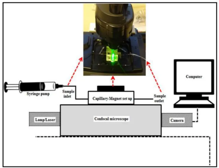
Figure 3. Experimental setup for in vitro magnetic localization of CM-ABRS SPIONs using a capillary–magnet system under a confocal microscope.

  1. RESULTS AND DISCUSSION

3.1.1 Optimization of carboxymethylation (CM-ABRS)

Parameter

Optimized value

Molar ratio (starch:CAA)

1:1.5

NaOH concentration

8 N

Reaction temperature

65°C

Reaction time

2.5 h

Degree of substitution (DS)

1.23 ± 0.01

The carboxymethylation process was optimized by systematically varying the reaction parameters. The optimized conditions resulted in efficient modification of Assam Bora rice starch, producing a high degree of substitution and demonstrating the suitability of the synthesized CM-ABRS for further nanoparticle formulation and biomedical applications.

The increase in DS confirmed successful introduction of carboxymethyl groups into the starch backbone.

3.1.2 FTIR and NMR analysis

FTIR spectra showed the appearance of characteristic carboxylate bands in CM‑ABRS, confirming etherification of ABRS. ¹H and ¹³C NMR spectra further verified the formation of CM‑ABRS through the appearance of new signals corresponding to carboxymethyl carbon atoms
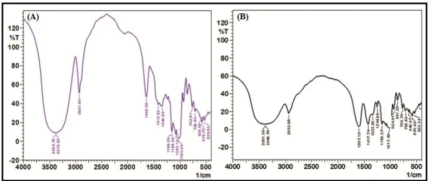
Figure 4. FT-IR spectra of: (A) ABRS, and (B) CM-ABRS

3.1.3 Thermal analysis

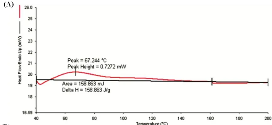
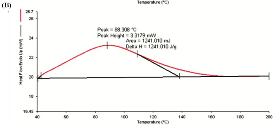
DSC analysis showed that carboxymethylation modified the thermal properties of ABRS by changing the gelatinization temperatures and reducing enthalpy (ΔH), indicating decreased crystallinity and improved molecular flexibility

Parameter

Value

Onset temperature

43.62°C

Peak temperature

67.24°C

Enthalpy (ΔH)

158.863 J/g

Figure 5. DSC Thermograms of: (A) CM-ABRS, and (B) ABRS

      1. Powder properties

Table 2. Comparative of powder properties of ABRS and CM-ABRS

Sr. No

Parameters

ABRS

CM-ABRS

1

Bulk Density(g/cm3)

0.387

0.476

2

Hausner’s Ratio (HR)

1.3576

1.122

3

Compressibility index (% CI)

26.341

10.11

4

Angle of repose (θ)

34.64

18.34

These values indicate excellent flow characteristics of the modified polymer.

3.1.5  XRD analysis

Native ABRS exhibited diffraction peaks at 15.10°, 17.67°, and 23.01° (2θ). CM‑ABRS showed reduced crystallinity, indicating structural modification of the starch matrix.
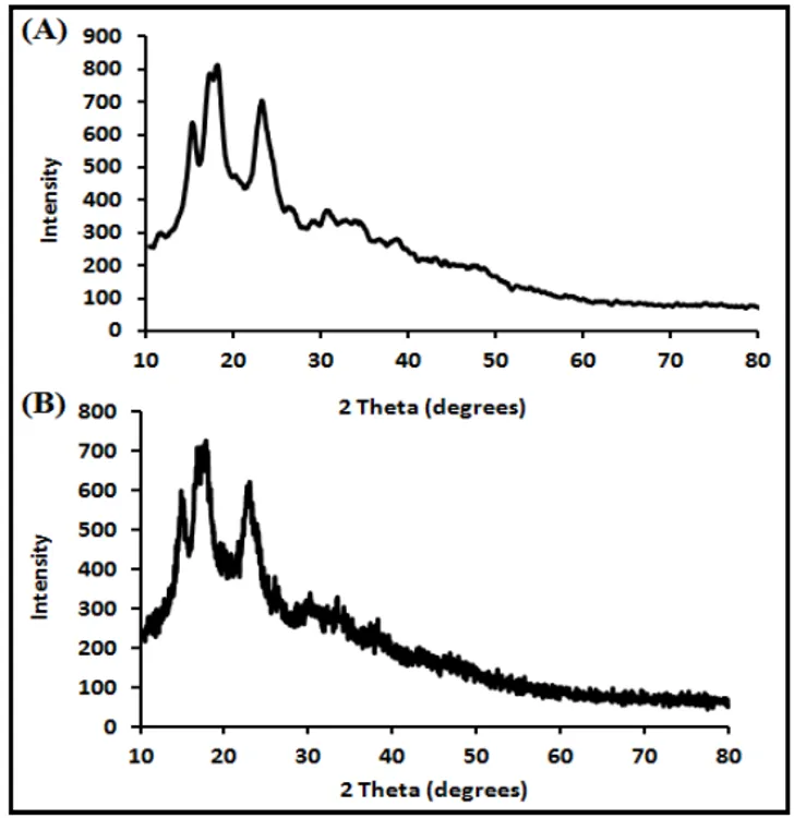
Figure 6. X-ray diffractogram of: (A) ABRS, (B) CM-ABRS

3.1.6 Swelling and solubility

At 65°C, CM‑ABRS exhibited:

  • Swelling index: 18.86 ± 0.68 g/g
  • Solubility: 79.22 ± 0.74%

The enhanced swelling behavior is advantageous for sustained drug release applications.

Table 3. Swelling Index and Solubility of ABRS and CM-ABRS

Temperature

ABRS SI (g/g)

CM-ABRS SI (g/g)

ABRS Solubility (%)

CM-ABRS Solubility (%)

30°C

2.80 ± 0.35

8.60 ± 0.54

1.78 ± 0.52

62.83 ± 0.64

55°C

7.21 ± 0.76

14.03 ± 0.80

11.80 ± 0.45

70.95 ± 0.71

65°C

11.81 ± 0.69

18.86 ± 0.68

27.47 ± 0.71

79.22 ± 0.74

75°C

15.01 ± 0.78

21.36 ± 0.87

22.04 ± 0.83

70.96 ± 0.34

CM-ABRS demonstrated substantially higher swelling and aqueous solubility than native starch.

3.1.7 Rheology

Table 4. Rheological and Mucoadhesive Properties

Parameter

Value

Flow behavior index (n)

0.45

Viscosity (Pa·s)

2.92

Consistency (g·s)

241.36 ± 1.25

Adhesiveness (g·s)

141.11 ± 1.60

Mucoadhesion (g·s)

6.12 ± 0.01

The CM-ABRS dispersion exhibited pseudoplastic non-Newtonian flow and improved gel consistency.

3.2 Synthesis of CM-ABRS-SPIONs

Table 5. Optimized CM-ABRS-SPION Formulation

Parameter

Optimized Value

Polymer concentration

0.8% w/v

Homogenization speed

25,000 rpm

Homogenization cycles

3

Particle size

203.7 ± 1.09 nm

PDI

0.027 ± 0.005

Effect of polymer concentration:

The optimum polymer concentration was 0.8% w/v, producing particles of 421.3 ± 5.82 nm with PDI 0.302 ± 0.013.

Effect of homogenization:

The best formulation was obtained at 25,000 rpm for 3 cycles.

Particle size: 203.7 ± 1.09 nm

PDI: 0.027 ± 0.005

The low PDI indicates a highly monodisperse nanoparticle population.

3.2.1 Formulation of CM-ABRS coated SPIONs (CM-ABRS-SPIONs)

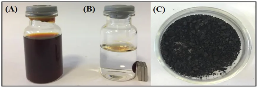
Figure 7. (A) Aqueous dispersion of CM-ABRS SPIONs, (B) CM-ABRS SPIONs attracted by magnet and (C) Lyophilized CM-ABRS SPIONs

3.2.3 TEM and zeta potential
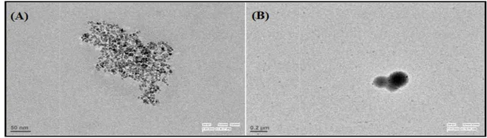
Figure 8. TEM micrograph of: (A) Pristine magnetite (B) CM-ABRS SPIONs

TEM images confirmed nearly spherical nanoparticles with uniform distribution. Zeta potential analysis demonstrated sufficient surface charge to maintain colloidal stability.
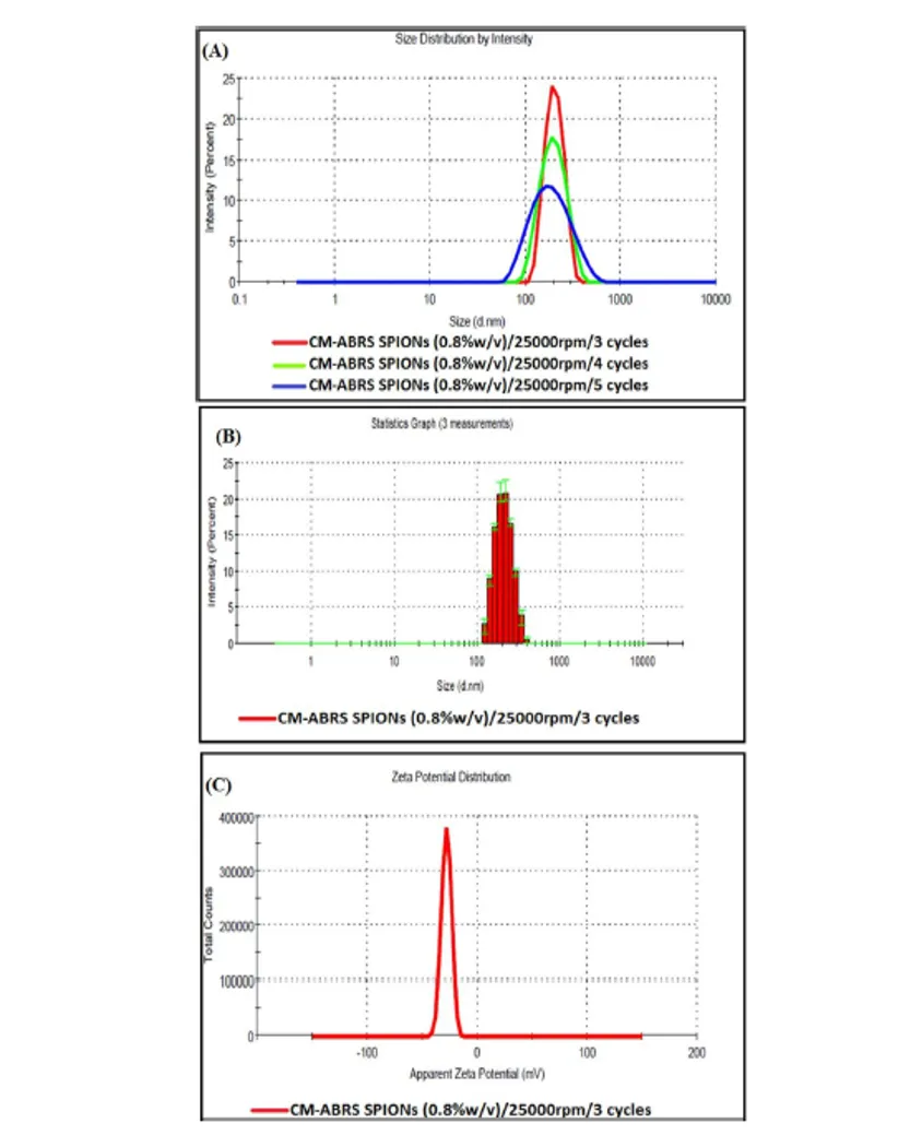
Figure 9. (A) Effect of homogenization cycles on particle size of CM-ABRS SPIONs

(B) Particle size distribution curve of optimized CM-ABRS SPIONs and

(C) Zeta potential distribution curve of optimized CM-ABRS SPIONs

3.2.4 Crystallographic properties of magnetite: X-Ray Diffraction

XRD analysis confirmed the cubic crystalline structure of Fe₃O₄ in both pure magnetite and CM-ABRS-SPIONs. The CM-ABRS coating preserved the crystal structure, indicating successful surface modification without affecting magnetite crystallinity.

3.2.5 Magnetic properties

VSM analysis showed superparamagnetic behavior with negligible remanence and coercivity, confirming the suitability of the nanoparticles for magnetic targeting applications.
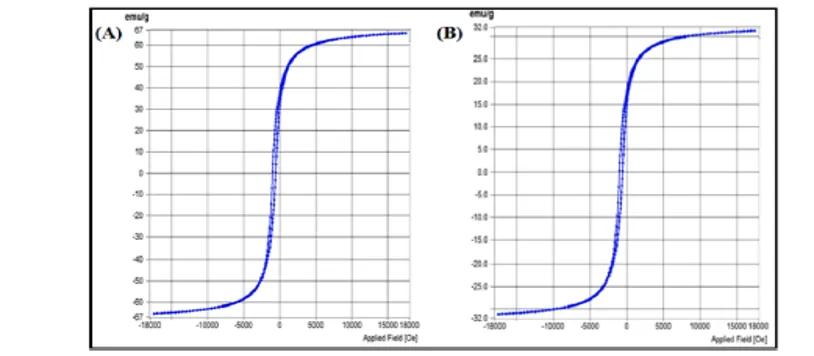
Figure 10. VSM results showing the M–H curves for: (A) Pristine magnetite, and (B) CM-ABRS SPIONs

3.3 In vitro magnetic localization

FITC-labeled red blood cells (RBCs) were used to visualize the in vitro localization of CM-ABRS SPIONs inside a microcapillary under an external magnetic field. The SPIONs appeared as black particles against the fluorescent RBC background. Upon application of the magnetic field, the nanoparticles accumulated along the bottom wall of the capillary due to the dominance of magnetic force over the hydrodynamic drag force (Figure 19).

The extent of SPION aggregation increased progressively with time and reached a steady state after 600 s, with no further significant increase in localization. The results demonstrate the effective magnetic responsiveness and targeting capability of CM-ABRS SPIONs, highlighting their potential as carriers for targeted drug delivery. Once localized at the target site, the drug can be released from the polymeric matrix in response to physiological or enzymatic stimuli.
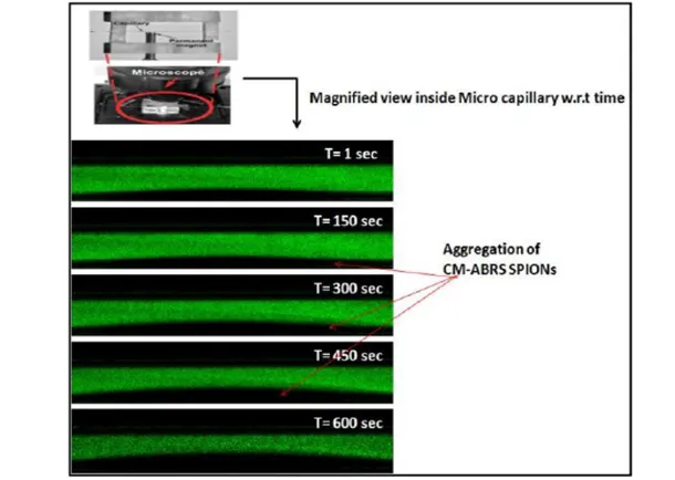
Figure 11. In vitro localization study of CM-ABRS SPIONs inside the micro capillary from t= 0 to 600s

4. CONCLUSION

A novel carboxymethyl Assam Bora rice starch (CM-ABRS) polymer was successfully synthesized and employed as a natural stabilizing agent for superparamagnetic iron oxide nanoparticles (SPIONs). The optimized CM-ABRS-SPIONs exhibited a nanoscale particle size (203.7 nm), excellent monodispersity (PDI 0.027), good colloidal stability, and superparamagnetic behavior. In vitro magnetic localization studies demonstrated efficient responsiveness of the nanoparticles to an external magnetic field, highlighting their potential for targeted anticancer drug delivery. The developed nanosystem combines the advantages of a biodegradable natural polymer with magnetically responsive nanoparticles and represents a promising platform for future targeted drug delivery and theranostic applications.

REFERENCES

  1. Akhtar, N., et al. (2022). SPIONs Conjugate Supported Anticancer Drug Delivery and Targeting. International Journal of Molecular Sciences.
  2. Vangijzegem, T., et al. (2023). Superparamagnetic Iron Oxide Nanoparticles: Formulation and Cancer Therapy Applications. Pharmaceutics.
  3. Musielak, M., et al. (2019). Superparamagnetic Iron Oxide Nanoparticles in Cancer Therapy. Journal of Controlled Release.
  4. Marekova, D. (2020). New Aspects of SPION-Based Nanocarriers in Drug Delivery. Anticancer Research.
  5. Ajith, S., et al. (2023). Nanoparticle-Based Materials in Anticancer Drug Delivery. Heliyon.
  6. Sun, L., et al. (2023). Smart Nanoparticles for Precise Cancer Therapy. Signal Transduction and Targeted Therapy.
  7. Catalano, E. (2019). Superparamagnetic Iron Oxide Nanoparticles Conjugated With Doxorubicin. arXiv.
  8. Gupta, R., et al. (2014). Synthesis and Evaluation of Anticancer Drug-Loaded SPIONs. Journal of Nanomedicine.
  9. Li, X., & Zhang, Y. (2015). Targeted Drug Delivery via SPIONs in Solid Tumors. International Journal of Nanomedicine.
  10. Singh, P., et al. (2016). Polymer-Coated SPIONs as Anticancer Delivery Vehicles. Nanomedicine Research Journal.
  11. Zhou, L., et al. (2017). Folate-Targeted SPIONs for Solid Tumor Therapy. Cancer Nanotechnology.
  12. Patel, S., et al. (2018). PEGylated SPIONs for Enhanced Anticancer Drug Delivery. Journal of Biomedical Nanotechnology.
  13. Kumar, M., et al. (2019). Dual Drug Co-Loaded SPIONs for Cancer Therapy. ACS Applied Nano Materials.
  14. Sharma, V., et al. (2020). Aptamer-Functionalized SPIONs in Targeted Therapy. Journal of Nanobiotechnology.
  15. Ahmed, Z., et al. (2021). PH-Responsive SPIONs for Controlled Anticancer Drug Release. International Journal of Pharmaceutics.
  16. Lee, D., et al. (2021). Impact of SPION Physicochemistry on Targeting Efficiency. Nanomedicine.
  17. Panda, Jnanranjan & Das, Dipanwita. (2025). Superparamagnetic iron oxide nanoparticle- based nanosystems for cancer theranostics. Global Translational Medicine. X. 1-20. 10.36922/gtm.8464.
  18. Mohammad, Faruq & Al-Lohedan, Hamad & Joshi, Payal & Obulapuram, Prasanna & CHAVALI, Murthy & Al-Balawi, Aiesha & Nikolova, Maria. (2022). Engineered superparamagnetic iron oxide nanoparticles for externally controlled hyperthermia, drug delivery, and therapeutic toxicity. Precision Nanomedicine. 5. 10.33218/001c.37603.
  19. Abdelmalek, Dorra & Bahloul, Nour & Bedchich, Kawther & Aifa, Sami & Messaoud, Mouna. (2026). Magnetic Iron Oxide Nanoparticles Conjugated to Anti-Cancer Drug for More Efficient Non-Small Cell Lung Cancer (NSCLC) Therapy. 10.5772/intechopen.1013211.
  20. Pucci, Carlotta & Degl'Innocenti, Andrea & Belenli Gümüş, Melike & Ciofani, Gianni. (2022). Superparamagnetic iron oxide nanoparticles for magnetic hyperthermia: Recent advancements, molecular effects, and future directions in the omics era. Biomaterials Science. 10. 2103-2121. 10.1039/D1BM01963E.
  21. Santhosh, Poornima & Ulrih, Nataša. (2013). Multifunctional Superparamagnetic Iron Oxide Nanoparticles: Promising Tools in Cancer Theranostics.. Cancer letters. 336. 10.1016/j.canlet.2013.04.032.
  22. Llamosa Perez, Daniel & Puentes, I. & Romero, G. & Saavedra Gaona, Indry & Vargas, C. & Rincón Ortiz, Rolando. (2023). Synthesis of superparamagnetic iron oxide nanoparticles coated with polyethylene glycol as potential drug carriers for cancer treatment. Journal of Nanoparticle Research.26. 10.1007/s11051-023-05900-5.
  23. Nedyalkova, Miroslava & Todorov, Boyan & Barazorda Ccahuana, Haruna & Madurga, Sergio. (2021). Iron oxide Nanoparticles in Anticancer Drug Delivery and Imaging Diagnostics. 10.1002/9781119754725.ch7.
  24. Fahim, Yosri & Hasani, Ibrahim & Ragab, Waleed. (2025). Promising biomedical applications using superparamagnetic nanoparticles. European Journal of Medical Research. 30. 441. 10.1186/s40001-025-02696-z.
  25. S, Nandisha & Sowbhagya, Sowbhagya. (2023). Nanomedicine & Nanotechnology Open Access Committed to Create Value for Researchers Superparamagnetic Iron Oxide Nanoparticles [SPION] and its Diversified Applications in the Medical Field: A Mini-Review Superparamagnetic Iron Oxide Nanoparticles [SPION] and its Diversified Applications in the Medical Field: A Mini-Review. Nanomedicine & Nanotechnology Open Access. 8. 10.23880/nnoa-16000248.
  26. Ajith, Saniha & Almomani, Fares & Elhissi, Abdelbary & Husseini, Ghaleb. (2023). Nanoparticle-based materials in anticancer drug delivery: Current and future prospects. Heliyon. 9. e21227. 10.1016/j.heliyon.2023.e21227.
  27. Douziech-Eyrolles, L., Marchais, H., Hervé, K., Munnier, E., Soucé, M., Linassier, C., Dubois, P., & Chourpa, I. (2007). Nanovectors for anticancer agents based on superparamagnetic iron oxide nanoparticles. International journal of nanomedicine, 2(4), 541–550.
  28. Mahmoudi, Morteza. (2011). Superparamagnetic iron oxide nanoparticles: Promises for diagnosis and treatment of cancer. International journal of molecular epidemiology and genetics. 2. 367-90.
  29. Kandasamy, Ganeshlenin & Maity, Dipak. (2015). Recent advances in superparamagnetic iron oxide nanoparticles (SPIONs) for in vitro and in vivo cancer nanotheranostics. International journal of pharmaceutics. 496. 10.1016/j.ijpharm.2015.10.058.
  30. Gupta, Jagriti & Prakash, Anand & Jaiswal, Manish & Agarrwal, Atanuu & Bahadur, D.. (2017). Superparamagnetic Iron Oxide-Reduced Graphene Oxide Nanohybrid-a Vehicle for Targeted Drug Delivery and Hyperthermia Treatment of Cancer. Journal of Magnetism and Magnetic Materials. 448. 10.1016/j.jmmm.2017.05.084.
  31. Annan, Emmanuel & Waheed, Sakariyau & Onifade, Isreal & Ruth, Adara & Adeleye, Oluwaseun & Chukwuma, Chiamaka & Ariyibi, Idowu & Onuche, Philip & Okafor, Chinaecherem & Ifijen, Ikhazuagbe. (2026). Iron Oxide Nanoparticles as Magnetic Nanocarriers for Targeted Delivery of Anticancer Biomolecules. Reviews and Advances in Chemistry. 15. 399-433.10.1134/S2634827625600380.
  32. Deka, A., Bardhan, P., Mandal, M., Kataki, R. (2022). Nano-biomaterials as a Potential Tool for Futuristic Applications. In: Hussain, C.M., Di Sia, P. (eds) Handbook of Smart Materials, Technologies, and Devices. Springer, Cham. https://doi.org/10.1007/978-3-030-84205-5_32
  33. Ahmad, Muhammad & Ali, Salman & Aslam, Husna & Atif, Ashfa & Ali, Ahmad & Muhammad, Rabiah & Abrar, Amna & Sohail, Muhammad & Uddin, Imad. (2025). SYNTHESIS AND CHARACTERIZATION OF IRON OXIDE NANO PARTICLES FOR TARGETED THERAPY. Journal of Medical & Health Sciences Review. 2. 10.62019/7gh1z833.
  34. Sandoval-Flores, Viviana & Ortega-Cardenas, J & Ramon-Gallegos, Eva & Gutiérrez-López, Gustavo & Ravelo Acuña, David & Yee-Madeira, H. & Mora-Escobedo, Rosalva. (2025). Characterization of superparamagnetic iron oxide nanoparticles and their effect on viability in cancer and non-cancerous cell-lines. Materials Research Express. 12. 10.1088/2053-1591/add098.
  35. P., Sathyadevi & Wang, Yun-Ming. (2019). Superparamagnetic Iron oxide Nanoparticulate System: Synthesis, Targeting, Drug Delivery and Therapy in Cancer. Dalton Transactions. 48. 10.1039/C9DT00459A.
  36. Lee N, Yoo D, Ling D, Cho MH, Hyeon T, Cheon J. Iron oxide based nanoparticles for multimodal imaging and magneto responsive therapy. Chem Rev. 2015;115:10637-10638. doi: 10.1021/acs.chemrev.5b00112.
  37. Gao J, Gu H, Xu B. Multifunctional magnetic nanoparticles: Design, synthesis, and biomedical applications. Acc Chem Res. 2009;42(8):1097-1107. doi: 10.1021/ar9000026
  38. Panda J, Ghouri K, Sarkar R, Tudu B. Fabrication and characterization of self-assembled zinc ferrite nanospheres for biomedical applications. Appl Phys A. 2022;128(4):1-9. doi: 10.1007/s00339-022-05456-6
  39. Hao R, Xing R, Xu Z, Hou Y, Gao S, Sun S. Synthesis, functionalization, and biomedical applications of multifunctional magnetic nanoparticles. Adv Mater. 2010;22:2729-2742. doi: 10.1002/adma.201000260
  40. Poller JM, Zaloga J, Schreiber E, et al. Selection of potential iron oxide nanoparticles for breast cancer treatment based on in vitro cytotoxicity and cellular uptake. Int J Nanomedicine. 2017;12:3207-3220. doi: 10.2147/IJN.S132369
  41. Panda J, Satapathy BS, Sarkar R, Tudu B. A  zinc ferrite nanodrug carrier for delivery of docetaxel: Synthesis, characterization, and in vitro tests on C6 glioma cells. J Microencapsul. 2022;39(2):136-144. doi: 10.1080/02652048.2022.2053757
  42. Hu Y, Mignani S, Majoral JP, Shen M, Shi X. Construction of iron oxide nanoparticle-based hybrid platforms for tumor imaging and therapy. Chem Soc Rev. 2018;47:1874-1900. doi: 10.1039/c7cs00657h
  43. Bray F, Laversanne M, Sung H, et al. Global cancer statistics 2022: GLOBOCAN estimates of incidence and mortality worldwide for 36 cancers in 185 countries. CA Cancer J Clin. 2024;74(3):229-263. doi: 10.3322/caac.21834
  44. Mitchell MJ, Billingsley MM, Haley RM, Wechsler ME, Peppas NA, Langer R. Engineering precision nanoparticles for drug delivery. Nat Rev Drug Discov. 2021;20:101-124. doi: 10.1038/s41573-020-0090-8
  45. Caster JM, Patel AN, Zhang T, Wang A. Investigational nanomedicines in 2016: A  review of nanotherapeutics currently undergoing clinical trials. Wiley Interdiscip Rev Nanomed Nanobiotechnol. 2017;9:1416-1425. doi: 10.1002/wnan.1416
  46. Panda J, Satapathay BS, Mishra A, Biswal B, Sahoo PK. Potential of Ferrite-Based Nanoparticles for Improved Cancer Therapy: Recent Progress and Challenges Ahead. Applications of Ferrites. London: Intech Open; 2023. p. 1-18. doi: 10.5772/intechopen.1002346
  47. Cui Y, Zhang M, Zeng F, Jin H, Xu Q, Huang Y. Dualtargeting magnetic PLGA nanoparticles for codelivery of paclitaxel and curcumin for brain tumor therapy. ACS Appl Mater Interfaces. 2016;8:32159-32169. doi: 10.1021/acsami.6b10175
  48. Shabatina TI, Vernya OI, Shabatin VP, Melnikov MY. Magnetic nanoparticles for biomedical purposes: Modern trends and prospects. Magnetochemistry. 2020;6:30. doi: 10.3390/magnetochemistry6030030
  49. Panda J, Satapathy BS, Majumder S, Sarkar R, Mukherjee B, Tudu B. Engineered polymeric iron oxide nanoparticles as potential drug carrier for targeted delivery of docetaxel to breast cancer cells. J Magn Magn Mater. 2019;485:165-173. doi: 10.1016/j.jmmm.2019.04.058
  50. Fatima H, Charinpanitkul T, Kim KS. Fundamentals to apply magnetic nanoparticles for hyperthermia therapy. Nanomaterials (Basel). 2021;11:1203. doi: 10.3390/nano11051203

Reference

  1. Akhtar, N., et al. (2022). SPIONs Conjugate Supported Anticancer Drug Delivery and Targeting. International Journal of Molecular Sciences.
  2. Vangijzegem, T., et al. (2023). Superparamagnetic Iron Oxide Nanoparticles: Formulation and Cancer Therapy Applications. Pharmaceutics.
  3. Musielak, M., et al. (2019). Superparamagnetic Iron Oxide Nanoparticles in Cancer Therapy. Journal of Controlled Release.
  4. Marekova, D. (2020). New Aspects of SPION-Based Nanocarriers in Drug Delivery. Anticancer Research.
  5. Ajith, S., et al. (2023). Nanoparticle-Based Materials in Anticancer Drug Delivery. Heliyon.
  6. Sun, L., et al. (2023). Smart Nanoparticles for Precise Cancer Therapy. Signal Transduction and Targeted Therapy.
  7. Catalano, E. (2019). Superparamagnetic Iron Oxide Nanoparticles Conjugated With Doxorubicin. arXiv.
  8. Gupta, R., et al. (2014). Synthesis and Evaluation of Anticancer Drug-Loaded SPIONs. Journal of Nanomedicine.
  9. Li, X., & Zhang, Y. (2015). Targeted Drug Delivery via SPIONs in Solid Tumors. International Journal of Nanomedicine.
  10. Singh, P., et al. (2016). Polymer-Coated SPIONs as Anticancer Delivery Vehicles. Nanomedicine Research Journal.
  11. Zhou, L., et al. (2017). Folate-Targeted SPIONs for Solid Tumor Therapy. Cancer Nanotechnology.
  12. Patel, S., et al. (2018). PEGylated SPIONs for Enhanced Anticancer Drug Delivery. Journal of Biomedical Nanotechnology.
  13. Kumar, M., et al. (2019). Dual Drug Co-Loaded SPIONs for Cancer Therapy. ACS Applied Nano Materials.
  14. Sharma, V., et al. (2020). Aptamer-Functionalized SPIONs in Targeted Therapy. Journal of Nanobiotechnology.
  15. Ahmed, Z., et al. (2021). PH-Responsive SPIONs for Controlled Anticancer Drug Release. International Journal of Pharmaceutics.
  16. Lee, D., et al. (2021). Impact of SPION Physicochemistry on Targeting Efficiency. Nanomedicine.
  17. Panda, Jnanranjan & Das, Dipanwita. (2025). Superparamagnetic iron oxide nanoparticle- based nanosystems for cancer theranostics. Global Translational Medicine. X. 1-20. 10.36922/gtm.8464.
  18. Mohammad, Faruq & Al-Lohedan, Hamad & Joshi, Payal & Obulapuram, Prasanna & CHAVALI, Murthy & Al-Balawi, Aiesha & Nikolova, Maria. (2022). Engineered superparamagnetic iron oxide nanoparticles for externally controlled hyperthermia, drug delivery, and therapeutic toxicity. Precision Nanomedicine. 5. 10.33218/001c.37603.
  19. Abdelmalek, Dorra & Bahloul, Nour & Bedchich, Kawther & Aifa, Sami & Messaoud, Mouna. (2026). Magnetic Iron Oxide Nanoparticles Conjugated to Anti-Cancer Drug for More Efficient Non-Small Cell Lung Cancer (NSCLC) Therapy. 10.5772/intechopen.1013211.
  20. Pucci, Carlotta & Degl'Innocenti, Andrea & Belenli Gümü?, Melike & Ciofani, Gianni. (2022). Superparamagnetic iron oxide nanoparticles for magnetic hyperthermia: Recent advancements, molecular effects, and future directions in the omics era. Biomaterials Science. 10. 2103-2121. 10.1039/D1BM01963E.
  21. Santhosh, Poornima & Ulrih, Nataša. (2013). Multifunctional Superparamagnetic Iron Oxide Nanoparticles: Promising Tools in Cancer Theranostics.. Cancer letters. 336. 10.1016/j.canlet.2013.04.032.
  22. Llamosa Perez, Daniel & Puentes, I. & Romero, G. & Saavedra Gaona, Indry & Vargas, C. & Rincón Ortiz, Rolando. (2023). Synthesis of superparamagnetic iron oxide nanoparticles coated with polyethylene glycol as potential drug carriers for cancer treatment. Journal of Nanoparticle Research.26. 10.1007/s11051-023-05900-5.
  23. Nedyalkova, Miroslava & Todorov, Boyan & Barazorda Ccahuana, Haruna & Madurga, Sergio. (2021). Iron oxide Nanoparticles in Anticancer Drug Delivery and Imaging Diagnostics. 10.1002/9781119754725.ch7.
  24. Fahim, Yosri & Hasani, Ibrahim & Ragab, Waleed. (2025). Promising biomedical applications using superparamagnetic nanoparticles. European Journal of Medical Research. 30. 441. 10.1186/s40001-025-02696-z.
  25. S, Nandisha & Sowbhagya, Sowbhagya. (2023). Nanomedicine & Nanotechnology Open Access Committed to Create Value for Researchers Superparamagnetic Iron Oxide Nanoparticles [SPION] and its Diversified Applications in the Medical Field: A Mini-Review Superparamagnetic Iron Oxide Nanoparticles [SPION] and its Diversified Applications in the Medical Field: A Mini-Review. Nanomedicine & Nanotechnology Open Access. 8. 10.23880/nnoa-16000248.
  26. Ajith, Saniha & Almomani, Fares & Elhissi, Abdelbary & Husseini, Ghaleb. (2023). Nanoparticle-based materials in anticancer drug delivery: Current and future prospects. Heliyon. 9. e21227. 10.1016/j.heliyon.2023.e21227.
  27. Douziech-Eyrolles, L., Marchais, H., Hervé, K., Munnier, E., Soucé, M., Linassier, C., Dubois, P., & Chourpa, I. (2007). Nanovectors for anticancer agents based on superparamagnetic iron oxide nanoparticles. International journal of nanomedicine, 2(4), 541–550.
  28. Mahmoudi, Morteza. (2011). Superparamagnetic iron oxide nanoparticles: Promises for diagnosis and treatment of cancer. International journal of molecular epidemiology and genetics. 2. 367-90.
  29. Kandasamy, Ganeshlenin & Maity, Dipak. (2015). Recent advances in superparamagnetic iron oxide nanoparticles (SPIONs) for in vitro and in vivo cancer nanotheranostics. International journal of pharmaceutics. 496. 10.1016/j.ijpharm.2015.10.058.
  30. Gupta, Jagriti & Prakash, Anand & Jaiswal, Manish & Agarrwal, Atanuu & Bahadur, D.. (2017). Superparamagnetic Iron Oxide-Reduced Graphene Oxide Nanohybrid-a Vehicle for Targeted Drug Delivery and Hyperthermia Treatment of Cancer. Journal of Magnetism and Magnetic Materials. 448. 10.1016/j.jmmm.2017.05.084.
  31. Annan, Emmanuel & Waheed, Sakariyau & Onifade, Isreal & Ruth, Adara & Adeleye, Oluwaseun & Chukwuma, Chiamaka & Ariyibi, Idowu & Onuche, Philip & Okafor, Chinaecherem & Ifijen, Ikhazuagbe. (2026). Iron Oxide Nanoparticles as Magnetic Nanocarriers for Targeted Delivery of Anticancer Biomolecules. Reviews and Advances in Chemistry. 15. 399-433.10.1134/S2634827625600380.
  32. Deka, A., Bardhan, P., Mandal, M., Kataki, R. (2022). Nano-biomaterials as a Potential Tool for Futuristic Applications. In: Hussain, C.M., Di Sia, P. (eds) Handbook of Smart Materials, Technologies, and Devices. Springer, Cham. https://doi.org/10.1007/978-3-030-84205-5_32
  33. Ahmad, Muhammad & Ali, Salman & Aslam, Husna & Atif, Ashfa & Ali, Ahmad & Muhammad, Rabiah & Abrar, Amna & Sohail, Muhammad & Uddin, Imad. (2025). SYNTHESIS AND CHARACTERIZATION OF IRON OXIDE NANO PARTICLES FOR TARGETED THERAPY. Journal of Medical & Health Sciences Review. 2. 10.62019/7gh1z833.
  34. Sandoval-Flores, Viviana & Ortega-Cardenas, J & Ramon-Gallegos, Eva & Gutiérrez-López, Gustavo & Ravelo Acuña, David & Yee-Madeira, H. & Mora-Escobedo, Rosalva. (2025). Characterization of superparamagnetic iron oxide nanoparticles and their effect on viability in cancer and non-cancerous cell-lines. Materials Research Express. 12. 10.1088/2053-1591/add098.
  35. P., Sathyadevi & Wang, Yun-Ming. (2019). Superparamagnetic Iron oxide Nanoparticulate System: Synthesis, Targeting, Drug Delivery and Therapy in Cancer. Dalton Transactions. 48. 10.1039/C9DT00459A.
  36. Lee N, Yoo D, Ling D, Cho MH, Hyeon T, Cheon J. Iron oxide based nanoparticles for multimodal imaging and magneto responsive therapy. Chem Rev. 2015;115:10637-10638. doi: 10.1021/acs.chemrev.5b00112.
  37. Gao J, Gu H, Xu B. Multifunctional magnetic nanoparticles: Design, synthesis, and biomedical applications. Acc Chem Res. 2009;42(8):1097-1107. doi: 10.1021/ar9000026
  38. Panda J, Ghouri K, Sarkar R, Tudu B. Fabrication and characterization of self-assembled zinc ferrite nanospheres for biomedical applications. Appl Phys A. 2022;128(4):1-9. doi: 10.1007/s00339-022-05456-6
  39. Hao R, Xing R, Xu Z, Hou Y, Gao S, Sun S. Synthesis, functionalization, and biomedical applications of multifunctional magnetic nanoparticles. Adv Mater. 2010;22:2729-2742. doi: 10.1002/adma.201000260
  40. Poller JM, Zaloga J, Schreiber E, et al. Selection of potential iron oxide nanoparticles for breast cancer treatment based on in vitro cytotoxicity and cellular uptake. Int J Nanomedicine. 2017;12:3207-3220. doi: 10.2147/IJN.S132369
  41. Panda J, Satapathy BS, Sarkar R, Tudu B. A  zinc ferrite nanodrug carrier for delivery of docetaxel: Synthesis, characterization, and in vitro tests on C6 glioma cells. J Microencapsul. 2022;39(2):136-144. doi: 10.1080/02652048.2022.2053757
  42. Hu Y, Mignani S, Majoral JP, Shen M, Shi X. Construction of iron oxide nanoparticle-based hybrid platforms for tumor imaging and therapy. Chem Soc Rev. 2018;47:1874-1900. doi: 10.1039/c7cs00657h
  43. Bray F, Laversanne M, Sung H, et al. Global cancer statistics 2022: GLOBOCAN estimates of incidence and mortality worldwide for 36 cancers in 185 countries. CA Cancer J Clin. 2024;74(3):229-263. doi: 10.3322/caac.21834
  44. Mitchell MJ, Billingsley MM, Haley RM, Wechsler ME, Peppas NA, Langer R. Engineering precision nanoparticles for drug delivery. Nat Rev Drug Discov. 2021;20:101-124. doi: 10.1038/s41573-020-0090-8
  45. Caster JM, Patel AN, Zhang T, Wang A. Investigational nanomedicines in 2016: A  review of nanotherapeutics currently undergoing clinical trials. Wiley Interdiscip Rev Nanomed Nanobiotechnol. 2017;9:1416-1425. doi: 10.1002/wnan.1416
  46. Panda J, Satapathay BS, Mishra A, Biswal B, Sahoo PK. Potential of Ferrite-Based Nanoparticles for Improved Cancer Therapy: Recent Progress and Challenges Ahead. Applications of Ferrites. London: Intech Open; 2023. p. 1-18. doi: 10.5772/intechopen.1002346
  47. Cui Y, Zhang M, Zeng F, Jin H, Xu Q, Huang Y. Dualtargeting magnetic PLGA nanoparticles for codelivery of paclitaxel and curcumin for brain tumor therapy. ACS Appl Mater Interfaces. 2016;8:32159-32169. doi: 10.1021/acsami.6b10175
  48. Shabatina TI, Vernya OI, Shabatin VP, Melnikov MY. Magnetic nanoparticles for biomedical purposes: Modern trends and prospects. Magnetochemistry. 2020;6:30. doi: 10.3390/magnetochemistry6030030
  49. Panda J, Satapathy BS, Majumder S, Sarkar R, Mukherjee B, Tudu B. Engineered polymeric iron oxide nanoparticles as potential drug carrier for targeted delivery of docetaxel to breast cancer cells. J Magn Magn Mater. 2019;485:165-173. doi: 10.1016/j.jmmm.2019.04.058
  50. Fatima H, Charinpanitkul T, Kim KS. Fundamentals to apply magnetic nanoparticles for hyperthermia therapy. Nanomaterials (Basel). 2021;11:1203. doi: 10.3390/nano11051203

Photo
Shivani Shettiwar
Corresponding author

Department of Pharmaceutical chemistry, Balwantrao Chavan College of Pharmacy, Naigaon, Nanded

Photo
Dr. Vishal Kshirsagar
Co-author

Department of Pharmaceutical chemistry, Balwantrao Chavan College of Pharmacy, Naigaon, Nanded

Photo
Sumera S. G.
Co-author

Department of Pharmaceutical chemistry, Balwantrao Chavan College of Pharmacy, Naigaon, Nanded

Shivani Shettiwar, Sumera S. G., Dr. Vishal Kshirsagar, Nano-Enabled Chemical Design of Anticancer Drug-incorporated Superparamagnetic Iron Oxide Systems, Int. J. of Pharm. Sci., 2026, Vol 4, Issue 8, 1454-1466. https://doi.org/10.5281/zenodo.21843158

More related articles
A Comprehensive Review on Herbal Hair Serum: Formu...
E. Hemalatha, A. Senthil, N. Sushmitha, R. Ramya, M. Mahalakshmi,...
Molecular Mechanisms Underlying Ulcerative Colitis...
Gaurav Budhlakoti, Vikas Bhatt, Arun Kumar Singh , Abhijeet Ojha,...
Related Articles
The Transformative Role of Artificial Intelligence in the Pharmaceutical Sector...
Prabal Pardeshi, Monika Madibone, Rutuja Pawar, Anjali Pawar, Rupali Pathre...
Non-Pharmacological and Lifestyle Modifications for The Management of Hypothyroi...
Aishwarya Mehtre, Mansi Kadbe, Nikita Dhamshetti, Shweta Shisode, Dr. Prajakta Kelgaonkar...
A Comprehensive Review on Herbal Hair Serum: Formulation and Evaluation Approach...
E. Hemalatha, A. Senthil, N. Sushmitha, R. Ramya, M. Mahalakshmi, V. Kathirnila...
More related articles
A Comprehensive Review on Herbal Hair Serum: Formulation and Evaluation Approach...
E. Hemalatha, A. Senthil, N. Sushmitha, R. Ramya, M. Mahalakshmi, V. Kathirnila...
Molecular Mechanisms Underlying Ulcerative Colitis: Recent Advances And Emerging...
Gaurav Budhlakoti, Vikas Bhatt, Arun Kumar Singh , Abhijeet Ojha, Jyoti Joshi ...
A Comprehensive Review on Herbal Hair Serum: Formulation and Evaluation Approach...
E. Hemalatha, A. Senthil, N. Sushmitha, R. Ramya, M. Mahalakshmi, V. Kathirnila...
Molecular Mechanisms Underlying Ulcerative Colitis: Recent Advances And Emerging...
Gaurav Budhlakoti, Vikas Bhatt, Arun Kumar Singh , Abhijeet Ojha, Jyoti Joshi ...