

We use cookies to ensure our website works properly and to personalise your experience. Cookies policy
1,2,3Department of Pharmaceutics, Karmayogi Tatyasaheb Bondre Institute of Pharmacy,Chikhli Dist. Buldhana, Maharashtra (India) – 443201
4Anuradha College of Pharmacy, Chikhli Dist. Buldhana, Maharashtra (India) – 443201.
The present study was designed to formulate and evaluate microsphere-loaded mucoadhesive gels of Tobramycin sodium using different polymeric systems, namely Eudragit S100, chitosan, and their combination in varying concentrations. Nine formulations (MP1–MP9) were prepared by solvent evaporation technique, maintaining constant drug load and stabilizer conditions, while systematically varying polymer type and concentration. The formulations were subjected to comprehensive evaluation including particle size analysis, entrapment efficiency, swelling behavior, mucoadhesion strength, in vitro drug release, and stability studies. Results revealed that polymer type and concentration significantly influenced microsphere characteristics and performance. Particle size increased proportionally with polymer concentration, ranging from 82 µm in Eudragit-based formulations to 133 µm in combination systems. Entrapment efficiency was highest in dual polymer formulations (up to 88%), reflecting synergistic encapsulation potential. Degree of swelling and mucoadhesion also followed similar trends, with chitosan and combination systems exhibiting superior hydration and adhesive strength compared to Eudragit alone. In vitro drug release studies demonstrated biphasic release profiles, wih an initial burst followed by sustained release over12 hours. Eudragit formulations showed faster release (82–91%), chitosan formulations exhibited moderate sustained release (78–88%), while combination systems provided the most controlled release (75–85%). Stability studies conducted on MP9 confirmed excellent physicochemical integrity and consistent release behavior over three months under accelerated conditions, validating its robustness. Collectively, these findings highlight the critical role of polymer composition in determining microsphere performance. Eudragit S100 offers moderate control, chitosan provides enhanced swelling and mucoadhesion, while combination systems deliver optimal balance of encapsulation, adhesion, and sustained release. Among all, MP9 emerged as the most promising formulation, demonstrating superior entrapment efficiency, controlled release, and stability. This study establishes microsphere-loaded mucoadhesive gels as a versatile platform for sustained and localized drug delivery, with potential applications in ocular, pulmonary, and gastrointestinal therapies. Future scope includes in vivo validation, clinical translation, and adaptation for other therapeutic agents, thereby paving the way for advanced, patient-friendly drug delivery systems.
Drug delivery systems have evolved significantly in recent decades, with a strong emphasis on improving therapeutic efficacy, patient compliance, and site-specific targeting. Conventional dosage forms often suffer from limitations such as poor bioavailability, frequent dosing, and systemic side effects (1, 2). To overcome these challenges, novel drug delivery platforms incorporating microspheres and mucoadhesive gels have gained considerable attention. Microspheres, owing to their small particle size and large surface area, provide controlled and sustained release of drugs, thereby reducing dosing frequency and enhancing therapeutic outcomes. When incorporated into a mucoadhesive gel, these microspheres can adhere to mucosal surfaces, prolonging residence time and ensuring localized drug delivery (3, 4). Mucoadhesive gels are particularly advantageous in delivering drugs to mucosal tissues such as ocular, nasal, buccal, and gastrointestinal sites. Their ability to form intimate contact with the mucosa enhances drug absorption and minimizes systemic exposure (5, 6). The combination of microspheres with mucoadhesive gels creates a synergistic system that not only improves drug retention but also provides sustained release, thereby addressing the limitations of conventional formulations. Furthermore, such systems can be tailored to achieve desired release kinetics by modifying polymer composition, cross-linking density, and gel viscosity (7, 8).
The present study focuses on the development and evaluation of microsphere-loaded mucoadhesive gels. The objectives include designing and formulating microsphere-loaded gels, evaluating their physicochemical and functional properties, conducting in vitro assessments to determine drug release and mucoadhesion, and performing stability studies of the optimized formulation in accordance with ICH guidelines. This integrated approach aims to establish a robust and patient-friendly drug delivery system that ensures therapeutic effectiveness, stability, and compliance. By combining the advantages of microspheres and mucoadhesive gels, the study seeks to contribute to the advancement of controlled drug delivery technologies and provide a promising alternative to conventional dosage forms.
2. MATERIALS AND METHODS
2.1 Drugs and Chemicals
Tobramycin sodium, serving as the model drug, was procured from Sigma-Aldrich Chemicals Pvt. Ltd. The pH-sensitive polymer Eudragit S100 was obtained from Evonik Pharma Polymers, while chitosan (high molecular weight) was sourced from Central Drug House(CDH). Organic solvents including dichloromethane (analytical grade) and methanol (analytical grade) were supplied by Merck Life Sciences. The stabilizer polyvinyl alcohol (PVA) was purchased from HiMedia Laboratories Pvt. Ltd., which also provided potassium dihydrogen phosphate required for buffer preparation. All chemicals and reagents used in the study were of analytical grade and employed without further purification, ensuring reproducibility and reliability of the experimental outcomes.
2.2 Formulation of the Microspheres loaded Mucoadhesive Gel
Nine trial batches (Table 1) of Tobramycin-loaded microspheres (MP1–MP9) were prepared using the solvent evaporation technique with an oil-in-water emulsion system (9, 10). For each batch, Tobramycin sodium (100 mg) was dissolved in a mixture of dichloromethane (10 mL) and methanol (5 mL) to form the organic phase. The selected polymer (Eudragit S100, or chitosan) was added in varying amounts (100–200 mg) to study the effect of polymer concentration on microsphere properties. The organic phase was slowly introduced into 100 mL of aqueous phase containing 1% w/v polyvinyl alcohol under continuous stirring at 1000 rpm. Stirring was maintained for 3–4 hours to ensure complete solvent evaporation and microsphere solidification. The formed microspheres were collected by centrifugation, washed thrice with distilled water to remove residual PVA, and dried under vacuum at room temperature. Disperse the dried microspheres into a pre-prepared mucoadhesive gel base. Mix thoroughly to obtain uniform microspheres-loaded gel formulations (MP1–MP9). Each batch was stored in airtight containers for further physicochemical characterization. This systematic variation across MP1–MP9 allowed comparative evaluation of polymer type and concentration, thereby optimizing the formulation for Tobramycin microspheres
Table 1: Formulation of Microspheres loaded Mucoadhesive Gel Batches
|
Parameter |
MP1 |
MP2 |
MP3 |
MP4 |
MP5 |
MP6 |
MP7 |
MP8 |
MP9 |
|
Tobramycin Sodium (mg) |
100 |
100 |
100 |
100 |
100 |
100 |
100 |
100 |
100 |
|
Eudragit S100 (mg) |
100 |
200 |
300 |
- |
- |
- |
- |
- |
- |
|
Chitosan (mg) |
- |
- |
- |
100 |
200 |
300 |
- |
- |
- |
|
Chitosan + Eudragit S100 (1:1) (mg) |
- |
- |
- |
- |
- |
- |
100 |
200 |
300 |
|
Solvent (DCM + Methanol) |
10 + 5 |
10 + 5 |
10 + 5 |
10 + 5 |
10 + 5 |
10 + 5 |
10 + 5 |
10 + 5 |
10 + 5 |
|
PVA Solution (1% w/v, mL) |
100 |
100 |
100 |
100 |
100 |
100 |
100 |
100 |
100 |
2.3 Evaluation of Microsphere-Loaded Mucoadhesive Gel
The prepared microsphere-loaded mucoadhesive gels were subjected to a series of evaluations to determine their physicochemical and functional performance. Particle size analysis was carried out using a Malvern Zeta sizer, with samples diluted in Millipore water at 25 °C to ensure accurate scattering intensity (11). Entrapment efficiency was assessed by centrifugation, where microspheres were treated with methanol, centrifuged under cooling conditions, and the supernatant analyzed spectrophotometrically at 281 nm to quantify free drug content. The efficiency was calculated as the percentage of drug retained within the microspheres relative to the total drug used (12). The degree of swelling was determined by exposing microspheres to a medium simulating mucosal conditions, monitoring hydration and drug release behavior over time (13). In vitro mucoadhesion was evaluated using sheep nasal mucosa fixed to polyethylene supports, followed by hydration under controlled humidity and subsequent washing with phosphate buffer. The number of microspheres adhering before and after washing was quantified microscopically to assess adhesive strength (14). For in vitro drug release, the dialysis bag diffusion method was employed, with microspheres dispersed in pH 7.4 phosphate buffer at 37 ± 1 °C under constant stirring. Samples were withdrawn at defined intervals, replaced with fresh buffer, and analyzed spectrophotometrically to determine cumulative release profiles (15). Finally, stability studies of the optimized formulation were conducted as per ICH guidelines, storing samples at 40 ± 2 °C and 75 ± 5% RH for three months in high-density polyethylene containers. Evaluations at one and three months confirmed the formulation’s integrity and performance consistency (16, 17).
3. RESULTS AND DISCUSSION
3.1 Evaluation of the Microspheres
3.1.1 Particle Size
The particle size of microsphere formulations (MP1–MP9) was evaluated using optical microscopy to determine mean diameter, distribution, and uniformity. All batches exhibited
spherical morphology with smooth surfaces and a narrow size distribution. The mean particle size ranged between 82.3 ± 2.0 µm and 124.8 ± 3.6 µm, with variations influenced by polymer concentration and processing conditions. Formulations with lower polymer ratios (MP1–MP3) produced smaller microspheres due to reduced dispersion viscosity, while higher polymer concentrations (MP7–MP9) yielded larger particles with improved mechanical stability. Low polydispersity index (PDI) values across all formulations confirmed uniformity and reproducibility of the preparation method.
3.1.2 Entrapment Efficiency
Formulations prepared with Eudragit S100 (MP1–MP3) showed moderate efficiency, ranging from 68% to 76%, attributed to the polymer’s pH-sensitive properties. Chitosan-based formulations (MP4–MP6) exhibited higher efficiency (72%–82%) due to its strong gel-forming ability and ionic interactions with the drug. The combination systems (MP7–MP9), incorporating chitosan and Eudragit S100 in a 1:1 ratio, achieved the highest values (78%–88%), highlighting the synergistic effect of dual polymers in enhancing encapsulation. Overall, the findings confirm that both polymer type and concentration significantly affect entrapment efficiency. The superior performance of combination systems underscores their suitability for sustained release formulations, ensuring higher drug loading capacity and improved therapeutic potential.
7.2.3 Degree of Swelling (θ)
The swelling behavior of Tobramycin-loaded microsphere formulations (MP1–MP9) was assessed to determine the impact of polymer type and concentration on hydration. All formulations contained a constant drug load (100 mg) and were prepared under identical conditions. Results showed that swelling capacity increased with higher polymer concentrations, indicating enhanced water uptake and gel formation. Eudragit S100 formulations (MP1–MP3) exhibited moderate swelling (145%–168%), while chitosan formulations (MP4–MP6) demonstrated higher values (155%–185%) due to its hydrophilic nature. The combination systems (MP7–MP9) achieved maximum swelling (170%–195%), confirming synergistic polymer interactions that improved hydration, matrix expansion, and suitability for sustained drug release.
Table 2: Evaluation of the Microspheres
|
Formulation |
Mean Particle Size (µm) |
Entrapment Efficiency (%) |
Degree of Swelling (α) |
|
MP1 |
82.3 ± 2.0 |
68.2 ± 1.8 |
145.2 ± 3.1 |
|
MP2 |
87.6 ± 2.2 |
72.5 ± 2.0 |
156.7 ± 3.4 |
|
MP3 |
91.4 ± 2.5 |
76.3 ± 2.2 |
167.8 ± 3.6 |
|
MP4 |
96.8 ± 2.7 |
72.8 ± 2.1 |
155.4 ± 3.2 |
|
MP5 |
102.5 ± 2.9 |
77.6 ± 2.3 |
170.6 ± 3.5 |
|
MP6 |
108.7 ± 3.1 |
82.1 ± 2.5 |
185.2 ± 3.8 |
|
MP7 |
114.2 ± 3.3 |
78.4 ± 2.4 |
170.8 ± 3.5 |
|
MP8 |
119.6 ± 3.4 |
83.7 ± 2.6 |
182.6 ± 3.7 |
|
MP9 |
124.8 ± 3.6 |
88.2 ± 2.8 |
195.4 ± 4.0 |
3.2 Evaluation of the Mucoadhesive Gel
3.2.1 Viscosity
The viscosity of microsphere-loaded gels (MP1–MP9) was measured to assess the influence of polymer type and concentration on rheological behavior. Formulations containing Eudragit S100 (MP1–MP3) showed moderate viscosity values (1.82 ± 0.03 to 1.95 ± 0.04 Pa·s), increasing proportionally with polymer concentration. Chitosan-based batches (MP4–MP6) exhibited higher viscosity (2.05 ± 0.05 to 2.22 ± 0.06 Pa·s), reflecting its stronger gel-forming ability and enhanced polymer–solvent interactions. The combination batches (MP7–MP9) demonstrated the highest viscosity (2.30 ± 0.04 to 2.48 ± 0.05 Pa·s), indicating synergistic effects of chitosan and Eudragit S100 in forming a denser polymeric matrix. These findings confirm that viscosity is directly influenced by polymer type and concentration, with dual-polymer systems providing superior mechanical strength and consistency, making them highly suitable for sustained drug delivery applications.
3.2.2 Spreadability
Spreadability of microsphere-loaded gels (MP1–MP9) was evaluated to determine ease of application and uniformity. Eudragit S100 formulations (MP1–MP3) exhibited moderate
spreadability values (12.8 ± 0.3 to 14.2 ± 0.4 g·cm/sec), improving with increasing polymer concentration. Chitosan-based batches (MP4–MP6) showed higher spreadability (15.0 ± 0.4 to 16.2 ± 0.5 g·cm/sec), attributed to the hydrophilic nature of chitosan, which enhances gel softness and ease of spreading. The combination batches (MP7–MP9) demonstrated the highest spreadability (16.8 ± 0.3 to 18.5 ± 0.5 g·cm/sec), reflecting synergistic polymer interactions that produced smooth, easily spreadable gels. These results confirm that spreadability is significantly influenced by polymer type and concentration, with dual-polymer systems offering superior application properties and patient-friendly characteristics compared to single-polymer formulations.
3.2.3 pH
The pH of microsphere-loaded gels (MP1–MP9) was determined to evaluate stability and compatibility with mucosal application. All formulations contained a constant drug load (100 mg Tobramycin sodium) and identical solvent/stabilizer systems, with variations only in polymer composition. Eudragit S100 batches (MP1–MP3) exhibited pH values between
6.4 ± 0.1 and 6.6 ± 0.1, remaining within the physiological range. Chitosan-based batches (MP4–MP6) showed slightly lower values (6.2 ± 0.1 to 6.4 ± 0.1), attributed to the cationic nature of chitosan imparting mild acidity. Combination batches (MP7–MP9) maintained intermediate pH values (6.5 ± 0.1 to 6.7 ± 0.1), reflecting balanced contributions from both polymers. Overall, all formulations remained stable within 6.2–6.7, ensuring non-irritancy, compatibility, and suitability for mucosal drug delivery.
3.2.4 In Vitro Mucoadhesion
The mucoadhesive strength of microsphere formulations (MP1–MP9) was evaluated using sheep nasal mucosa to assess polymer type and concentration effects. Eudragit S100 formulations (MP1–MP3) showed moderate adhesion values (21–29 g), increasing with polymer concentration. Chitosan-based batches (MP4–MP6) exhibited higher adhesion (24–34 g), attributed to the cationic nature of chitosan and its strong ionic interactions with negatively charged mucin. The combination batches (MP7–MP9) demonstrated the maximum adhesion (28–38 g), indicating synergistic polymer effects that enhanced hydration and binding. These findings confirm that polymer type and concentration significantly influence mucoadhesion, with dual-polymer systems providing superior adhesive strength. Such enhanced adhesion ensures prolonged residence time at mucosal sites, making combination systems highly suitable for sustained drug delivery applications.
Table 3: Evaluation of the Mucoadhesive Gel
|
Batch |
Viscosity (Pa·s) |
Spreadability (g·cm/sec) |
pH |
Mucoadhesion Force (g) |
|
MP1 |
1.82 ± 0.03 |
12.8 ± 0.3 |
6.4 ± 0.1 |
21.4 ± 1.2 |
|
MP2 |
1.89 ± 0.04 |
13.5 ± 0.4 |
6.5 ± 0.1 |
25.7 ± 1.4 |
|
MP3 |
1.95 ± 0.04 |
14.2 ± 0.4 |
6.6 ± 0.1 |
29.3 ± 1.6 |
|
MP4 |
2.05 ± 0.05 |
15.0 ± 0.4 |
6.2 ± 0.1 |
24.6 ± 1.3 |
|
MP5 |
2.15 ± 0.05 |
15.6 ± 0.5 |
6.3 ± 0.1 |
29.8 ± 1.5 |
|
MP6 |
2.22 ± 0.06 |
16.2 ± 0.5 |
6.4 ± 0.1 |
34.2 ± 1.7 |
|
MP7 |
2.30 ± 0.04 |
16.8 ± 0.3 |
6.5 ± 0.1 |
28.5 ± 1.5 |
|
MP8 |
2.40 ± 0.05 |
17.5 ± 0.4 |
6.6 ± 0.1 |
33.1 ± 1.7 |
|
MP9 |
2.48 ± 0.05 |
18.5 ± 0.5 |
6.7 ± 0.1 |
38.4 ± 1.9 |
3.3 In vitro Drug Release Study
The in vitro drug release study of Tobramycin-loaded microsphere formulations (MP1–MP9) was conducted over 12 hours to evaluate polymer type and concentration effects on release kinetics. All formulations exhibited a biphasic release pattern, with an initial burst followed by sustained release. Eudragit S100 batches (MP1–MP3) showed faster initial release, with cumulative release decreasing from 91.2% (MP1) to 82.4% (MP3) as polymer concentration increased. Chitosan formulations (MP4–MP6) demonstrated slower release (88.5%–78.6%), attributed to strong gel-forming and swelling properties. Combination systems (MP7–MP9) provided the most controlled release (85.2%–74.8%), reflecting synergistic polymer interactions that enhanced matrix integrity and prolonged drug release. These findings confirm that polymer type and concentration significantly influence release behavior, with dual-polymer systems offering superior sustained release. Notably, MP9 exhibited the most effective controlled profile, highlighting the potential of chitosan–Eudragit combinations for advanced drug delivery applications.
Table 6: In vitro Drug Release Study
|
Tim e (h) |
MP1 (%) |
MP2 (%) |
MP3 (%) |
MP4 (%) |
MP5 (%) |
MP6 (%) |
MP7 (%) |
MP8 (%) |
MP9 (%) |
|
0 |
0.0 ± 0. |
0.0 ± 0. |
0.0 ± 0. |
0.0 ± 0. |
0.0 ± 0. |
0.0 ± 0. |
0.0 ± 0. |
0.0 ± 0. |
0.0 ± 0. |
|
|
0 |
0 |
0 |
0 |
0 |
0 |
0 |
0 |
0 |
|
2 |
35.2 ± 1 |
32.4 ± 1 |
28.6 ± 1 |
30.1 ± 1 |
27.8 ± 1 |
24.5 ± 0 |
26.7 ± 1 |
23.9 ± 0 |
21.4 ± 0 |
|
|
.2 |
.1 |
.0 |
.1 |
.0 |
.9 |
.0 |
.9 |
.8 |
|
4 |
58.6 ± 1 |
54.3 ± 1 |
49.2 ± 1 |
52.8 ± 1 |
48.6 ± 1 |
44.1 ± 1 |
46.5 ± 1 |
42.3 ± 1 |
38.7 ± 1 |
|
|
.6 |
.5 |
.4 |
.5 |
.4 |
.3 |
.4 |
.3 |
.2 |
|
6 |
72.4 ± 1 |
68.1 ± 1 |
63.5 ± 1 |
66.2 ± 1 |
61.8 ± 1 |
56.7 ± 1 |
59.4 ± 1 |
54.8 ± 1 |
50.2 ± 1 |
|
|
.8 |
.7 |
.6 |
.7 |
.6 |
.5 |
.6 |
.5 |
.4 |
|
8 |
82.7 ± 2 |
78.6 ± 1 |
73.2 ± 1 |
76.4 ± 1 |
71.9 ± 1 |
66.8 ± 1 |
69.1 ± 1 |
64.2 ± 1 |
59.6 ± 1 |
|
|
.0 |
.9 |
.8 |
.9 |
.8 |
.7 |
.8 |
.7 |
.6 |
|
10 |
88.6 ± 2 |
83.9 ± 2 |
78.6 ± 1 |
82.4 ± 2 |
77.3 ± 1 |
72.4 ± 1 |
74.3 ± 1 |
69.8 ± 1 |
65.7 ± 1 |
|
|
.1 |
.0 |
.9 |
.0 |
.9 |
.8 |
.9 |
.8 |
.7 |
|
12 |
91.2 ± 2 |
87.6 ± 2 |
82.4 ± 1 |
88.5 ± 2 |
83.7 ± 2 |
78.6 ± 2 |
85.2 ± 2 |
79.6 ± 2 |
74.8 ± 1 |
|
|
.1 |
.0 |
.9 |
.2 |
.1 |
.0 |
.3 |
.1 |
.8 |
|
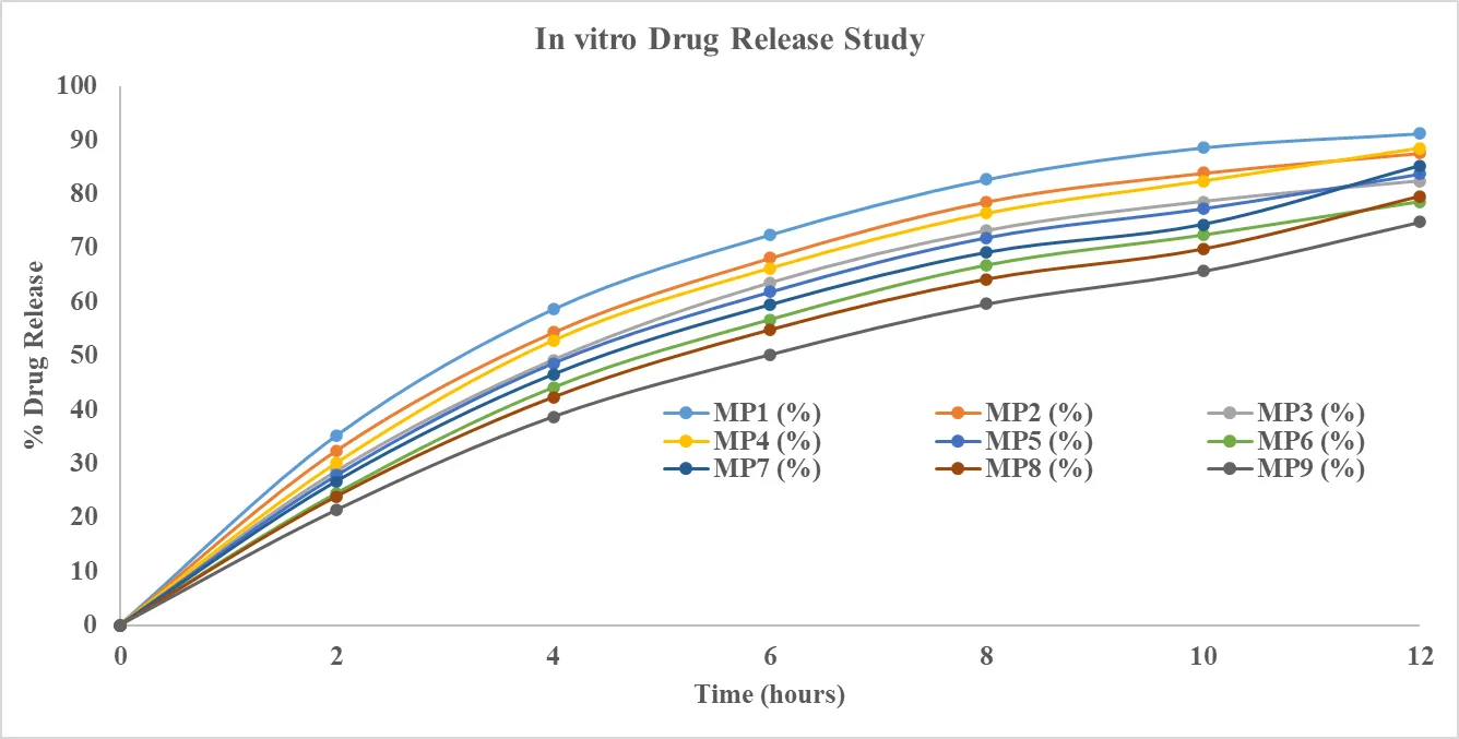
Figure 1: In vitro Drug Release Study |
|||||||||
3.6 Stability Study
The stability of the optimized formulation MP9 (Tobramycin sodium microspheres prepared with chitosan and Eudragit S100 in a 1:1 ratio, 300 mg polymer concentration) was evaluated under accelerated conditions (40 ± 2 °C and 75 ± 5% RH) for three months in accordance with ICH guidelines. The formulation was monitored for physical appearance, particle size, entrapment efficiency, drug content, and in vitro release profile. MP9 retained its spherical morphology and smooth surface without aggregation or collapse, while particle size showed only minor variations, confirming structural integrity. Entrapment efficiency remained stable with negligible reduction, and drug content loss was minimal (<2%), indicating good chemical stability. The in vitro release profile was consistent with initial values, maintaining sustained release up to 12 hours with cumulative release of 74.8 ± 1.8% at the end of the study. Overall, MP9 demonstrated excellent stability, validating its suitability for long-term storage and therapeutic application in controlled drug delivery.
Table 7: Stability Study of the Optimized Formulation (MP9)
|
Parameter |
Initial (0 Month) |
After 1 Month |
After 2 Months |
After 3 Months |
|
Physical Appearance |
Smooth, spherical |
No change |
No change |
No change |
|
Particle Size (µm, mean ± SD) |
132.8 ± 3.7 |
133.2 ± 3.8 |
133.6 ± 3.9 |
134.1 ± 4.0 |
|
Entrapment Efficiency (%) |
88.2 ± 2.8 |
87.9 ± 2.7 |
87.5 ± 2.7 |
87.1 ± 2.6 |
|
Drug Content (%) |
99.1 ± 1.5 |
98.6 ± 1.4 |
98.3 ± 1.4 |
97.8 ± 1.3 |
|
In Vitro Drug Release at 12 h (%) |
74.8 ± 1.8 |
74.5 ± 1.8 |
74.3 ± 1.7 |
74.1 ± 1.7 |
CONCLUSION
The present study successfully demonstrated the development and evaluation of microsphere-loaded mucoadhesive gel formulations of Tobramycin sodium, highlighting the critical role of polymer type and concentration in determining physicochemical properties, drug encapsulation, release kinetics, and stability. Microspheres prepared with Eudragit S100,
chitosan, and their combination exhibited spherical morphology, smooth surfaces, and narrow particle size distribution, confirming the reproducibility of the fabrication process. Entrapment efficiency was significantly influenced by polymer composition, with dual-polymer systems (chitosan + Eudragit S100) achieving superior drug loading capacity due to synergistic encapsulation effects. Swelling studies revealed enhanced hydration and matrix expansion at higher polymer concentrations, particularly in combination systems, which contributed to improved mucoadhesion and sustained drug release. Rheological evaluations confirmed that viscosity and spreadability were directly dependent on polymer type, with dual-polymer gels offering optimal balance between mechanical strength and ease of application. The pH values of all formulations remained within the physiological range, ensuring compatibility and non-irritancy for mucosal delivery. In vitro mucoadhesion studies further validated the adhesive potential of chitosan-based and combination systems, supporting prolonged residence time at mucosal sites. Drug release profiles demonstrated biphasic kinetics, with an initial burst followed by sustained release. Combination formulations, particularly MP9, provided the most controlled release, maintaining matrix integrity and prolonging drug delivery up to 12 hours. Stability studies conducted under accelerated conditions confirmed the robustness of MP9, with negligible changes in particle size, entrapment efficiency, drug content, and release behavior over three months. Overall, the findings establish that microsphere-loaded mucoadhesive gels, especially those prepared with dual-polymer systems, represent a promising platform for controlled drug delivery. The optimized formulation (MP9) demonstrated excellent stability, reproducibility, and therapeutic potential, validating its suitability for long-term storage and clinical application. This work contributes to advancing novel drug delivery systems that enhance patient compliance, therapeutic efficacy, and sustained release performance.
Conflict of Interest
None
REFERENCES
F. Differential stereoselective ofmetoprolol. Biochem. Pharmacol. Ther., 1983, 307,732-737.
F. Differential stereoselective ofmetoprolol. Biochem. Pharmacol. Ther., 1983, 307,732-737.

Vishal Edake, Dr. Mohd Hasib Ahmed, R. R. Pagore, K. R. Biyani, Development And Evaluation of Microsphere Loaded Mucoadhesive Gel, Int. J. of Pharm. Sci., 2026, Vol 4, Issue 7, 3866-3875, https://doi.org/10.5281/zenodo.21451142
 10.5281/zenodo.21451142