

We use cookies to ensure our website works properly and to personalise your experience. Cookies policy
Shambhunath Institute of Pharmacy, Jhalwa Prayagraj.
The present study is focused on development and evaluation of Tapentadol loaded nanosphere by using solvent evaporation method to improve drug encapsulation and increase sustained drug release. Nanospheres were prepared using Eudragit RS-100. UV spectrophotometric analysis confirms the drug purity. SEM analysis shows the spherical particle with smooth surface. EE% ranges from 70.8 to 79.2. Percentage yield of nanosphere was 82%. DLS shows the particle mostly below 260nm. Zeta potential was obtained -30.1mV. F2 formulation shows the best in-vitro release
Nano drug delivery systems (NDDS) are one of the research areas. Nanotechnology is the science of building up individual molecules, atoms or compounds into structures to produce new materials with new properties and is the spectacular development that has helped drug delivery.[1] It is the study of tiny structures that range in size from 1 to 100 nanometers.[2] Less variation in plasma levels (and therefore less side effects) will occur due to the longer half-life of the medication in the bloodstream as a result of nano DDS. These Nano DDS contain different kinds of nanocarriers ranging from solid lipid nanoparticles to micelles, nanogels, dendrimers, polymersomes, liposomes, carbon nanotubes, nanocrystals, silica nanoparticles, nanocapsules and nanospheres.[3][4]
Tapentadol IP is a novel centrally acting opioid, with noradrenalin reuptake inhibition and µ-opioid receptor agonistic properties. Due to its dual mode of action, it is an effective analgesic in the treatment of acute, chronic and neuropathic pain.[5] When compared to non-steroidal anti-inflammatory medicines (NSAIDs), its adverse effect profile is better. But, a high first pass digestion (68%) makes Tapentadol poorly bioavailable. Nadkarni reports that about 97% of the molecule is metabolized and none of the metabolites has an analgesic effect. Moreover, Tapentadol’s short half-life (4 hours) and fast elimination from the body because of the clearance (1530±177 ml/min) result in low patient compliance. Due to its hydrophilic nature and low oral bioavailability, less is able to permeate the blood-brain barrier (BBB) and enter the central nervous system (CNS) where it has its effect. Moreover, it has a variety of negative side effects such as constipation, drowsiness, severe nausea, stomach disorder and hallucinations etc. due to its widespread dispersion in the body.[6]
The main objective of formulation of nanosphere:
MATERIALS
Tapentadol IP was obtained from Digital Vision, H.P. as a gift sample. Eudragit RS-100, Polyvinyl Alcohol, Ethyl Acetate and Ethanol was procured from college lab.
METHOD
Preparation of Tapentadol IP loaded nanosphere:
The nanosphere was prepared by using emulsion solvent evaporation method in which Tapentadol IP was dissolved in ethanol and Eudragit RS-100 was dissolved in 10 ml of ethyl acetate with continuous stirring at magnetic stirrer at room temperature until clear solution was formed. After that both these mixture are mixed by using micropipette to form a primary emulsion, put the emulsion on probe sonicator for 15 minutes for the stable emulsion formation (Pulse mode sonication). This emulsion was then added drop wise in the 25 ml of 2% PVA with a proper mixing. Then the obtained solution was homogenised by homogenizer for about 15 minutes at 12000rpm. After this the solution was kept aside for 4-6 hrs for solvent evaporation at room temp. Centrifuge the solution by using cold centrifugation for 15 min at 15000 rpm followed by washing and drying using freeze dryer. Then the nanospheres was collected and stored in a closed glass container.[6]
UV Spectroscopy of Tapentadol IP in Ethanol:
Tapentadol hydrochloride exhibits a characteristic maximum absorption value of UV spectrophotometric analysis (λmax) in a suitable solvent at around 271–272 nm. A simple, accurate and reliable UV method can be applied following Beer-Lambert's Law over a valid concentration range.[7]
Characterization of Drug Loaded Nanosphere:
Scanning Electron Microscopy (SEM): Form and surface properties of the nanospheres produced were determined with scanning electron microscopy (SEM). The nanospheres are diluted in water, applied to the glass slide and allowed to dry at room temperature (25°C) to form the thin layer. The size and shape were determined by scanning electron microscopy (SEM) and the results were obtained.[8]
Zeta Potential: It is used to measure the electrical potential and surface charge characteristics of nanospheres. Both the dispersion media and the particle's composition have an impact on them. Particle aggregation and charge stability are also measured with it. The zetasizer is used to determine it.[9]
EE%: The entrapment efficacy %EE of nanosphere, the formulation washed twice with distilled water to remove any surface absorb drug after that nanosphere was centrifuged at 7000rpm for 1 hour and the sphere and supernatant were separated. The absorbance of the entrapped drug was determined by UV visible spectrophotometer at 272nm.[10]
EE%=Total drug-Free drugTotal drug×100

Particle Size: The size of the particles plays an important role in the characterization of nanospheres, influencing drug loading, stability, cellular uptake, and release behavior. Dynamic light scattering (DLS) is a commonly used technique for determining the hydrodynamic diameter of nanoparticles. Generally, the smaller and more evenly distributed nanospheres exhibit improved therapeutic efficacy and bioavailability.[11]
PDI: A nanosphere formulation's homogeneity of particle size distribution is indicated by the polydispersity index (PDI). PDI values of < 0.3 are considered to be narrow with good formulation quality and stability. Lower PDI means a more consistent performance in delivering medication, due to less aggregation.[12]
FTIR: Using an FT-IR spectrophotometer, FT-IR analysis can be used to assess the chemical integrity and potential chemical interaction between medication and polymer.[13]
In-vitro release: An in vitro drug release study of drug loaded nanospheres can be performed using the dialysis bag diffusion method. Using this method, a dialysis membrane tube filled with phosphate buffer solution is kept at 37°C while being shaken at a speed of 120 rpm.[14]
RESULT & DISCUSSION
UV spectroscopy:
λmax of Tapentadol IP was obtained 272 nm under UV spectroscopy which is shown below:
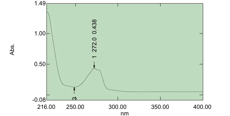
Figure: UV Spectroscopy Image of Tapentadol IP
Calibration Curve of Tapentadol IP
The absorbance data points underwent a linear regression analysis, which produced a straight line to aid in the determination of the medication amount based on linear equation. A regression value of 0.99 was obtained.
Table: Statistical data for calibration curve
|
S.No. |
Parameters |
Values |
|
1. |
λmax |
272nm |
|
2. |
Slope |
0.00862 |
|
3. |
R 2 |
0.99 |
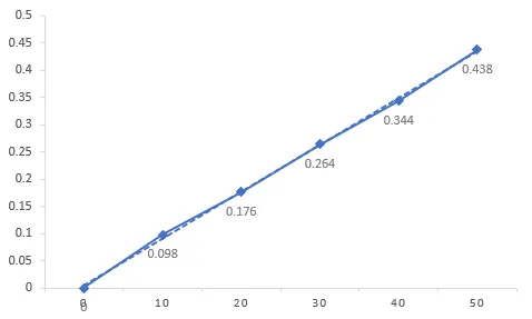
Figure: Calibration Curve of Tapentadol IP
Surface Morphology:
Using scanning electron microscope surface morphology of the Tapentadol IP nanosphere formulation was assessed. Using double-sided sticky tape, the sample was immediately placed onto the SEM sample holder, and scanning electron microscopy pictures were captured at 11mm x 3000 SE magnifications at an acceleration voltage of 15 kV. The nanosphere's SEM picture is displayed in below figure.
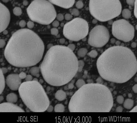
Figure: SEM of Tapentadol IP nanosphere
Percentage Drug Entrapment Efficacy:
The nanospheres had entrapment efficacy between 70.8 to 79.2. The highest drug entrapment efficacy was 79.2.
Percentage Yield:
The reported percentage yield was obtained 82% which shows that the nanosphere contains a good amount of medicament.
F2 selected as optimized batch.
Particle size:
The Dynamic light scattering (DLS) method was used to analyse the mean particle size of nanospheres. These nanospheres minimum average diameter were reported to 203 nm & maximum average diameter to 254 nm.
Particle Size Analysis (DLS):
Table: Data of particle size of different batches
|
Batch |
Trial 1 (nm) |
Trial 2 (nm) |
Trial 3 (nm) |
Mean ± SD |
|
F1 |
208 |
215 |
219 |
214 ± 5.57 |
|
F2 |
242 |
251 |
254 |
249 ± 6.24 |
|
F3 |
203 |
210 |
220 |
211 ± 8.54 |
PDI:
Table: Data of Polydispersity Index
|
Batch |
PDI |
|
F1 |
0.24 |
|
F2 |
0.29 |
|
F3 |
0.18 |
Table: Data of Polydispersity Index
Particle size increased with increase in polymer concentration. PDI <0.3 confirms uniform distribution.
Zeta Potential
The majority of the nanosphere particles in the formulation had this charge, as seen by the plot's peak at -30.1 mV, indicating a strong affinity between the particles.
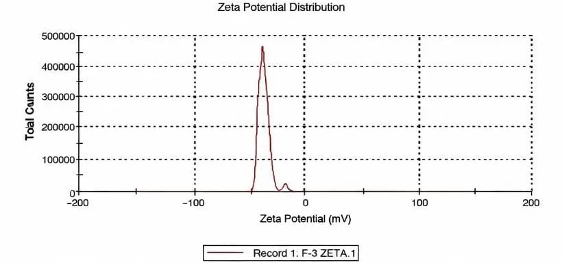
Figure: Zeta potential of Tapentadol nanosphere
FTIR study:
According to FTIR spectroscopy analysis, showed that the presence of characteristics functional group in the sample and there is no significant chances in characteristics peak of Tapentadol, indicates the chemical stability and absence of drug-polymer interaction.The characteristics peak of Tapentadol -OH/ -NH, aromatic, C-N are present. Peak 3452.84 cm-1 shows the presence of O-H & N-H stretching, 1635.32 cm-1 shows the aromatic C=C / N-H blending, 1384.34 cm-1 indicates C-N stretching/ CH3 blending & 539.75 cm-1 shows the aromatic ring deformation, these peaks confirms the compatibility of Tapentadol with PVA and Eudragit RS-100 and Tapentadol is stable in nanosphere with these polymers.
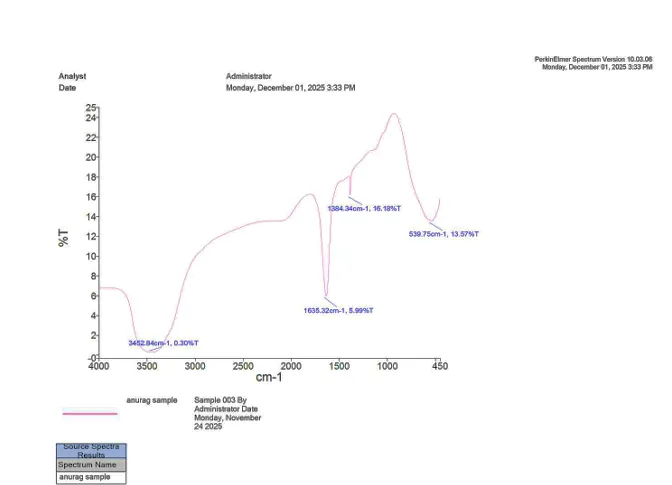
Figure: FTIR of Tapentadol IP loaded nanosphere
In-vitro Drug release:
In an in-vitro dissolution test, Tapentadol loaded nanosphere released the medication for upto 08 hrs. The drug release increased in proportion to time, indicating that the medication will remain in body for a longer period of time and release continuously over time.
Medium: pH 6.8 Phosphate Buffer
Speed: 100 rpm
Temp: 37±0.5°C
Dissolution Data
Table: Statistical data of dissolution study
|
Time (Hrs) |
F1 (%) |
F2(%) |
F3(%) |
|
1 |
24 |
14 |
27 |
|
2 |
38 |
26 |
41 |
|
4 |
53 |
46 |
57 |
|
6 |
67 |
62 |
69 |
|
8 |
79 |
75 |
78 |
Table: Statistical table of F2 for % drug release
|
Time |
% Drug Release |
|
0 |
0 |
|
1 |
14 |
|
2 |
26 |
|
4 |
46 |
|
6 |
62 |
|
8 |
75 |

Figure: Graph of F2 drug release
CONCLUSION
Tapentadol-loaded nanospheres were successfully developed using the solvent evaporation method with Eudragit RS-100. The prepared formulation exhibited satisfactory particle size, high entrapment efficiency, good stability, and sustained drug release characteristics. Among all formulations, F2 demonstrated the most favorable release profile and physicochemical properties. The findings suggest that the developed nanosphere system may serve as a promising approach for prolonged delivery of Tapentadol and improved therapeutic effectiveness.
REFERENCES


Anurag Dwivedi, Sagar Bansal, Sandeep Pandey, Mohammad Sarfraz, Development and Evaluation of Tapentadol Nanospheres by using Solvent Evaporation Method, Int. J. of Pharm. Sci., 2026, Vol 4, Issue 7, 3847-3854, https://doi.org/10.5281/zenodo.21450959
 10.5281/zenodo.21450959