
We use cookies to ensure our website works properly and to personalise your experience. Cookies policy
Tataysaheb kore collage of pharmacy warnanagar.
Background: Metabolic syndrome (MetS) is a multifactorial metabolic disorder characterized by obesity, dyslipidemia, insulin resistance, hyperglycemia, and oxidative stress, leading to an increased risk of cardiovascular diseases and type 2 diabetes mellitus. Polyherbal formulations have gained considerable attention due to their multi-target therapeutic potential and favorable safety profile.Objective: The present study aimed to evaluate the anti-metabolic syndrome activity of a polyherbal suspension containing hydroethanolic extracts of Tecomella undulata stem bark and Camellia sinensis leaves in high-fat diet (HFD)-induced metabolic syndrome in Wistar rats.Methods: Hydroethanolic extracts of T. undulata and C. sinensis were formulated into a polyherbal suspension. Metabolic syndrome was induced in male Wistar rats by feeding a high-fat diet for eight weeks. Animals were treated with the polyherbal formulation at doses of 200 mg/kg and 400 mg/kg for four weeks. Orlistat (10 mg/kg) served as the standard drug. Body weight, adiposity indices, serum lipid profile, fasting blood glucose, oral glucose tolerance, hepatic and renal biochemical markers, oxidative stress parameters, and histopathological changes were evaluated.Results: HFD-fed rats exhibited significant weight gain, dyslipidemia, hyperglycemia, oxidative stress, and hepatic steatosis. Treatment with the polyherbal formulation significantly reduced body weight, total cholesterol, triglycerides, LDL-cholesterol, fasting blood glucose, and lipid peroxidation levels while increasing HDL-cholesterol and endogenous antioxidant enzyme activities. The formulation also improved oral glucose tolerance, hepatic and renal function markers, and attenuated histopathological alterations in liver tissue. The higher dose (400 mg/kg) demonstrated effects comparable to the standard drug, orlistat.Conclusion: The polyherbal suspension of Tecomella undulata and Camellia sinensis exhibited significant protective effects against HFD-induced metabolic syndrome through anti-obesity, antihyperlipidemic, antihyperglycemic, antioxidant, and hepatoprotective mechanisms. These findings suggest its potential as a promising herbal therapeutic approach for the management of metabolic syndrome.
Metabolic syndrome (MetS) is a complex cluster of interconnected metabolic abnormalities that includes central obesity, dyslipidemia, hyperglycemia, insulin resistance, and hypertension. It has emerged as one of the most significant global public health challenges due to its increasing prevalence and strong association with cardiovascular diseases, type 2 diabetes mellitus (T2DM), non-alcoholic fatty liver disease (NAFLD), and premature mortality. According to the World Health Organization (WHO), the prevalence of metabolic syndrome has increased dramatically over the past few decades as a result of rapid urbanization, sedentary lifestyles, excessive caloric intake, and unhealthy dietary habits. The coexistence of these metabolic disturbances significantly increases the risk of morbidity and mortality worldwide.
Insulin resistance and visceral obesity are considered the primary pathological mechanisms involved in the development of metabolic syndrome. Excessive accumulation of adipose tissue leads to the release of free fatty acids, pro-inflammatory cytokines, and reactive oxygen species (ROS), resulting in chronic low-grade inflammation and oxidative stress. These pathological changes contribute to impaired glucose metabolism, abnormal lipid profiles, endothelial dysfunction, and hepatic steatosis. Consequently, the management of metabolic syndrome requires therapeutic interventions capable of targeting multiple metabolic pathways simultaneously.
Current treatment strategies primarily focus on lifestyle modifications and pharmacological management of individual components of metabolic syndrome. Drugs such as statins, metformin, antihypertensive agents, and anti-obesity medications are commonly prescribed. However, long-term use of these medications is often associated with adverse effects, high treatment costs, poor patient compliance, and polypharmacy-related complications. Furthermore, no single synthetic drug effectively addresses all pathological aspects of metabolic syndrome. Therefore, there is a growing interest in exploring alternative therapeutic approaches that are safe, cost-effective, and capable of exerting multi-targeted pharmacological actions.
Medicinal plants have been widely utilized in traditional systems of medicine for the prevention and treatment of metabolic disorders. Polyherbal formulations, which combine two or more medicinal plants, have gained considerable attention due to their synergistic therapeutic effects. The combination of multiple herbs allows simultaneous modulation of different pathological pathways while minimizing toxicity and enhancing overall efficacy. Several phytoconstituents, including flavonoids, polyphenols, alkaloids, terpenoids, and saponins, have demonstrated significant antioxidant, anti-inflammatory, antihyperlipidemic, and antihyperglycemic activities, making them promising candidates for the management of metabolic syndrome.
Tecomella undulata (Sm.) Seem., commonly known as Rohida or Desert Teak, belongs to the family Bignoniaceae and is widely distributed in the arid regions of India. Traditionally, the plant has been used for the treatment of liver disorders, obesity, diabetes, inflammation, and various metabolic diseases. Phytochemical investigations have revealed the presence of naphthoquinones, flavonoids, iridoid glycosides, phenolic compounds, and triterpenoids that possess potent antioxidant, hepatoprotective, hypolipidemic, and antidiabetic activities. Previous studies have reported that extracts of T. undulata effectively reduce serum lipid levels, improve glucose metabolism, and protect against oxidative stress-induced tissue damage.
Camellia sinensis (L.) Kuntze, commonly known as green tea, is one of the most widely consumed beverages worldwide. It is rich in catechins, particularly epigallocatechin gallate (EGCG), which exhibits strong antioxidant and anti-inflammatory properties. Numerous experimental and clinical studies have demonstrated the beneficial effects of C. sinensis in obesity, dyslipidemia, insulin resistance, and cardiovascular disorders. Green tea polyphenols have been reported to enhance lipid metabolism, reduce body weight gain, improve insulin sensitivity, and suppress oxidative stress through multiple molecular mechanisms. Additionally, catechins modulate key metabolic signaling pathways, including AMP-activated protein kinase (AMPK), thereby contributing to improved metabolic homeostasis.
The combination of Tecomella undulata and Camellia sinensis is expected to provide complementary pharmacological actions against metabolic syndrome. While T. undulata primarily contributes hepatoprotective, hypolipidemic, and antidiabetic effects, C. sinensis offers potent antioxidant, anti-obesity, and insulin-sensitizing activities. The synergistic interaction between these medicinal plants may result in enhanced therapeutic efficacy through simultaneous modulation of multiple pathogenic mechanisms involved in metabolic syndrome.
High-fat diet (HFD)-induced metabolic syndrome in Wistar rats is a well-established experimental model that closely mimics the pathological features observed in humans. Prolonged consumption of a high-fat diet leads to obesity, dyslipidemia, hyperglycemia, insulin resistance, oxidative stress, and hepatic steatosis, making it an ideal model for evaluating potential therapeutic interventions. Therefore, the present study was designed to investigate the protective effects of a polyherbal suspension containing hydroethanolic extracts of Tecomella undulata and Camellia sinensis against HFD-induced metabolic syndrome in Wistar rats. The study aimed to evaluate its effects on body weight, lipid profile, glucose metabolism, hepatic and renal function, oxidative stress biomarkers, and histopathological alterations. The findings of this investigation may provide scientific evidence supporting the development of a safe and effective herbal therapeutic approach for the management of metabolic syndrome.
MATERIALS AND METHODS
Plant Materials
The stem bark of Tecomella undulata (Sm.) Seem. and leaves of Camellia sinensis (L.) Kuntze were procured from authenticated herbal suppliers. The plant materials were cleaned, shade-dried, powdered, and stored in airtight containers until further use.
Chemicals and Reagents
All chemicals and reagents used in the study were of analytical grade. Ethanol, sodium carboxymethyl cellulose (Na-CMC), Tween 80, methyl paraben, propyl paraben, and other laboratory reagents were procured from authorized suppliers. Commercial diagnostic kits were used for biochemical estimations.
Table 1. Chemicals, Reagents and Diagnostic Kits Used in the Study
|
Reagent / Material |
Source |
|
Ethanol (95% v/v) AR |
Merck Specialities, Mumbai, India |
|
Methanol HPLC Grade |
Merck Specialities, Mumbai, India |
|
Sodium Carboxymethyl Cellulose (Na-CMC) |
Loba Chemie Pvt. Ltd., Mumbai, India |
|
Tween 80 (Polysorbate 80) |
SD Fine-Chem Ltd., Mumbai, India |
|
Methyl Paraben and Propyl Paraben |
SD Fine-Chem Ltd., Mumbai, India |
|
Ascorbic Acid (L-Ascorbic Acid) |
Sigma-Aldrich, Bengaluru, India |
|
Citric Acid / Sodium Citrate Buffer |
SD Fine-Chem Ltd., Mumbai, India |
|
Folin–Ciocalteu Reagent |
Merck Specialities, Mumbai, India |
|
Aluminium Chloride (Anhydrous) |
SD Fine-Chem Ltd., Mumbai, India |
|
Gallic Acid Standard |
Sigma-Aldrich, Bengaluru, India |
|
Quercetin Standard |
Sigma-Aldrich, Bengaluru, India |
|
Catechin Standard |
Sigma-Aldrich, Bengaluru, India |
|
Epigallocatechin Gallate (EGCG, ≥95% HPLC) |
Sigma-Aldrich, Bengaluru, India |
|
Lapachol Reference Standard |
Sigma-Aldrich, Bengaluru, India |
|
DPPH, ABTS and FRAP Reagents |
Sigma-Aldrich, Bengaluru, India |
|
Orlistat (Standard Drug) |
Cipla Ltd., Mumbai, India |
|
Cholesterol |
Sigma-Aldrich, Bengaluru, India |
|
Cholic Acid |
Sigma-Aldrich, Bengaluru, India |
|
Lard / Vanaspati (for HFD) |
Local Procurement (Food Grade) |
|
Casein |
Nutrilab India Pvt. Ltd., Pune, India |
|
Mineral and Vitamin Mix (AIN-93) |
Nutrilab India Pvt. Ltd., Pune, India |
|
Lipid Profile Diagnostic Kits |
Erba Mannheim Diagnostics, India |
|
Liver Function Test (LFT) Kits |
Erba Mannheim Diagnostics, India |
|
Renal Function Test (RFT) Kits |
Erba Mannheim Diagnostics, India |
|
Glucose Estimation Kits |
Span Diagnostics Ltd., Surat, India |
|
Insulin ELISA Kit (Rat Specific) |
Krishgen Biosystems, Mumbai, India |
|
TNF-α, IL-6 and CRP ELISA Kits |
Krishgen Biosystems / Diaclone, India |
|
MDA Assay Reagents |
Cayman Chemical, USA |
|
GSH Assay Reagents |
SRL Pvt. Ltd., Mumbai, India |
|
SOD Assay Reagents |
SRL Pvt. Ltd., Mumbai, India |
|
CAT Assay Reagents |
SRL Pvt. Ltd., Mumbai, India |
|
10% Neutral Buffered Formalin |
In-house Preparation |
|
Hematoxylin Stain |
SRL Pvt. Ltd., Mumbai, India |
|
Eosin Stain |
Loba Chemie Pvt. Ltd., Mumbai, India |
Experimental Animals
Healthy adult male Wistar rats weighing 180–220 g were obtained from a registered animal house facility. Animals were housed under standard laboratory conditions (temperature 25 ± 2°C, relative humidity 55 ± 5%, and 12 h light/dark cycle) with free access to standard pellet diet and water. The experimental protocol was approved by the Institutional Animal Ethics Committee (IAEC) and conducted according to CPCSEA guidelines.
Table 2. Instruments and Equipment Used in the Study
|
Instrument / Equipment |
Make / Model |
|
Analytical Balance (0.1 mg Readability) |
Mettler Toledo / Sartorius |
|
Soxhlet Extraction Apparatus and Glassware |
Borosil Glass Works Ltd., Mumbai, India |
|
Rotary Vacuum Evaporator |
Buchi R-100 (Switzerland) / Rotavac, IKA |
|
Vacuum Oven / Hot Air Oven |
Macro Scientific Works, Delhi, India |
|
UV–Visible Spectrophotometer |
Shimadzu UV-1900 / UV-1800 |
|
HPLC System with Photodiode Array Detector |
Shimadzu LC-2030C 3D / Waters |
|
HPTLC System |
CAMAG, Switzerland |
|
Digital pH Meter |
Eutech Instruments / Hanna Instruments |
|
Brookfield Viscometer |
Brookfield LVDV-II+ Pro, USA |
|
Optical Microscope with Eyepiece Micrometer |
Olympus CX-23 / Nikon |
|
Zeta Sizer / Dynamic Light Scattering Analyzer |
Malvern Zetasizer Nano ZS |
|
Refrigerated Cooling Centrifuge |
REMI C-24BL / Eppendorf 5810R |
|
ELISA Microplate Reader |
BioTek Epoch / Thermo Multiskan |
|
Automated Biochemistry Analyzer |
Erba Chem-7 / Transasia XL-180 |
|
Glucometer |
Roche Diagnostics Accu-Chek Active Strips |
|
Non-Invasive Blood Pressure Analyzer |
IITC Life Science / BIOPAC Systems |
|
Rotary Microtome |
Leica RM-2125 / Erma Inc. |
|
Oral Gavage Needles and Animal Handling Tools |
Standard Laboratory Equipment |
Preparation of Hydroethanolic Extracts
The powdered plant materials were subjected to hydroethanolic extraction using ethanol:water (70:30 v/v). The extraction process was carried out by maceration with intermittent shaking for 72 h. The extracts were filtered and concentrated under reduced pressure using a rotary evaporator. The concentrated extracts were further dried and stored in airtight containers for further studies.

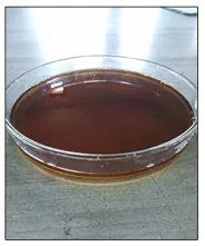
Fig No. 1: Hydro-ethanolic Extraction
Preliminary Phytochemical Screening
The hydroethanolic extracts of Tecomella undulata and Camellia sinensis were subjected to preliminary qualitative phytochemical screening for the detection of alkaloids, flavonoids, phenolics, tannins, saponins, glycosides, terpenoids, and carbohydrates using standard procedures.
Table 3. Qualitative Phytochemical Screening Tests Applied
|
Phytoconstituent Class |
Qualitative Tests Applied |
|
Alkaloids |
Mayer’s Test, Dragendorff’s Test, Wagner’s Test, Hager’s Test |
|
Flavonoids |
Shinoda Test (Mg + Conc. HCl), Alkaline Reagent Test, Lead Acetate Test |
|
Tannins and Phenolic Compounds |
Ferric Chloride Test, Gelatin Test, Lead Acetate Test |
|
Saponins |
Foam Test, Haemolytic Test |
|
Cardiac Glycosides |
Keller–Killiani Test, Legal’s Test |
|
Anthraquinone Glycosides |
Borntrager’s Test, Modified Borntrager’s Test |
|
Steroids and Triterpenoids |
Liebermann–Burchard Test, Salkowski Test |
|
Carbohydrates and Reducing Sugars |
Molisch’s Test, Fehling’s Test, Benedict’s Test |
|
Proteins and Amino Acids |
Biuret Test, Ninhydrin Test |
|
Quinones and Naphthoquinones |
Sulphuric Acid Test, Sodium Hydroxide (NaOH) Test |
|
Iridoids |
Trim–Hill Test |
|
Fixed Oils and Fats |
Spot Test, Saponification Test |
Determination of Total Phenolic Content (TPC)
The total phenolic content of the extracts was determined using the Folin–Ciocalteu reagent method. Gallic acid was used as the standard, and the results were expressed as milligrams of gallic acid equivalents (mg GAE/g extract).
Determination of Total Flavonoid Content (TFC)
The total flavonoid content was estimated using the aluminum chloride colorimetric method. Quercetin was used as the reference standard, and the results were expressed as milligrams of quercetin equivalents (mg QE/g extract).
In-vitro Antioxidant Activity
The antioxidant activity of the extracts was evaluated using the DPPH free radical scavenging assay. Different concentrations of extracts were tested, and percentage inhibition was calculated in comparison with the control.
Formulation of Polyherbal Suspension
The polyherbal suspension was prepared by incorporating hydroethanolic extracts of Tecomella undulata and Camellia sinensis into an aqueous vehicle containing sodium carboxymethyl cellulose as a suspending agent. Tween 80 was used as a wetting agent, while methyl paraben and propyl paraben were incorporated as preservatives. The formulation was mixed thoroughly to obtain a homogeneous suspension.
Table 4. Composition of Polyherbal Suspension
|
Component |
Functional Role |
Quantity |
|
Tecomella undulata Extract |
Active Ingredient |
q.s. (per dose) |
|
Camellia sinensis Extract |
Active Ingredient |
q.s. (per dose) |
|
Sodium Carboxymethyl Cellulose (Na-CMC) |
Suspending Agent |
1.0% w/v |
|
Tween 80 |
Wetting Agent |
0.5% w/v |
|
Methyl Paraben |
Preservative |
0.18% w/v |
|
Propyl Paraben |
Preservative |
0.02% w/v |
|
Citric Acid / Sodium Citrate |
Buffering Agent (pH 4.5) |
q.s. |
|
Ascorbic Acid |
Antioxidant Stabilizer |
0.1% w/v |
|
Orange Syrup |
Flavouring Agent |
5.0% v/v |
|
Stevia Extract |
Natural Sweetener |
0.05% w/v |
|
Distilled Water |
Vehicle |
q.s. to 100 mL |
Evaluation of Polyherbal Suspension
The prepared suspension was evaluated for the following parameters:
Organoleptic Characteristics
Colour, odour, appearance, and homogeneity were evaluated visually.
pH Determination
The pH of the formulation was measured using a calibrated digital pH meter.
Particle Size Analysis
Particle size distribution was determined using a particle size analyzer.
Zeta Potential
Zeta potential was measured to assess the physical stability of the formulation.
Acute Oral Toxicity Study
Acute oral toxicity was performed according to OECD Guideline 423. Female Wistar rats were administered a single oral dose of the polyherbal formulation and observed for signs of toxicity, behavioural changes, and mortality for 14 days.
Induction of Metabolic Syndrome
Metabolic syndrome was induced by feeding rats with a high-fat diet (HFD) continuously for eight weeks. The high-fat diet consisted of standard pellet diet supplemented with cholesterol, lard, and other dietary components capable of inducing obesity, dyslipidemia, and insulin resistance.
Experimental Design
Animals were randomly divided into five groups (n = 6):
Group I: Normal Control (Standard Diet)
Group II: HFD Control
Group III: HFD + Orlistat (10 mg/kg)
Group IV: HFD + Polyherbal Formulation Low Dose (200 mg/kg)
Group V: HFD + Polyherbal Formulation High Dose (400 mg/kg)
Following induction of metabolic syndrome, treatment was continued for four weeks.
Table 5. Composition of High-Fat Diet (HFD)
|
Ingredient |
Composition (% w/w) |
Functional Role |
|
Lard (Saturated Fat Source) |
32.0 |
Primary Fat Source |
|
Soybean Oil |
3.0 |
Essential Fatty Acids (PUFA Source) |
|
Casein |
20.0 |
Protein Source |
|
Sucrose / Corn Starch |
15.0 |
Carbohydrate Source |
|
Maltodextrin |
5.0 |
Carbohydrate Source |
|
Cellulose (Fibre) |
5.0 |
Dietary Fibre / Bulk Agent |
|
Cholesterol |
1.0 |
Cholesterol Supplementation |
|
Cholic Acid |
0.25 |
Bile Acid Supplementation |
|
AIN-93M Vitamin Mix |
1.0 |
Vitamin Source |
|
AIN-93M Mineral Mix |
3.5 |
Mineral Source |
|
DL-Methionine |
0.30 |
Sulphur-Containing Amino Acid |
|
Choline Bitartrate |
0.25 |
Lipotropic Agent |
|
Vanaspati / Palm Oil |
13.7 |
Additional Saturated Fat Source |
|
Total |
100.0 |
~5.2 kcal/g; 60% kcal from Fat |
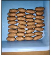
Fig No. 2: Prepared High-Fat Diet
Table 6. Experimental Design and Animal Grouping
|
Group |
Diet |
Treatment |
Number of Animals |
|
Group I – Normal Control (NC) |
Standard Pellet Diet ad libitum |
0.5% Na-CMC Vehicle, 2 mL/kg, p.o., Once Daily |
n = 6 |
|
Group II – HFD Control |
High-Fat Diet ad libitum |
0.5% Na-CMC Vehicle, 2 mL/kg, p.o., Once Daily |
n = 6 |
|
Group III – HFD + Standard (Orlistat) |
High-Fat Diet ad libitum |
Orlistat 10 mg/kg, p.o., Once Daily |
n = 6 |
|
Group IV – HFD + PHF Low Dose (PHF-LD) |
High-Fat Diet ad libitum |
Polyherbal Suspension 200 mg/kg, p.o., Once Daily |
n = 6 |
|
Group V – HFD + PHF High Dose (PHF-HD) |
High-Fat Diet ad libitum |
Polyherbal Suspension 400 mg/kg, p.o., Once Daily |
n = 6 |
Evaluation Parameters
Body Weight Measurement
Body weight of animals was recorded weekly throughout the experimental period.
Assessment of Visceral Fat and Adiposity Index
At the end of the study, visceral fat was collected and weighed. Adiposity index and Lee index were calculated.
Serum Lipid Profile
Blood samples were collected, and serum was analyzed for:
Fasting Blood Glucose
Fasting blood glucose levels were measured using standard biochemical methods.
Oral Glucose Tolerance Test (OGTT)
OGTT was performed after overnight fasting. Glucose solution was administered orally, and blood glucose levels were recorded at predetermined intervals.
Liver Function Tests
Serum levels of:
were determined using diagnostic kits.
Renal Function Tests
The following renal biomarkers were estimated:
Assessment of Oxidative Stress Markers
Liver homogenates were analyzed for:
Relative Organ Weight
Relative liver weight was calculated at the end of the study.
Histopathological Examination
Liver tissues were fixed in 10% formalin, processed, stained with hematoxylin and eosin (H&E), and examined under a light microscope for histopathological changes.
Statistical Analysis
Data were expressed as Mean ± SEM. Statistical analysis was performed using one-way analysis of variance (ANOVA) followed by Tukey’s multiple comparison test. A value of p < 0.05 was considered statistically significant.
RESULTS AND DISCUSSION
Extractive Yield of Plant Materials
Hydroethanolic extraction of Tecomella undulata stem bark and Camellia sinensis leaves yielded appreciable quantities of dried extracts. The extraction process effectively concentrated bioactive phytoconstituents, indicating the suitability of hydroethanolic solvent systems for extracting both polar and moderately non-polar compounds. The observed yield may be attributed to the presence of phenolics, flavonoids, glycosides, and other secondary metabolites in the selected plant materials.
Table 7. Extractive Yield of Hydroethanolic Extracts
|
Plant Material |
Solvent System |
Extraction Method |
Extractive Yield (% w/w) |
Appearance of Extract |
|
Tecomella undulata Stem Bark |
Hydroethanolic (70:30 v/v) |
Cold Maceration (72 h) |
11.42 ± 0.65 |
Dark Brown, Sticky Mass |
|
Camellia sinensis Leaves |
Hydroethanolic (70:30 v/v) |
Cold Maceration (72 h) |
24.86 ± 0.92 |
Greenish-Brown, Hygroscopic Mass |
Preliminary Phytochemical Screening
Qualitative phytochemical screening confirmed the presence of flavonoids, phenolic compounds, tannins, glycosides, saponins, and terpenoids in both extracts. These phytoconstituents are well known for their antioxidant, antihyperlipidemic, anti-inflammatory, and antidiabetic activities. The presence of these bioactive compounds supports the traditional medicinal use of both plants and provides a scientific basis for their evaluation against metabolic syndrome.
Table 8. Preliminary Qualitative Phytochemical Screening of Hydroethanolic Extracts
|
Phytoconstituent Class |
T. undulata Extract |
C. sinensis Extract |
|
Alkaloids |
– |
+++ |
|
Flavonoids |
+++ |
+++ |
|
Phenolics |
+++ |
+++ |
|
Tannins |
++ |
+++ |
|
Saponins |
++ |
++ |
|
Cardiac Glycosides |
– |
– |
|
Anthraquinone Glycosides |
– |
– |
|
Steroids |
++ |
+ |
|
Triterpenoids |
+++ |
+ |
|
Naphthoquinones |
+++ |
– |
|
Iridoids |
++ |
– |
|
Carbohydrates |
+ |
++ |
|
Proteins / Amino Acids |
+ |
++ |
|
Reducing Sugars |
+ |
++ |
|
Fixed Oils / Fats |
+ |
+ |
Note: (+++ = abundantly present, ++ = moderately present, + = present in traces, − = absent)
Total Phenolic and Flavonoid Content
The extracts exhibited considerable amounts of total phenolic and flavonoid contents. Phenolic compounds are recognized as potent antioxidants capable of scavenging free radicals and reducing oxidative stress. Similarly, flavonoids contribute to improved lipid metabolism, glucose homeostasis, and protection against inflammatory responses. The high phenolic and flavonoid content observed in the present study may have contributed significantly to the biological activity of the polyherbal formulation.
Table 9. Total Phenolic Content (TPC) and Total Flavonoid Content (TFC) of Hydroethanolic Extracts
|
Extract |
Total Phenolic Content (mg GAE/g Extract) |
Total Flavonoid Content (mg QE/g Extract) |
|
Tecomella undulata Extract |
138.5 ± 6.2 |
41.2 ± 2.1 |
|
Camellia sinensis Extract |
294.6 ± 8.4 |
87.3 ± 3.2 |
DPPH Free Radical Scavenging Activity
Both plant extracts demonstrated significant DPPH radical scavenging activity, indicating strong antioxidant potential. The antioxidant activity can be attributed to the presence of polyphenolic compounds, particularly catechins from Camellia sinensis and flavonoids present in Tecomella undulata. The ability of the formulation to neutralize free radicals may play an important role in reducing oxidative stress associated with metabolic syndrome.
Table 10. DPPH Free Radical Scavenging Activity of Hydroethanolic Extracts
|
Test Substance |
IC₅₀ (µg/mL) |
|
Ascorbic Acid (Standard) |
9.8 ± 0.4 |
|
Camellia sinensis Extract |
22.6 ± 1.1 |
|
Tecomella undulata Extract |
56.4 ± 2.3 |
Evaluation of Polyherbal Suspension
The prepared polyherbal suspension exhibited acceptable organoleptic characteristics, good homogeneity, satisfactory pH, and desirable physical stability. Particle size analysis and zeta potential studies indicated the formation of a stable suspension system suitable for oral administration. The formulation remained physically stable throughout the study period without significant sedimentation or phase separation.
Table 11. Physicochemical Evaluation of the Polyherbal Suspension
|
Parameter |
Observed Value |
Inference |
|
Colour |
Uniform Brown |
Acceptable |
|
Odour |
Characteristic, Herbal |
Acceptable |
|
Taste |
Mildly Bitter-Sweet |
Acceptable |
|
Appearance |
Homogeneous, No Grittiness or Caking |
Acceptable |
|
pH (25 ± 2°C) |
4.52 ± 0.05 |
Within Target Range (4.5 ± 0.2) |
|
Sedimentation Volume (F) at 24 h |
0.92 ± 0.02 |
Excellent Flocculation Behaviour |
|
Redispersibility (Number of Inversions) |
5–6 |
Acceptable (≤ 10 Inversions) |
|
Viscosity at 20 rpm (cP) |
480 ± 18 |
Pseudoplastic (Thixotropic) Flow Behaviour |
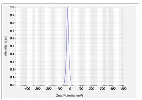
Fig. No. 3: Zeta Potential
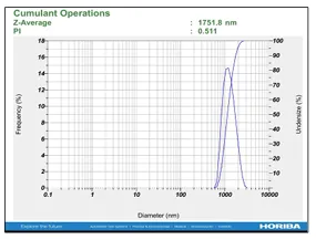
Fig. No. 4: Particle Size
Effect on Body Weight
Rats fed a high-fat diet showed a significant increase in body weight compared with the normal control group, confirming successful induction of obesity and metabolic syndrome. Treatment with the polyherbal formulation significantly reduced body weight gain in a dose-dependent manner. The higher dose group demonstrated a greater reduction in body weight, comparable to the standard drug orlistat.
The anti-obesity activity may be attributed to the ability of Camellia sinensis catechins to enhance energy expenditure, promote fatty acid oxidation, and inhibit lipid absorption, while Tecomella undulata contributes through modulation of lipid metabolism and antioxidant activity.
Table 12. Effect of Polyherbal Formulation on Body Weight of Wistar Rats
|
Group |
Day 0 (g) |
Week 8 (g) |
Week 12 (g) |
|
I – Normal Control |
198.5 ± 4.2 |
286.4 ± 5.8 |
298.7 ± 6.1 |
|
II – HFD Control |
196.8 ± 3.9 |
405.3 ± 7.6 |
428.6 ± 8.9 |
|
III – HFD + Orlistat (10 mg/kg) |
199.2 ± 4.6 |
407.8 ± 7.2 |
338.5 ± 6.8 |
|
IV – HFD + PHF-LD (200 mg/kg) |
197.6 ± 4.1 |
404.6 ± 7.9 |
372.4 ± 7.5 |
|
V – HFD + PHF-HD (400 mg/kg) |
198.4 ± 4.5 |
406.1 ± 7.4 |
348.2 ± 7.1 |
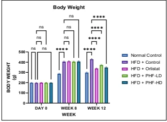
Fig. No. 5: Body Weight
Effect on Visceral Fat, Adiposity Index and Lee Index
A marked increase in visceral fat accumulation, adiposity index, and Lee index was observed in HFD-treated animals. Administration of the polyherbal formulation significantly reduced these obesity-related parameters. Reduction of visceral adiposity is particularly important because visceral fat is strongly associated with insulin resistance, inflammation, and cardiovascular complications.
The observed effect suggests that the formulation effectively prevented abnormal fat deposition and improved overall metabolic status.
Table 13. Effect of Polyherbal Formulation on Visceral Fat Mass, Adiposity Index and Lee Index
|
Group |
Visceral Fat (g) |
Adiposity Index (%) |
Lee Index |
|
I – Normal Control |
4.18 ± 0.21 |
1.42 ± 0.08 |
298.5 ± 4.6 |
|
II – HFD Control |
12.86 ± 0.58 |
3.00 ± 0.12 |
352.8 ± 5.3 |
|
III – HFD + Orlistat |
5.92 ± 0.32 |
1.75 ± 0.09 |
312.6 ± 4.9 |
|
IV – HFD + PHF-LD |
8.45 ± 0.41 |
2.27 ± 0.10 |
328.4 ± 5.1 |
|
V – HFD + PHF-HD |
6.42 ± 0.36 |
1.84 ± 0.10 |
316.8 ± 4.8 |
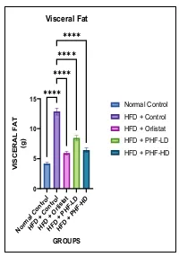
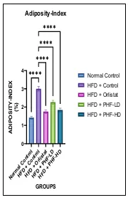
Fig. No. 6: Visceral Fat Fig. No. 7: Adiposity-index
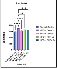
Fig. No. 8: Lee index
Effect on Serum Lipid Profile
High-fat diet feeding resulted in significant dyslipidemia characterized by elevated total cholesterol, triglycerides, and LDL-cholesterol levels with a reduction in HDL-cholesterol. Treatment with the polyherbal formulation significantly improved all lipid parameters.
The antihyperlipidemic activity may be explained by inhibition of cholesterol synthesis, enhancement of lipid catabolism, reduction of intestinal lipid absorption, and improved hepatic lipid metabolism. Green tea catechins have been reported to suppress HMG-CoA reductase activity and stimulate fatty acid oxidation, while Tecomella undulata possesses documented hypolipidemic activity.
Table 14. Effect of Polyherbal Formulation on Serum Lipid Profile in HFD-Induced Wistar Rats
|
Group |
TC (mg/dL) |
TG (mg/dL) |
HDL-C (mg/dL) |
LDL-C (mg/dL) |
|
I – Normal Control |
94.2 ± 3.6 |
85.4 ± 3.1 |
53.6 ± 1.9 |
23.5 ± 1.8 |
|
II – HFD Control |
198.6 ± 6.8 |
183.5 ± 5.7 |
24.2 ± 1.4 |
137.7 ± 4.9 |
|
III – HFD + Orlistat |
128.4 ± 4.9 |
112.3 ± 4.2 |
46.8 ± 1.7 |
59.1 ± 2.7 |
|
IV – HFD + PHF-LD |
162.8 ± 5.6 |
143.9 ± 4.8 |
36.4 ± 1.5 |
97.6 ± 3.8 |
|
V – HFD + PHF-HD |
134.6 ± 5.2 |
118.7 ± 4.4 |
45.2 ± 1.6 |
65.7 ± 2.9 |
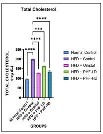
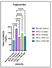
Fig. No. 9: Total Cholesterol Fig. No. 10: Triglycerides
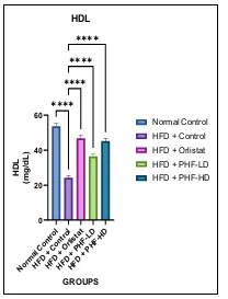
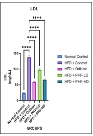
Fig. No. 11: HDL Fig. No. 12: LDL
Effect on Fasting Blood Glucose
The HFD control group showed significantly elevated fasting blood glucose levels compared with normal animals. Treatment with the polyherbal formulation significantly lowered blood glucose levels in a dose-dependent manner.
This effect may be associated with improved insulin sensitivity, enhanced glucose uptake, and reduced hepatic glucose production. Polyphenols and flavonoids present in the formulation may contribute to glucose regulation through modulation of insulin signaling pathways.
Table 15. Effect of Polyherbal Formulation on Fasting Blood Glucose Levels in HFD-Induced Wistar Rats
|
Group |
Day 0 (mg/dL) |
Week 8 (mg/dL) |
Week 12 (mg/dL) |
|
I – Normal Control |
88.4 ± 2.5 |
91.2 ± 2.7 |
91.6 ± 2.8 |
|
II – HFD Control |
89.6 ± 2.7 |
138.2 ± 4.2 |
143.8 ± 4.6 |
|
III – HFD + Orlistat |
90.2 ± 2.8 |
139.4 ± 4.4 |
104.6 ± 3.3 |
|
IV – HFD + PHF-LD |
89.8 ± 2.6 |
137.6 ± 4.3 |
125.4 ± 3.8 |
|
V – HFD + PHF-HD |
90.5 ± 2.9 |
139.8 ± 4.5 |
108.4 ± 3.5 |
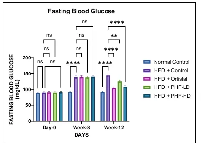
Fig. No. 13: Fasting Blood Glucose
Oral Glucose Tolerance Test (OGTT)
The HFD control group exhibited impaired glucose tolerance, whereas treatment with the polyherbal formulation significantly improved glucose utilization and reduced postprandial glucose levels.
Improvement in OGTT suggests enhanced peripheral glucose uptake and restoration of insulin responsiveness. The results indicate the potential of the formulation to prevent insulin resistance associated with metabolic syndrome.
Table 16. Effect of Polyherbal Formulation on Oral Glucose Tolerance Test (OGTT) in HFD-Induced Wistar Rats
|
Group |
0 min (mg/dL) |
30 min (mg/dL) |
60 min (mg/dL) |
120 min (mg/dL) |
AUC₀–₁₂₀ |
|
I – Normal Control |
88.2 ± 2.7 |
128.6 ± 4.2 |
118.4 ± 3.8 |
95.2 ± 3.0 |
12,420 ± 318 |
|
II – HFD Control |
142.5 ± 4.5 |
232.4 ± 6.8 |
218.7 ± 6.2 |
180.6 ± 5.4 |
22,850 ± 538 |
|
III – HFD + Orlistat |
105.2 ± 3.4 |
162.4 ± 5.1 |
148.3 ± 4.6 |
112.5 ± 3.7 |
15,250 ± 396 |
|
IV – HFD + PHF-LD |
124.7 ± 3.9 |
195.4 ± 5.8 |
178.2 ± 5.3 |
138.4 ± 4.2 |
18,640 ± 462 |
|
V – HFD + PHF-HD |
107.5 ± 3.5 |
170.2 ± 5.3 |
152.6 ± 4.7 |
118.7 ± 3.8 |
15,940 ± 412 |
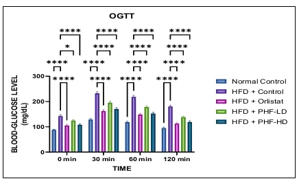
Fig. No. 14: Oral glucose tolerance test (OGTT)
Effect on Liver Function Parameters
Significant elevations in serum AST, ALT, ALP, and bilirubin levels were observed in HFD-fed animals, indicating hepatic injury and steatosis. Administration of the polyherbal formulation significantly reduced these biomarkers toward normal levels.
The hepatoprotective effect may be attributed to antioxidant and anti-inflammatory properties of the phytoconstituents present in both plant extracts. Reduction in liver enzyme levels indicates preservation of hepatocellular integrity and improved liver function.
Table 17. Effect of Polyherbal Formulation on Liver Function Parameters in HFD-Induced Wistar Rats
|
Group |
AST (U/L) |
ALT (U/L) |
ALP (U/L) |
Total Bilirubin (mg/dL) |
|
I – Normal Control |
46.2 ± 2.1 |
34.8 ± 1.9 |
142.6 ± 4.8 |
0.42 ± 0.04 |
|
II – HFD Control |
108.4 ± 3.8 |
96.5 ± 3.4 |
248.3 ± 7.2 |
1.18 ± 0.08 |
|
III – HFD + Orlistat |
62.6 ± 2.6 |
52.8 ± 2.3 |
168.4 ± 5.2 |
0.58 ± 0.05 |
|
IV – HFD + PHF-LD |
82.4 ± 3.1 |
72.6 ± 2.8 |
198.5 ± 6.1 |
0.86 ± 0.07 |
|
V – HFD + PHF-HD |
66.5 ± 2.7 |
56.4 ± 2.4 |
174.6 ± 5.4 |
0.62 ± 0.05 |
Fig. No. 15: AST Fig. No. 16: ALT
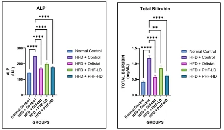
Fig. No. 17: ALP Fig. No. 18: Total Bilirubin
Effect on Renal Function Parameters
HFD-induced metabolic syndrome significantly increased serum urea, creatinine, and uric acid levels, suggesting impaired renal function. Treatment with the polyherbal formulation significantly improved renal biomarkers.
The nephroprotective activity may be due to attenuation of oxidative stress, reduction of systemic inflammation, and improved metabolic control.
Table 18. Effect of Polyherbal Formulation on Renal Function Parameters in HFD-Induced Wistar Rats
|
Group |
Urea (mg/dL) |
Creatinine (mg/dL) |
Uric Acid (mg/dL) |
|
I – Normal Control |
30.4 ± 1.4 |
0.52 ± 0.04 |
1.46 ± 0.08 |
|
II – HFD Control |
58.7 ± 2.3 |
0.96 ± 0.07 |
2.94 ± 0.14 |
|
III – HFD + Orlistat |
38.6 ± 1.7 |
0.66 ± 0.05 |
1.74 ± 0.10 |
|
IV – HFD + PHF-LD |
47.8 ± 1.9 |
0.78 ± 0.06 |
2.18 ± 0.12 |
|
V – HFD + PHF-HD |
40.2 ± 1.8 |
0.68 ± 0.05 |
1.82 ± 0.11 |
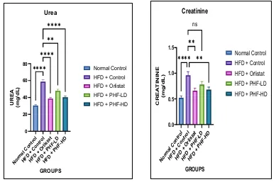
Fig. No. 19: Urea Fig. No. 20: Creatinine
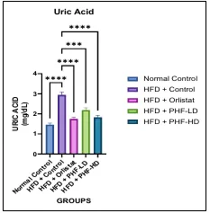
Fig. No. 21: Uric Acid
Effect on Oxidative Stress Markers
Oxidative stress plays a crucial role in the pathogenesis of metabolic syndrome. HFD-fed rats showed significantly elevated malondialdehyde (MDA) levels and decreased endogenous antioxidant enzymes including GSH, SOD, and CAT.
Treatment with the polyherbal formulation significantly reduced lipid peroxidation and restored antioxidant defense mechanisms. The strong antioxidant activity may be attributed to catechins, flavonoids, and phenolic compounds present in the formulation. Restoration of antioxidant status contributes to protection against cellular and tissue damage.
Table 19. Effect of Polyherbal Formulation on Hepatic Oxidative Stress Markers in HFD-Induced Wistar Rats
|
Group |
MDA (nmol/mg) |
GSH (µg/mg) |
SOD (U/mg) |
CAT (U/mg) |
|
I – Normal Control |
1.48 ± 0.09 |
12.62 ± 0.48 |
28.5 ± 1.2 |
58.4 ± 2.3 |
|
II – HFD Control |
5.86 ± 0.24 |
4.85 ± 0.22 |
11.6 ± 0.6 |
21.8 ± 1.0 |
|
III – HFD + Orlistat |
2.42 ± 0.12 |
10.46 ± 0.41 |
24.2 ± 1.1 |
48.7 ± 2.1 |
|
IV – HFD + PHF-LD |
3.58 ± 0.18 |
8.42 ± 0.35 |
19.4 ± 0.9 |
38.6 ± 1.7 |
|
V – HFD + PHF-HD |
2.64 ± 0.14 |
10.08 ± 0.39 |
23.1 ± 1.1 |
45.8 ± 2.0 |
Fig. No. 22: MDA Fig. No. 23: GSH
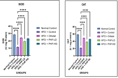
Fig. No. 24: SOD Fig. No. 25: CAT
Relative Organ Weight
Animals receiving a high-fat diet exhibited increased liver weight due to hepatic fat accumulation. Treatment with the polyherbal formulation significantly reduced liver weight, indicating attenuation of hepatic steatosis and improved lipid metabolism.
Table 20. Effect of Polyherbal Formulation on Relative Liver Weight in HFD-Induced Wistar Rats
|
Group |
Relative Liver Weight (% of Body Weight) |
|
I – Normal Control |
3.04 ± 0.12ᵃᵃᵃ |
|
II – HFD Control |
4.62 ± 0.18 |
|
III – HFD + Orlistat |
3.28 ± 0.13*** |
|
IV – HFD + PHF-LD |
3.72 ± 0.15** |
|
V – HFD + PHF-HD |
3.34 ± 0.14*** |
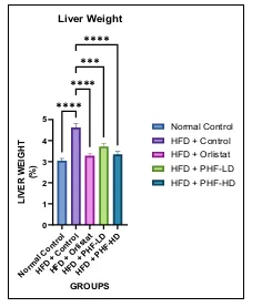
Fig. No. 26: Liver Weight
Histopathological Evaluation
Histopathological examination of liver tissues from the normal control group showed normal hepatic architecture. In contrast, HFD-fed animals exhibited marked hepatic steatosis, fatty degeneration, inflammatory cell infiltration, and hepatocellular damage.
Treatment with the polyherbal formulation markedly improved liver histology, with reduced fat accumulation and restoration of normal hepatic architecture. The higher dose group demonstrated substantial protection comparable to orlistat treatment.
The histopathological findings strongly support the biochemical results and confirm the protective effect of the formulation against HFD-induced hepatic injury.
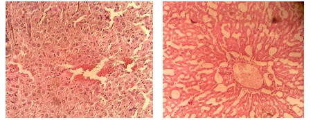
Fig. No. 27: Normal Control Fig. No. 28 : HFD Control
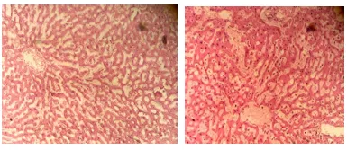
Fig. No. 29: HFD + Orlistat Fig. No. 30 : HFD + PHF-LD
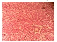
Fig. No. 31: HFD + PHF-HD
DISCUSSION
Metabolic syndrome is a multifactorial disorder characterized by the coexistence of obesity, insulin resistance, dyslipidemia, oxidative stress, and organ dysfunction, all of which significantly increase the risk of cardiovascular disease and type 2 diabetes mellitus. In the present study, chronic administration of a high-fat diet successfully induced metabolic syndrome in Wistar rats, as evidenced by significant increases in body weight, visceral adiposity, serum lipid levels, fasting blood glucose, oxidative stress markers, and hepatic and renal dysfunction. These findings confirm the suitability of the HFD-induced rat model for evaluating potential therapeutic interventions against metabolic syndrome.
The polyherbal formulation developed from hydroethanolic extracts of Tecomella undulata and Camellia sinensis demonstrated significant protective effects against multiple pathological components of metabolic syndrome. Preliminary phytochemical screening revealed the presence of flavonoids, phenolic compounds, tannins, triterpenoids, naphthoquinones, iridoids, and other bioactive constituents. Furthermore, both extracts exhibited substantial total phenolic and flavonoid contents and demonstrated considerable antioxidant activity in the DPPH assay, indicating their potential to combat oxidative stress-related metabolic disturbances.
One of the most notable findings of the study was the significant reduction in body weight gain, visceral fat accumulation, adiposity index, and Lee index observed following treatment with the polyherbal formulation. Excess adipose tissue, particularly visceral fat, is recognized as a major contributor to insulin resistance and chronic inflammation. The observed anti-obesity activity may be attributed to the ability of catechins present in Camellia sinensis to enhance thermogenesis, stimulate fatty acid oxidation, inhibit adipogenesis, and improve energy expenditure. Simultaneously, phytoconstituents present in Tecomella undulata may contribute to improved lipid utilization and metabolic regulation.
Dyslipidemia is a major component of metabolic syndrome and is characterized by elevated total cholesterol, triglycerides, and LDL-cholesterol together with reduced HDL-cholesterol levels. The present study demonstrated that the polyherbal formulation significantly improved serum lipid profiles in HFD-fed rats. Reduction in total cholesterol, triglycerides, and LDL-cholesterol accompanied by an increase in HDL-cholesterol suggests improved lipid metabolism and reduced cardiovascular risk. These effects may result from inhibition of cholesterol biosynthesis, suppression of intestinal lipid absorption, stimulation of fatty acid oxidation, and activation of AMP-activated protein kinase (AMPK)-mediated metabolic pathways.
Hyperglycemia and impaired glucose tolerance observed in the HFD control group indicate the development of insulin resistance, a central pathogenic feature of metabolic syndrome. Treatment with the polyherbal formulation significantly reduced fasting blood glucose levels and improved oral glucose tolerance. The enhanced glucose handling observed in treated animals may be associated with improved insulin sensitivity, increased peripheral glucose uptake, and reduced hepatic glucose production. Polyphenols and catechins are known to regulate glucose metabolism through activation of insulin signaling pathways and enhancement of GLUT-4 translocation, thereby facilitating glucose utilization.
Oxidative stress is considered a critical factor in the progression of obesity-related metabolic disorders. High-fat diet feeding resulted in increased lipid peroxidation as evidenced by elevated hepatic MDA levels and significant depletion of endogenous antioxidant enzymes including GSH, SOD, and CAT. Administration of the polyherbal formulation markedly reduced oxidative stress and restored antioxidant defense systems. These findings indicate that the formulation effectively neutralized reactive oxygen species and protected tissues from oxidative damage. The strong antioxidant activity observed may be attributed to the synergistic effects of flavonoids, phenolic compounds, catechins, triterpenoids, and naphthoquinones present in both plant extracts.
Metabolic syndrome frequently leads to hepatic steatosis and liver dysfunction due to excessive lipid accumulation and oxidative stress. Significant elevations in AST, ALT, ALP, bilirubin, and relative liver weight observed in the HFD control group confirmed the development of hepatic injury. Treatment with the polyherbal formulation significantly normalized these parameters and reduced liver enlargement. Histopathological examination further demonstrated attenuation of fatty degeneration, inflammatory infiltration, and hepatocellular damage. These findings indicate substantial hepatoprotective activity and suggest improved hepatic lipid metabolism and preservation of hepatocellular integrity.
Similarly, renal dysfunction induced by chronic metabolic stress was evidenced by elevated serum urea, creatinine, and uric acid levels in HFD-fed animals. Treatment with the polyherbal formulation significantly improved these renal biomarkers, indicating nephroprotective activity. Improvement in renal function may be attributed to reduced oxidative stress, improved glycemic control, and attenuation of systemic inflammation.
Among the tested doses, the higher dose of the polyherbal formulation (400 mg/kg) consistently produced greater improvements across all evaluated parameters and exhibited efficacy comparable to the standard drug Orlistat. The superior activity observed at the higher dose suggests a dose-dependent pharmacological response and highlights the therapeutic potential of the formulation.
Collectively, the findings of the present study demonstrate that the polyherbal formulation exerts a comprehensive protective effect against high-fat diet-induced metabolic syndrome through multiple mechanisms, including anti-obesity, antihyperlipidemic, antihyperglycemic, antioxidant, hepatoprotective, and nephroprotective actions. The synergistic interaction between Tecomella undulata and Camellia sinensis appears to be responsible for the broad-spectrum therapeutic efficacy observed. These results provide strong scientific evidence supporting the use of this polyherbal formulation as a promising multi-target herbal intervention for the management of metabolic syndrome and its associated complications.
CONCLUSION
The present study demonstrated that the polyherbal suspension containing hydroethanolic extracts of Tecomella undulata and Camellia sinensis possesses significant protective effects against high-fat diet-induced metabolic syndrome in Wistar rats. Chronic administration of the high-fat diet successfully induced obesity, dyslipidemia, hyperglycemia, hepatic dysfunction, renal impairment, oxidative stress, and histopathological alterations, closely resembling the clinical manifestations of metabolic syndrome in humans.
Treatment with the polyherbal formulation produced marked improvements in body weight, visceral adiposity, lipid profile, fasting blood glucose, oral glucose tolerance, liver function markers, renal function parameters, and oxidative stress biomarkers. The formulation significantly reduced serum total cholesterol, triglycerides, LDL-cholesterol, fasting blood glucose, and lipid peroxidation while enhancing HDL-cholesterol levels and endogenous antioxidant defenses. Histopathological findings further confirmed the protective effect of the formulation by demonstrating substantial restoration of normal hepatic architecture and reduction of fatty degeneration and inflammatory changes.
The observed pharmacological effects may be attributed to the synergistic action of bioactive phytoconstituents, including flavonoids, phenolic compounds, catechins, and other antioxidant molecules present in both plant extracts. These compounds collectively contribute to antihyperlipidemic, antihyperglycemic, antioxidant, hepatoprotective, and anti-obesity activities, thereby targeting multiple pathological pathways involved in metabolic syndrome.
Among the tested doses, the higher dose (400 mg/kg) exhibited superior therapeutic efficacy and produced effects comparable to the standard drug, orlistat. The absence of toxicity in acute oral toxicity studies further supports the safety profile of the formulation.
Overall, the findings of the present investigation suggest that the polyherbal formulation of Tecomella undulata and Camellia sinensis represents a promising multi-target herbal therapeutic approach for the management of metabolic syndrome. Further studies involving detailed mechanistic investigations, chronic toxicity evaluation, and clinical trials are warranted to establish its therapeutic potential and facilitate its future development as a safe and effective herbal intervention for metabolic disorders.
REFERENCES

Asifa Yasin Mulani, Sahil Muneer Sherkar, Dr Kiran Patil, Ajit Patil, Evaluation Of Polyherbal Formulation on High Fat Diet Induced Meatbolic Syndrome in Wistar Rat, Int. J. of Pharm. Sci., 2026, Vol 4, Issue 6, 5172-5195, https://doi.org/10.5281/zenodo.20769821
 10.5281/zenodo.20769821