

We use cookies to ensure our website works properly and to personalise your experience. Cookies policy
Dr. Shivajirao Kadam College of Pharmacy Kasabe Digraj, MS, India.416305.
NSAIDs are great for arthritic pain but can have some unwanted complications like ulcers, bleeding kidney issues, and a higher chance of a heart complications. NSAID can even cause more stress on your body by making more reactive oxygen species. To overcome this problem therapeutic option is herbal medicine. Medicinal plants have intrinsic active substances to heal sickness or relieve pain. Leaves of Vitex negundo in the Verbenaceae family which show anti arthritic activity was used for preparation of ethosomes. In order to successfully formulate the ethosomes, a cold method was effectively used. 400 milligrams of soya lecithin, 14 milliliters of ethanol, and 200 milligrams of polyethylene glycol 4000 are the components that make up the ideal formulation. Transmission electron microscopy images exposed the circular shape then clarified the formation of ethosomes. Particle size, zeta potential was found to be 162.3 nm and -29 mV respectively. Entrapment efficiency of optimized batch was found to be 78.5%. Finally, prepared ethosomal gel system significantly enhance skin penetration and having sustained release for prolong duration of time.
Medicinal plants have intrinsic active substances to heal sickness or relieve pain. It is commonly known that conventional remedies and medicinal plants are used as therapeutic agents to preserve health in the majority of underdeveloped nations. Historically, herbs have been regarded as safe and have been used by the general people as well as practitioners of traditional medicine all over the globe for the purpose of treating a broad range of disorders[1,2]. Rheumatoid Arthritis treatment generally includes the use of DMARDs, or disease-modifying antirheumatic medications, include sulfasalazine, methotrexate, leflunomide, and hydroxychloro. These medications help improve joint function and slow down the damage process. Other treatment options include Corticosteroids, biological response modifiers and non-steroidal anti-inflammatory medications COX. To reduce pain and inflammation. Despite advancements in RA therapies, concerns about safety and effectiveness continues to restrict their usage. Cyclooxygenase-2 inhibitors may lead to severe cardiovascular side effects. NSAIDs are great for arthritic pain but can have some unwanted complications like ulcers, bleeding kidney issues, and a higher chance of a heart complications. NSAID can even cause more stress on your body by making more reactive oxygen species. Both NSAIDs and COX-2 inhibitors work well but come with risks like GI problems, kidney issues, and heart complications[3]. The viable epidermis plays a crucial role in skin barrier function, primarily through intercellular lipid channels and various partitioning activities. Depending on their solubility, medications can diffuse across the stratum corneum and migrate from one layer to another [4]. Once a substance gets through the corneum layer, there is no considerable extra barrier to its penetration into the remaining epidermal layers. The concentration gradient almost diminishes in the dermal layer once circulation starts. The systemic circulation works as a reservoir or "sink" for the medication [5]. Recently, introduced a new type of vesicular carrier known as ethosomes. These systems are simple to produce, non-irritating, and primarily composed of phospholipids and ethanol, which are common ingredients in medicinal formulations. Here we concentrate on the encapsulating of medicines in lipid vesicles created from phospholipids (liposomes) which have been proved to facilitate transport of pharmaceuticals into and over epidermis [6-9]. Several strategies have been explored to enhance drug penetration across the skin, including the use of lipid vesicles known as liposomes, which are made up of phospholipids. While liposomes show promise in facilitating the transport of drugs into and through the skin [10], they have limitations such as small size, low entrapment efficiency, and negative zeta potential. To address these issues, improved lipid carriers called ethosomes were developed. Ethosomes are composed of ethanol, phospholipids, and water, with ethanol concentrations ranging from 20-50% [11]. Ethanol is recognized for its ability to enhance permeation. Research demonstrated that lipid vesicular systems could coexist with high ethanol concentrations, leading to the creation of ethosomes [12]. The fundamental distinction between liposomes and ethosomes resides in their makeup. The increased ethanol content in ethosomes boosts their skin penetration capabilities. This high ethanol content can disrupt the skin lipid bilayer, allowing the vesicles to penetrate the stratum corneum by passing through small openings created in the disturbed stratum corneum lipids. Additionally, the characteristics of ethosomal formulations may be adjusted by changing the proportions of ingredients [13]. Because of the higher ethanol concentration, ethosomal lipid membranes are more loosely packed than those of conventional vesicles, while maintaining equivalent stability [14]. Ethosomes are particularly effective for delivering a wide range of proteins and peptides and are commonly given in gel or cream form for patient comfort [15].
MATERIAL AND METHOD
Material:
Soya lecithin, Carbopol 934, Glycerine, Triethanolamine were purchased from Loba chemie Pvt. Ltd. Mumbai; Ethanol, Methanol were purchased from Gemini Associates, Goa, and Polyethylene glycol 4000, Propyl Paraben, Methyl Paraben were obtained from Research Lab Fine Chem. Mumbai. Every other substance and reagent was of analytical quality.
Plant Material:
The fresh leaves of Vitex negundo Linn. were obtained from a local area Kasabe Digraj and authenticated by a Dept. of Botany, Dr. Patangarao Kadam College Sangli. The herbarium was deposited in the Dept. of Botany Dr. Patangarao Kadam College Sangli for future reference.
Method:
Plant Material Processing:
The fresh leaves of Vitex negundo were collected and thoroughly rinsed. Leaves air dried in the shade and then roughly ground in a mixer. They were then stored in separate airtight containers at room temperature for future use.
Plant Extraction:
Using a Soxhlet apparatus, continuous hot solvent extraction was carried out for two hours at 50 °C with a solvent:drug ratio of 10:1 (mL/g). After that, the menstruum was filtered. Using a rotary evaporator set at 40 °C, the filtrate was evaporated to obtain the residue.
Preliminary Phytochemical Studies:
The extract was subjected to several tests to identify various compounds, including proteins, amino acids, glycosides, carbohydrates, alkaloids (using the Dragendorff and Mayer tests), phenols, tannins, flavonoids (using the Shinado test), and anthraquinones (using the Borntrager test)[16].
Formulation Development of Ethosome of Vitex negundo:
Ethosomes produced utilizing cold technique. Soya lecithin, PEG-4000, and Vitex negundo leaf extract dissolved in ethanol. Obtained mixture was adjusted by dropwise introduction of double distilled water with continued stirring at 700 rpm for 2 hours and temperature is maintained 30 °C to facilitate ethosomes production. The obtained ethosomes were sonicated for fifteen minutes. To avoid structural injury of ethosomes, applied sonication round each of 5 min by using probe sonication. Variable concentrations of soya lecithin, PEG 4000, and ethanol were utilized, the resulting ethosomal suspension was refrigerated for later use[17].
Evaluation of the prepared Ethosomes:
Vesicle size, size distribution, and zeta potential:
The zeta potential, vesicle size and size distribution, of extract-loaded ethosomes were analyzed using a particle size analyzer (HORIBA PS-100). The samples were made by agitating them for three minutes then diluting them with 0.5% w/v deionized water. Results were shown using the average and standard deviation of measurements taken from distinct batches of samples.
Morphology:
Using a glass rod, the ethosomal dispersion was applied to the glass slide. By observing the ethosomal solution under a 100X optical microscope, the formation of vesicles was verified. Images of vesicles were taken. Shapes of the ethosomes were assessed through TEM. The optimized batch of ethosomes was diluted tenfold and gently placed on a carbon coated copper grid for one minute before carefully removing any excess with filter paper. The staining agent was 1% phosphotungstic acid solution. Utilizing an acceleration voltage of 200 kV, the TEM apparatus captured images, which were subsequently subjected to analysis through imaging viewer software.
Entrapment Efficiency (?) of Ethosome:
The ? of ethosomes are utilizing by centrifugation technique. In tube 5 millilitre of freshly prepared ethosomal formulation placed. For thirty minutes, a cooling centrifuge at (Remi Instrument) used to whirl the centrifuge tube at 15,000 revolutions per minute. After that, the ethosomal formulation was centrifuged. Each time, 1ml of un-entrapped drug suspension supernatant was collected and spectrophotometrically analysed at 256 nm with a UV spectrophotometer (SHIMDZU UV -1900).
To calculate the percentage entrapment efficiency, the following formula was used:
% Entrapment Efficiency =Initial drug - final drugInitial drug
In-Vitro Diffusion Study:
The diffusion technique was employed to achieve in-vitro release of Vitex negundo extract loaded ethosomes. These experiments were conducted using Franz diffusion cells. In the donor compartment, varying batches of Vitex negundo extract ethosomal formulation, were placed. The receptor filled with PBS at pH 6.8 as receiving medium. A semi-permeable cellulose membrane, previously soak in PBS pH 6.8 for 24 hours, separates the donor and receptor compartments. For 8 hours at 37°C, the solution in the receiving compartment was agitated with a magnetic stirrer at a rate of 50 rpm, 3mL samples were obtained and replaced with new phosphate buffer solution on a regular basis to maintain sink condition. The collected samples were diluted and subsequently analysed for drug release using a UV-Spectrophotometer.
Anti-Inflammatory Activity:
The anti-inflammatory activity of prepared ethosomal suspension was determined using the protein denaturation method. The reaction mixture totaled 5ml includes 0.2ml egg albumin, 2.8ml phosphate buffer pH 6.4, and 2ml of prepared ethosomal suspension with concentrations of 10, 20, 50, and 100 ?g/ml. The test solution and control blank ethosomal suspension were incubated at 37°C in a biological oxygen demand (BOD) incubator for 15 minutes, followed by a 5-minute heating period at 70°C. The absorbance was then measured at 660 nm with UV spectroscopy, using the vehicle as a blank. Protein inhibition is then estimated using the formula below:[18]
Inhibition = AC- AT/AC x100
Where,
AC - Absorbance of control solution,
AT - Absorbance of test sample.
Table 1: Formulation of Ethosomes
|
Ingredients |
E1 |
E2 |
E3 |
E4 |
E5 |
E6 |
E7 |
E8 |
E9 |
|
Vitex negundo extract |
1 |
1 |
1 |
1 |
1 |
1 |
1 |
1 |
1 |
|
Soya Lecithin |
350 |
350 |
350 |
400 |
400 |
400 |
450 |
450 |
450 |
|
Ethanol |
12 |
12 |
12 |
14 |
14 |
14 |
16 |
16 |
16 |
|
PEG 4000 |
200 |
200 |
200 |
200 |
200 |
200 |
200 |
200 |
200 |
|
Distilled Water |
Q.S to 40 ml |
||||||||
Formulation of Ethosomal Gel:
Carbopol 934 (1%, 1.5 %, 2%, and 2.5%) added in adequate amount of water as a gelling agent and allowed for complete soaking. The previously prepared Ethosomal formulation was incorporated into the gel with continuously stirring. remaining volume adjusted by water. Propylene glycol and triethanolamine added in above preparation with continuous stirring by using mechanical stirrer until uniform gel is formed methyl and propyl paraben was added as preservative. 1?rbopol gel showed better appearance due to this 1% concentration of Carbopol is selected as an optimum concentration for incorporation of ethosomes.
Table 2: Composition of Ethosomal Gel
|
Sr. No. |
Ingredients |
Quantity Taken |
Role |
|
1 |
Carbopol 934 |
1 gm |
Gelling Agent |
|
2 |
Triethanolamine |
0.01 ml |
pH Modifier |
|
3 |
Propylene Glycol |
10 ml |
Plasticizer |
|
3 |
Methyl Paraben |
0.01w/w |
Preservative |
|
4 |
Propyl Paraben |
0.02w/w% |
Preservative |
|
5 |
Distilled Water |
QS |
Vehicle |
Evaluation of Ethosomal Gel:
Visual Appearance:
Visually observed. It should have uniform consistency having slightly characteristic smell and glassy appearance
pH Determination:
A digital pH monitor was used to determine the pH. One of the most crucial elements of the formulation process is pH.
Spreadability:
Spreadability is the important concept of topically applicable formulation. Spreadability means ability to spread. Taken one gram of sample and kept it on the sample plate then another plate was kept on the same plate which was previously tied with the thread which was attached to weight. The weight was kept on the plate and time required for sliding for 5cm distance by upper plate was measured.
S=M ×LT
Where,
S= Spreadability,
M=Mass,
L=length, and
T=Time
Viscosity:
Viscosity of ethosomal gel of Vitex negundo was determined by Brookfield Viscometer (DV III) model using spindle no S-64 at 100 rpm The results were recorded after the viscometer shows a stable number.
Drug Content:
Drug content refers to the amount of active pharmaceutical ingredient (API) present in a formulation. It is a critical parameter in stability studies, as it ensures that the formulation delivers the correct dosage throughout its shelf life. To determine drug content, samples are typically analysed using methods like UV spectroscopy. 1gm of prepared gel was added into the volumetric flask of 50 ml and volume was made up by using the methanol, then sonicated to dissolve the gel and to get clear solution. Solution was then filtered and diluted with methanol. At 256 nm wavelength the solution was scanned to get drug concentration, methanol was used as a reference. Following equation was then used to get drug content.
Drug Content = Practical yield / theoretical yield*100
In-Vitro Diffusion Study:
The diffusion technique was employed to achieve in-vitro release of vitex negundo extract loaded ethosomal gel. These experiments were conducted using Franz diffusion cells. A semi-permeable cellulose membrane, previously soak in PBS pH 6.8 for 24 hours, separates the donor and receptor compartments. In the donor compartment, ethosomal gel formulation, were placed. The receptor compartment filled up with PBS at pH 6.8 as the receiving medium. For 24 hr hours at 37°C, the solution in the receiving compartment was agitated with the magnetic stirrer at a rate of 50 rpm, 3mL samples were obtained and replaced with new phosphate buffer solution on a regular basis to maintain sink condition. The collected samples were diluted and subsequently analysed for drug release using a UV-Spectrophotometer.
Release kinetics:
Several mathematical models were fitted to the gathered data to establish the active component's release pattern from the herbal gel.
Anti-Inflammatory Assay (HRBC Membrane Stabilization Test):
Ten milliliters of fresh human blood will be drawn into heparinized centrifuge tubes, spun for ten minutes at 3000 rpm, and then three times with an equal volume of normal saline solution. After measuring the blood volume, it will be reconstituted in 10% v/v normal saline. One milliliter of formulation and one milliliter of 10% red blood cell suspension made up the reaction mixture. There will be saline in the control. The usual drug (positive control) will be aspirin. The samples will be centrifuged for five minutes at 2500 rpm after being incubated for thirty minutes at 56 °C. The absorbance of the supernatant will then be measured at 560 nm. This is how the % membrane stabilization activity is going to be calculated.
Percentage of haemolysis = OD of test /OD of control x 100.
Percent of protection = 1 - OD of test / OD of control x 100.
"OD of control" refers to the absorbance of the negative control, whereas "OD of test" refers to the optical density or absorbance of the test sample[19].
Stability:
Stability in pharmaceutical preparations refers to the ability of specific formulation within a given container-closing system to maintain its physical, microbiological, chemical and therapeutic properties throughout its shelf life. To evaluate stability, the product is subjected to stress conditions such as varying humidity and temperature. For instance, a three-month stability study of a prepared ethosomal gel was conducted at room temperature (25°C ± 2°C). During this period, the gel's appearance, drug content, and pH were assessed at 30-day intervals.
RESULT AND DISCUSSION
Phytochemical Studies:
The tests indicate that carbohydrates, alkaloids, flavonoids, cardiac glycosides, and saponins are present in the extract. However, proteins are absent. This information is crucial for understanding the chemical composition of the extract and its potential biological activities.
Ethosomes:
Table 3: Vesicle size, Size Distribution, and % CR and Entrapment efficiency
|
Batch code |
Formulation response |
|||
|
Particle Size nm |
PDI |
% CR |
Entrapment efficiency |
|
|
E1 |
143.9 |
0.378 |
89.29% |
69.01 |
|
E2 |
130.2 |
0.403 |
94.48% |
70.44 |
|
E3 |
127.8 |
0.233 |
98.15% |
67.62 |
|
E4 |
173.7 |
0.637 |
88.22% |
74.94 |
|
E5 |
162.3 |
0.351 |
92.65% |
78.5 |
|
E6 |
158.4 |
0.357 |
94.18% |
71.45 |
|
E7 |
293 |
0.593 |
85.78% |
82.94 |
|
E8 |
227 |
0.726 |
87.00% |
88.71 |
|
E9 |
218 |
0.570 |
91.12% |
83.86 |
Particle Size:
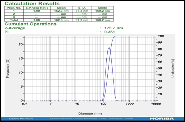
Fig. 1: Particle Size (Batch E5)
Zeta Potential:
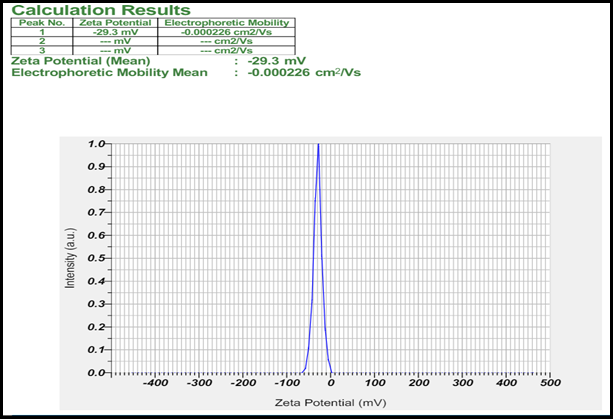
Fig. 2: Zeta Potential (Batch E5)
Morphology:
Present TEM image of a batch no. E5 Ethosomes synthesized using the cold method the TEM image reveals that the ethosomes exhibit a smooth and spherical configuration. (Fig. no.3 A and B).
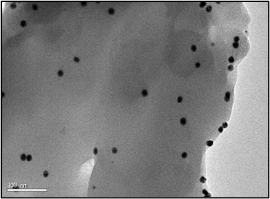
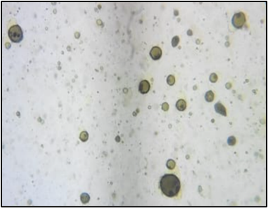
Fig. 3: A) TEM image B) Microscopic Image of Ethosomes
In-vitro Diffusion Study of Ethosomes:
The drug release patterns across various ethosomal batches were characterized by plotting a graph depicting the percentage of drug released over time. After 8 hours, the cumulative drug release percentage for ethosomal loaded batch coded as E5 was determined to be 92.65. (Fig. 4)
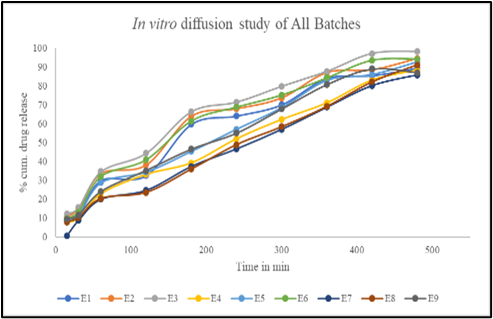
Fig. 4: In vitro Diffusion Study of All Batches
Anti-Inflammatory Activity:
The anti-inflammatory activity carried out by protein denaturation study shows that the ethosomal suspension (Batch E5) of concentration 100 ug/ml showed % protein inhibition of 59.30%, which proves that prepared formulation has good anti-inflammatory activity. (Table 4)
Table 4: Present Protein inhibition of optimized batch E5
|
Sample |
Concentration |
% Inhibition |
|
Blank |
- |
- |
|
Test1 |
10ug/ml |
22.65% |
|
Test2 |
20ug/ml |
32.89% |
|
Test3 |
50ug/ml |
44.48% |
|
Test4 |
100ug/ml |
59.30% |
Evaluation of Vitex negundo Loaded Ethosomal Gel:
Physical Appearance:
The observations for ethosomal gel are as follows: The colour of the gel is greenish, and it has a characteristic odour. The consistency of the gel is semisolid, and it is homogenous in nature. These characteristics confirm the desired physical properties of the Vitex negundo loaded ethosomal gel.
pH determination:
Ideal pH suitable for skin delivery is slightly acidic. Ethosomal Gel's pH was found to be 5.51.
Spreadability:
Length of slide = 20 cm.
Length slide travel = 5 cm
Time = 10 sec
Weight taken = 30 gm
Formula for Spreadability
= M * L / T
= 30 * 5 / 10
= 15 gm.cm/sec
It shows that it has good Spreadability and the sample can easily spread on the applied area. Spreadability is directly proportional to the permeation.
Viscosity Analysis:
Viscosity analysis was done by using spindle no S64 at 100 RPM and it found 3292.7 cp.
Drug Content:
The drug content of vitex negundo loaded ethosomal gel was found to be 99.1% using the formula
Drug content = Practical Yield /Theoretical yield *100
Practical yield =9.91
Theoretical yield in 1gram of gel =10 mg.
In-vitro Diffusion Study of Ethosomal Gel:
Optimized batch of ethosomes was loaded in carbopol gel base and the drug release patterns of ethosomal gel were characterized by plotting a graph depicting the percentage of drug released over time. After 12 hours, the cumulative drug release percentage for ethosomal-loaded Gel was determined to be 94.89%.
The Kinetic Profile of In-Vitro Drug Release of Ethosomal Gel" compares the drug release kinetics for an ethosomal gel formulation using various models. The Zero Order Kinetic model, with an R?2; value of 0.9764, suggests a nearly constant drug release rate over time. The First Order Kinetic model has a lower R?2; value of 0.7926, indicating a weaker fit. The Higuchi Model, which describes diffusion-based release, has an R?2; value of 0.8979. The Korsmeyer Peppas model shows the best fit with an R?2; value of 0.9788, and an n (slope) value of 0.401, indicating Fickian diffusion. Thus, the drug release from the ethosomal gel is best described by the Korsmeyer Peppas model and follows a Fickian diffusion mechanism. (Fig. 5)
Release Kinetic of Ethosomal Gel:
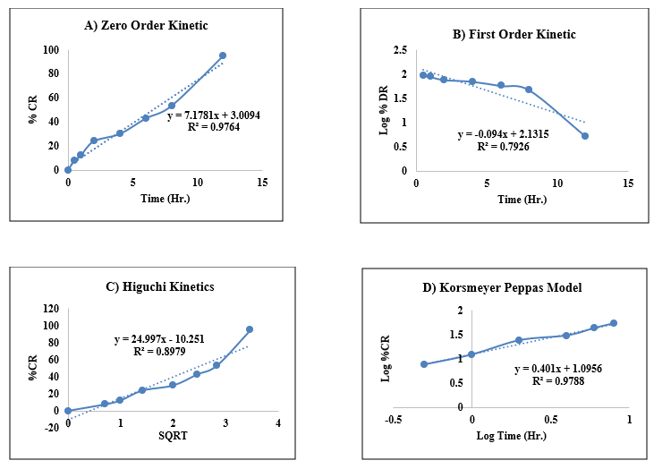
Fig. 5: Models graphs of drug release kinetics profiles A) Zero order kinetics B) First order kinetics C) Higuchi kinetics D) Korsmeyer Peppas Equation.
Comparative Drug Release Study between Ethosomal Gel and normal Gel:
The results of a comparative drug release study between ethosomal gel and normal gel over a 24-hour period. At the start of the study (0 hours), both gels showed a 0% cumulative release of the drug. After 0.5 hours, the ethosomal gel released 7.62% of the drug, whereas the normal gel released 4.08%. At the 1-hour mark, the ethosomal gel's drug release increased to 12.2%, while the normal gel released 7.69%. By 2 hours, the ethosomal gel had a cumulative release of 23.88%, compared to 15.53% for the normal gel. At the 4-hour point, the ethosomal gel released 29.91% of the drug, while the normal gel released 23.42%. After 6 hours, the ethosomal gel's release was 42.74%, and the normal gel's release was 33.75%. At 8 hours, the cumulative release from the ethosomal gel reached 53.27%, whereas the normal gel had released 41.22%. By 12 hours, the ethosomal gel had released 94.89% of the drug, significantly more than the 69.89% released by the normal gel. Finally, after 24 hours, the ethosomal gel showed a 95.13% cumulative release, compared to 73.03% for the normal gel. (Fig. no. 6)
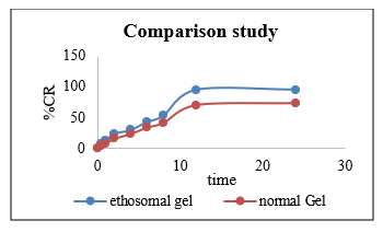
Fig. 6: Comparative Drug Release Study between Ethosomal Gel and normal Gel
Anti-Inflammatory Activity:
Human erythrocyte membranes may be shielded against hypotonic solution-induced lysis by using extracts at concentrations between 20 and 100 µg/mL. At 100 µg/mL, ET was shown to block 48.87% of RBC hemolysis, whereas aspirin inhibited 93.45% of hemolysis at the same dose. Because the membranes of human red blood cells and lysosomal membrane components are similar, the ability of a drug to suppress hypotonicity-induced human red blood cell (HRBC) membrane lysis may be used as a gauge of its anti-inflammatory properties. As shown in Figures 7A and 7B, the study's findings showed that sample ET may considerably and dose-dependently prevent HRBC haemolysis.
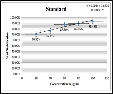
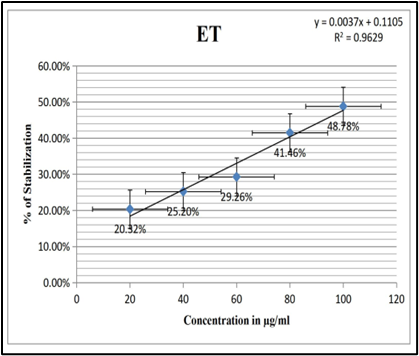
Fig. 7: A) In vitro Anti-Inflammatory Activity of Standard (HRBC) and B) In vitro Anti-Inflammatory Activity of Ethosome (HRBC)
Stability Study:
The stability assessment of the Vitex negundo loaded ethosomal Gel involve the visual appearance, pH and Drug content over a storage period of the 3-months , the results indicate that there were no notable alterations observed. After one month, there were no changes observed in the pH, appearance, or drug content of the formulation. At the end of the second month, the stability study results remained consistent with no changes in pH, appearance, or drug content. By the third month, there were still no changes in pH and appearance; however, the drug content was noted to be 98%.
CONCLUSION
The study presents an ethosomal gel loaded with Vitex negundo extract as an effective alternative to conventional NSAIDs for arthritic pain. Ethosomes offer superior skin penetration and sustained release, mitigating the adverse effects of traditional NSAIDs and COX-2 inhibitors. The ethosomes, prepared via the cold method, showed good stability, a particle size of 162.3 nm, and an encapsulation efficiency of 78.5%. In-vitro diffusion studies revealed a 92.65% drug release over 8 hours, and significant anti-inflammatory activity was observed. The ethosomal gel formulation demonstrated ideal physical properties, including a pH of 5.51, good spreadability, and a viscosity of 3292.7 cp, with a 94.89% drug release after 12 hours, outperforming normal gel. Comparative studies showed the ethosomal gel had a 95.13% release after 24 hours, versus 73.03% for normal gel. This innovative formulation offers improved anti-inflammatory effects and sustained release, suggesting its potential as an alternative arthritis treatment.
ACKNOWLEDGEMENTS: The authors wish to acknowledge the support and contributions that facilitated the completion of this work.
Conflict of interest statement: The authors declared no conflict of interest.
REFERENCES


Parag Shiralkar, Arati Shinde, Dipak Bhingardeve*, Shrwani Motugade, Tanuja Kadam, Godfrey Mathews, Sagar Jadhav, Formulation and Characterization of Ethosomal Gel Containing Vitex Negundo Extract for Treatment of Arthritis, Int. J. of Pharm. Sci., 2025, Vol 3, Issue 2, 991-1002. https://doi.org/10.5281/zenodo.14865943
 10.5281/zenodo.14865943