

We use cookies to ensure our website works properly and to personalise your experience. Cookies policy
Department of Pharmaceutics, J.K.K. Nattraja College of Pharmacy, Kumarapalayam – 638 183, Namakkal District, Tamil Nadu, India. Affiliated to The Tamil Nadu Dr. M.G.R. Medical University, Chennai.
This study aimed to design and thoroughly evaluate an in situ ophthalmic gelling system for ciprofloxacin using natural polymers, specifically pectin and gellan gum. Our main goal was to solve the persistent issues associated with standard eye drops, such as fast precorneal drainage, low bioavailability, and the need for frequent dosing. To start, we completed preformulation testing covering everything from identification and solubility curves to melting points and Fourier Transform Infrared Spectroscopy (FTIR). The results verified that ciprofloxacin was pure and completely compatible with both natural polymers. Subsequently, we prepared four distinct batches (F1–F4) by varying the polymer ratios. Every batch was evaluated for appearance, clarity, pH levels, viscosity profiles, gelling capacity, drug uniformity, in vitro release kinetics, and sterility. Before gelation, the liquids maintained a comfortable pH range of 6.2–6.7. Once gelation occurred in the simulated tear fluid, the pH shifted to a highly safe physiological range of 6.4–6.9. Viscosity tests revealed pseudoplastic and shear-thinning behaviors. Among the group, formulation F1 stood out as the optimized blend, demonstrating near-instantaneous and durable gelation upon exposure to simulated tear fluid (pH 7.4). During our 12-hour in vitro release analysis, F1 exhibited a highly controlled, sustained delivery pattern, successfully releasing 77.33% of the drug by the 8th hour. Finally, standard autoclave sterilization was successful, with no microbial or fungal growth detected over a two-week incubation period. Overall, this ion-sensitive in situ gel drastically extended ocular residence time and offered a stable and reliable platform for treating bacterial eye infections
Delivering therapeutic agents to target eye tissues remains a substantial challenge in modern pharmaceutics, primarily because the eye is exceptionally well defended. For decades, classic topical options, such as standard aqueous eye drops, dense ointments, and suspensions, have been the default treatments for ocular infections. However, these traditional formats are clinically inefficient. Most eye drops suffer from poor ocular bioavailability, frequently losing over 95% of the active dose before penetrating the cornea. This massive loss occurs because the eye rapidly drains foreign fluids via a high tear turnover rate (roughly 1–3 µL/min), paired with nasolacrimal drainage and defensive blinking reflexes. To compensate for these anatomical hurdles, patients must frequently apply drops throughout the day. This constant redosing cycle is far from ideal; it regularly causes localized irritation, fluctuates drug concentrations in the eye, and ultimately frustrates patients, leading to poor patient compliance.(1,2)To overcome these limitations, Novel Drug Delivery Systems (NDDS) have become a major focal point in formulation research. The core concept behind NDDS is simple: to engineer a system that delivers the right amount of drug over an extended timeframe, thereby reducing both localized side effects and systemic absorption. Among these strategies, in situ gelling platforms have emerged as a particularly effective solution. These formulations exist as free-flowing liquid drops at room temperature, making them easy and comfortable to administer as normal eye drops. However, upon contact with the conjunctival sac, they undergo a rapid phase change, transitioning from a fluid sol to a viscoelastic gel matrix that adheres to the ocular surface.(3,4)Formulation scientists typically trigger this sol-to-gel transition using three main environmental cues: temperature-responsive systems (which leverage polymers like poloxamers that solidify upon warming to the eye temperature), pH-responsive systems (which use polymers like Carbopol that transition when moving from the slightly acidic environment of the formulation to the neutral environment of the eye), and ion-activated systems. The third mechanism is the cornerstone of the current study. Ion-activated systems take advantage of the native cations (such as Na+, K+, and Ca2+) present in the human tear film. When these ions encounter specific anionic polysaccharides, the polymer chains are cross-linked almost instantly. We selected two natural polymers for this task: pectin and gellan. Pectin relies on its galacturonic acid units to form a stable network when it binds to calcium ions, following the well-documented 'egg-box' structural model. Gellan gum forms a complex three-dimensional web through cation-driven aggregation of double helices. By combining both polymers, we aimed to create a formula with a low initial viscosity for smooth dropping but strong mechanical gel strength once instilled. This dual-polymer shield resists the intense shear force of blinking, extends the drug's contact time, and ensures a steady release of Ciprofloxacin Hydrochloride to fight aggressive bacterial eye infections.(5,6)
2. MATERIALS AND METHODS
2.1 Materials
We used Ciprofloxacin as our broad-spectrum antibiotic agent. For the ion-responsive polymeric matrix, we sourced high-grade Pectin and Gellan Gum. Benzalkonium Chloride was chosen to serve as the formulation’s antimicrobial preservative, while Sodium Chloride was added to fine-tune the osmotic tonicity. To stabilize the pH, we set up a buffering system using Disodium Hydrogen Phosphate and Potassium Dihydrogen Phosphate, using pure Distilled Water as our universal liquid vehicle. We ensured that all acquired reagents were of strict analytical grade.(7)
2.2 Preformulation and Characterization Studies
Before starting the actual formulation steps, we performed a preformulation profile to confirm the identity, purity, and chemical behavior of the raw drug. First, we noted the sensory qualities, including color, odor, and texture. Next, we performed static solubility tests by mixing the drug with several distinct solvents: distilled water, 0.1 N Hydrochloric acid, and pure ethanol. We checked the crystalline integrity of the drug by measuring its melting point inside a digital capillary apparatus, while a calibrated digital pH meter provided the precise baseline acidity of a 1% w/v aqueous solution. To confirm that our drug and polymers would play well together over time, we analyzed their chemical compatibility using Fourier Transform Infrared Spectroscopy (FTIR).(8)
2.3 Preparation of the Ion-Activated In-Situ Gel
We manufactured trial batches using a structured aqueous induction approach. First, we carefully dispersed gellan gum into distilled water and heated it to 70–80°C under steady stirring to ensure that the polymer chains were completely uncoiled and hydrated. Once fully dissolved, the solution was allowed to cool to room temperature. In a separate container, we allowed the target concentration of pectin to hydrate thoroughly in distilled water. Meanwhile, we dissolved Ciprofloxacin Hydrochloride in a small baseline volume of water along with benzalkonium chloride, tonicity modifiers, and phosphate buffers. Next, we slowly poured the active drug solution into the blended polymer phases while keeping a magnetic stirrer running continuously. We carefully adjusted the pre-gelation pH to sit comfortably between 6.2 and 6.7 using small drops of either 0.1 N NaOH or 0.1 N HCl. Finally, we brought the solution to its total volume and sterilized the entire batch in an autoclave at 121°C for 15 min.(9)
Table 1. Composition of In-Situ Gel Batches (per 10 mL)
|
Batch ID
|
Gellan Gum (g)
|
Pectin (g)
|
Ciprofloxacin (g)
|
Distilled Water (mL) |
|
F1 |
0.02 |
0.01 |
0.03 |
10 |
|
F2 |
0.02 |
0.03 |
0.03 |
10 |
|
F3 |
0.06 |
0.01 |
0.03 |
10 |
|
F4 |
0.06 |
0.03 |
0.03 |
10 |
2.4 Evaluation Parameters
1. Visual Appearance and Clarity: Inspected under intense cross-lighting.
2. pH Determination: Logged before and after gelation using a digital pH meter.
3. Rheological Profiling: Tracked dynamic viscosity across multiple rotation speeds using a Brookfield viscometer.
4. Gelling Capacity Index: Graded phase transition speed in Simulated Tear Fluid (STF, pH 7.4).
5. In-Vitro Drug Release Kinetics: Performed using multi-compartment diffusion cells across an 8-12 hour horizon.
6. Sterility Testing: Evaluated in SCDM and FTM media over 14 straight incubation days.(10)
3. RESULTS AND DISCUSSION
3.1 Preformulation and Characterization Analysis
Our baseline organoleptic checks confirmed that the Ciprofloxacin Hydrochloride raw material was uniform, odorless, intensely bitter, and presented as a pale yellow crystalline powder. Solubility profiling confirmed that the drug dissolved perfectly in distilled water and cleared completely in acidic media (0.1 N HCl), though it proved highly resistant to dissolving in pure ethanol.(11)
Table 2. Physio-chemical Analysis of Raw Drug Component
|
Parameter |
Compendial Literature Range |
Observed Empirical Value |
|
Melting Point (°C) |
255–257°C |
256°C |
|
Organoleptic Physical Form |
Crystalline Powder |
Concordant Crystalline Powder |
|
Odor / Taste Profiles |
Odorless / Bitter |
Odorless / Bitter |
3.2 Formulation Evaluation Parameters and Rheology
Visual inspections showed that batches F1 and F2 achieved near-flawless clarity with no visible particles or haziness. Viscosity tracking revealed a direct relationship with the polymer concentrations. Dynamic rheological testing showed that every single batch possessed pseudoplastic, shear-thinning thixotropic properties. This behavior is ideal for eye drops: when the eyelid blinks and applies shear stress, the formulation's viscosity drops instantly to reduce friction and irritation, as illustrated below in Figure 1.(12)
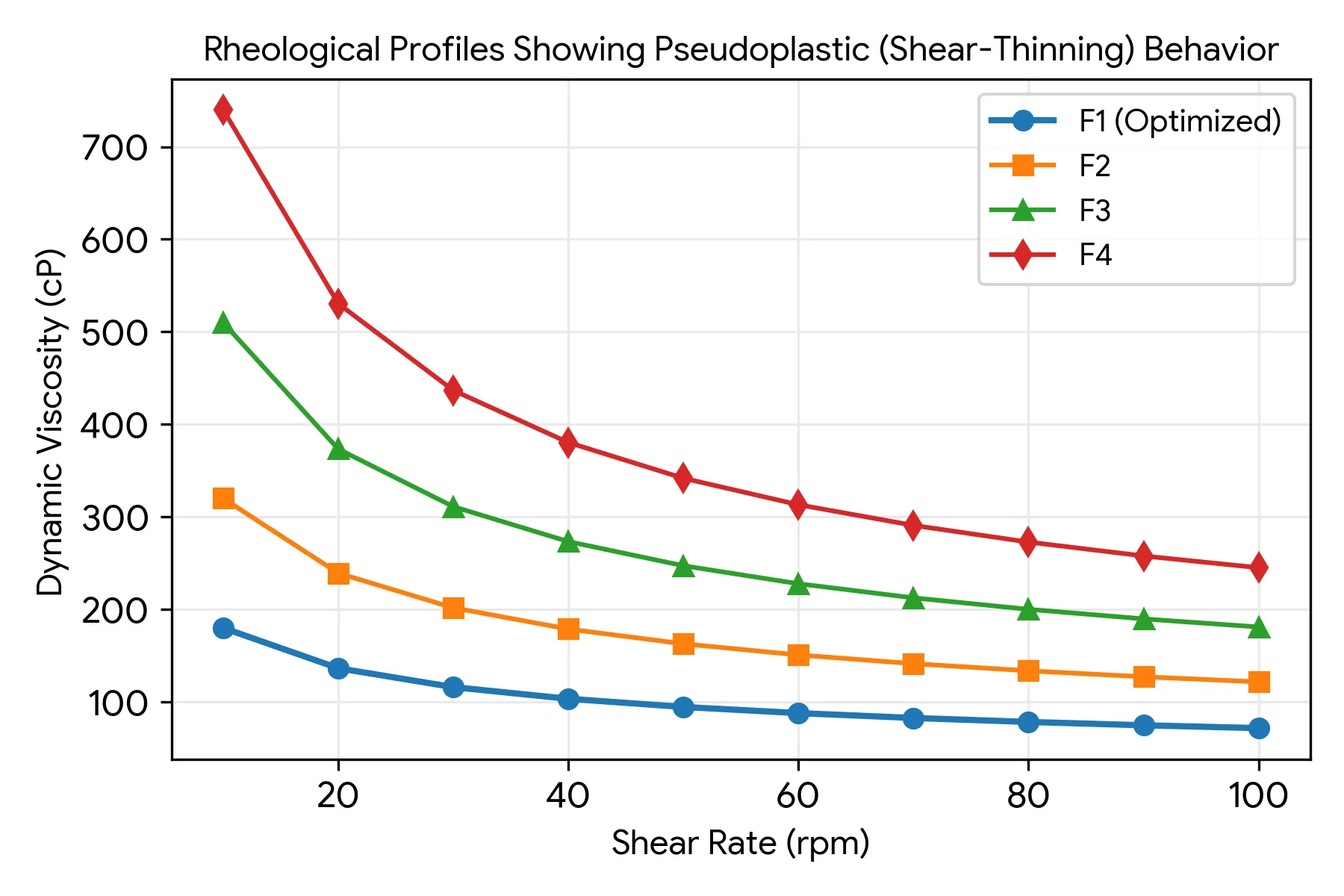
Figure :1 Rheological Profiles Showing Pseudoplastic (Shear-Thinning) Behavior of Formulations F1-F4.
Table :3 pH, Viscosity, and Clarity Profiles of Formulated Batches
|
Batch ID
|
Pre-Gelation pH
|
Post-Gelation pH
|
Viscosity (cP)
|
Visual Appearance
|
Clarity Index
|
|
F1
|
6.5
|
6.9
|
180
|
Pale Yellow Solution |
Excellent / Fully Clear |
|
F2
|
6.7
|
6.7
|
320
|
Pale Yellow Solution |
Excellent / Fully Clear |
|
F3
|
6.6
|
6.5
|
510
|
Slightly Hazy Sol
|
Slightly Translucent
|
|
F4
|
6.2
|
6.4
|
740
|
Slightly Hazy Sol |
Slightly Translucent |
3.3 FTIR spectra mixture of drug and polymers:
The FTIR spectrum of the mixed sample produced a broad peak around 3400 cm1, which was associated with O-H stretching vibrations of hydroxyl groups present in the polymer structure. The highest point at 2920 cm1 is an indication of aliphatic C–H stretching. C=O stretching of the ester or carboxylic groups is indicated by a strong band of approximately 1730 cm1. The peaks observed at approximately 1630 cm1 signify vibrations of carboxylate ions, and absorption bands were recorded between 1100 and 1600. 1050. These specific spikes confirm the presence of polymer functional groups and indicate a lack of major interaction between drug and polymers.(13)
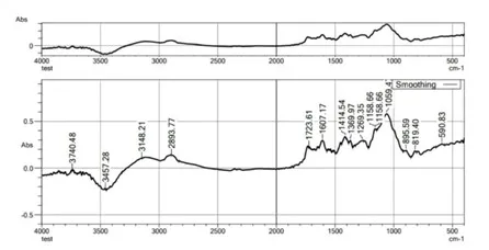
Figure :2 FTIR spectra mixture of drug and polymers
Table :4 FTIR spectra mixture of drug and polymers
|
S.No
|
Peak Position (cm⁻¹) |
Observed Functional Group
|
Interpretation
|
|
1
|
~3400–3450
|
O–H stretching
|
Broad peak indicating hydroxyl groups of polymers (pectin/gellan gum) |
|
2
|
~2920
|
C–H stretching
|
Aliphatic CH stretching vibration of polymer backbone |
|
3 |
~1720–1740 |
C=O stretching |
Carbonyl group of ester or carboxylic acid |
|
4 |
~1630–1650 |
COO⁻ stretching |
Asymmetric stretching of carboxylate ions |
|
5 |
~1450 |
C–H bending |
CH₂ bending vibration |
|
6 |
~1380 |
C–H deformation |
Methyl group bending vibration |
|
7 |
~1250 |
C–O stretching |
Ester C–O vibration |
|
8]\ |
~1100–1150 |
C–O–C stretching |
Glycosidic linkage of polysaccharide |
|
9 |
~1020–1050 |
C–O stretching |
Alcoholic C–O vibration |
|
10 |
~820–850 |
C–H bending |
Fingerprint region confirming polysaccharide structure |
3.4 UV Spectrophotometric Analysis
3.4.1 Preparation of Stock Solution
Ciprofloxacin Drug (10 mg) was accurately weighed and transferred in 100 ml volumetric flask and dissolved in a small amount of distilled water by shaking gently and volume was made up to 100 ml distilled water in volumetric flask. The resultant solution was concentration 100 μg/ml was formed. Then 10 ml of this solution was taken in 100 ml of volumetric flask and volume made u with water to from stock solution.(14,15)
3.4.2 Preparation of Calibration Curve
Table :5 Calibration data of Ciprofloxacin at 273 nm.
|
s.no |
Concentration(µg/ml) |
Absorbance at 273nm |
|
1 |
10 |
0.248 |
|
2 |
20 |
0.459 |
|
3 |
30 |
0.648 |
|
4 |
40 |
0.848 |
|
5 |
50 |
1.001 |
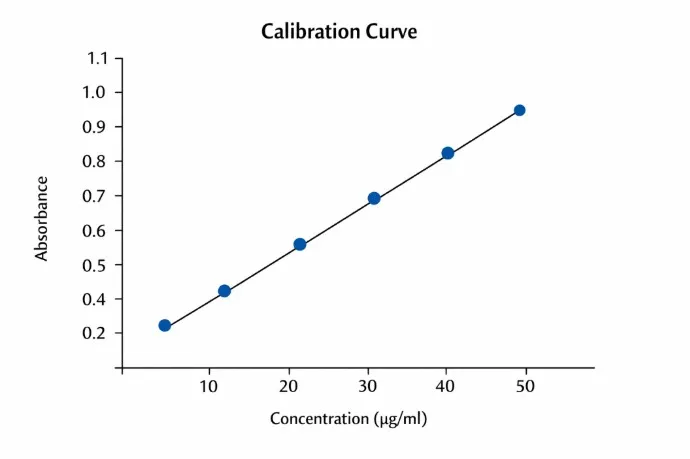
Figure :3 Standard Calibration Curve Of Ciprofloxacin in Distilled Water
3.5 Gelling Capacity
The gelling capacity was analyzed by of the prepared formulation was determined by placing a drop of the formulation in a vial containing 2 ml of freshly prepared simulated tear fluid and visually observed.(16)
Table :6 Coding of Gelling Capacity
|
S.no |
Observation |
Coding |
|
1 |
No elation |
_ |
|
2 |
Gelation occurred in few minutes and remained for few hour |
+ |
|
3 |
Gelation occurred in immediate, remained for few hour |
++ |
|
4 |
Gelation immediate, and for extended period |
+++ |
|
5 |
Very stiff gel |
++++ |
Table :7 Coding of Gelling Capacity Of formulation
|
S.no |
Formulation |
Coding |
|
1 |
F1 |
+++ |
|
2 |
F2 |
++ |
|
3 |
F3 |
+ |
|
4 |
F4 |
++++ |
3.6 Melting Point
The Melting point of Ciprofloxacin HCl was studied in the mid of 290-300°C which was found to be within the limits of the declared scale i.e 273°C indicated the authenticity of drug.(17)
Table :8 Melting Point of formulation
|
S.no |
Formulation |
Observed melting point |
|
1 |
F1 |
267 |
|
2 |
F2 |
256 |
|
3 |
F3 |
278 |
|
4 |
F4 |
289 |
3.7 In-Vitro Drug Release Kinetics
The in-vitro dissolution profiles highlighted a clear advantage for our optimized in-situ gel (F1) over classic, fast-draining eye drops. In an environment mimicking human tears (pH 7.4), the ciprofloxacin migrated steadily out of the cross-linked polymer web. During the first hour, F1 delivered a controlled burst release of 21.19%, reaching a cumulative release of 77.33% by hour 8. The mathematical modeling confirmed that this profile fits zero-order release kinetics closely, as mapped out in Figure 2.(18,19)
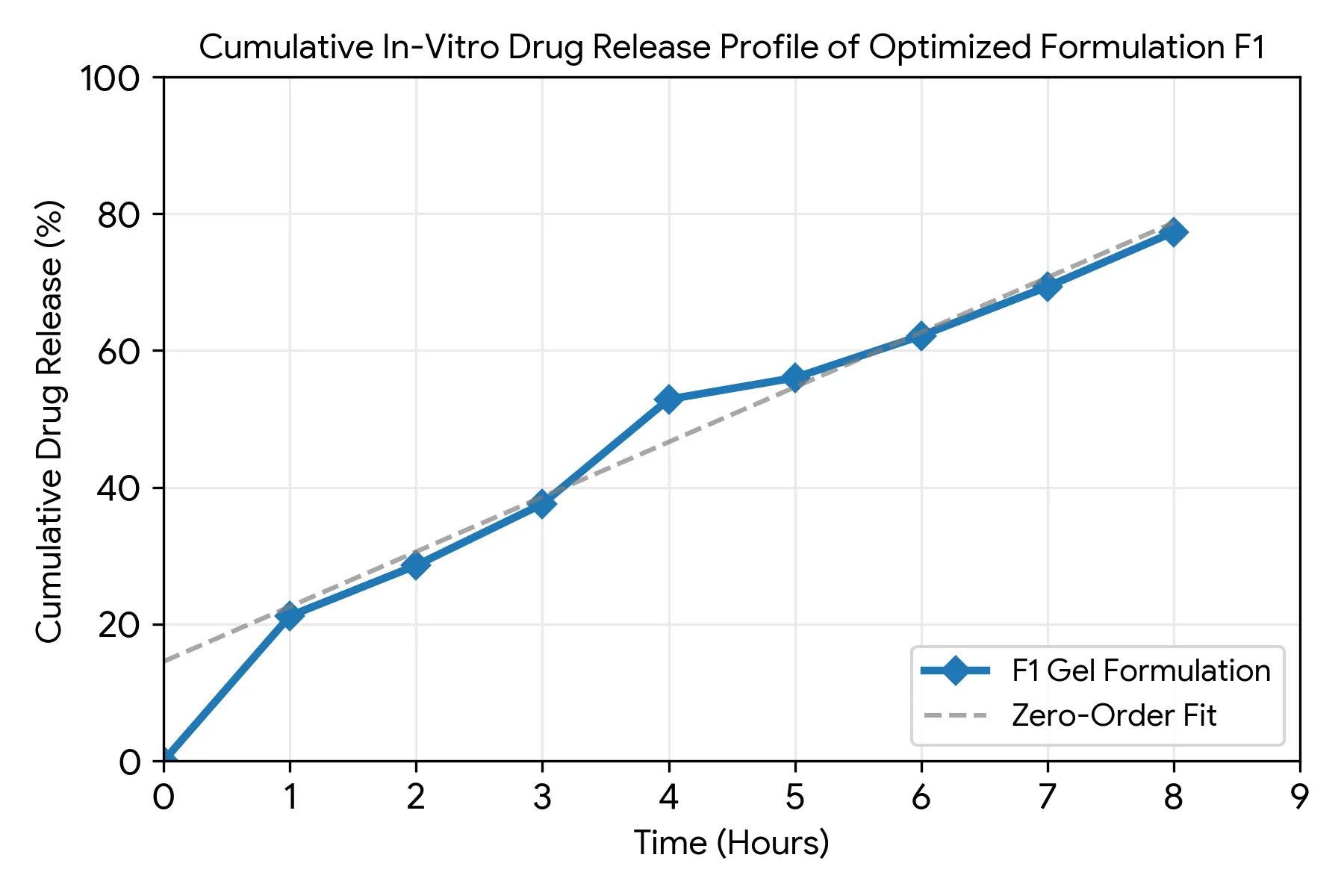
Figure :4 Cumulative In-Vitro Drug Release Profile and Zero-Order Kinetic Modeling for Optimized Formulation F1.
Table :9 In-Vitro Cumulative Dissolution Kinetics for Optimized Formulation F1
|
Sampling Interval (Hours)
|
Receptor Medium Volume (mL)
|
Spectrophotometric Absorbance (273 nm)
|
Cumulative Drug Release (%)
|
|
1 |
10 mL |
0.193 |
21.19% |
|
2 |
10 mL |
0.235 |
28.58% |
|
3 |
10 mL |
0.286 |
37.57% |
|
4 |
10 mL |
0.373 |
52.87% |
|
5 |
10 mL |
0.391 |
56.03% |
|
6 |
10 mL |
0.426 |
62.17% |
|
7 |
10 mL |
0.468 |
69.33% |
|
8 |
10 mL |
0.512 |
77.33% |
3.5 Sterility Evaluation
Throughout the 14-day incubation window in both SCDM and FTM media, we observed no cloudiness, sediment, or visible colony growth, proving optimal sterility.(20)
CONCLUSION
In this study, we successfully developed a responsive, ion-activated ciprofloxacin ophthalmic in-situ gel using a combination of natural pectin and gellan gum. The optimized formulation, F1, behaves like a fluid liquid in the bottle for easy drop application, but transforms into a stable gel matrix the moment it touches simulated tear fluid, presenting an ideal choice for sustained ocular therapeutics.
REFERENCES


Srinath. R, Joe lithiya. D, Subramani. C, Naveen Kumar. S, Eswaramoorthi. M, Formulation And Evaluation of An In-Situ Ophthalmic Gel of Ciprofloxacin Using Natural Polymers Pectin and Gellan Gum, Int. J. of Pharm. Sci., 2026, Vol 4, Issue 8, 5181-5189, https://doi.org/10.5281/zenodo.22203859
 10.5281/zenodo.22203859