

We use cookies to ensure our website works properly and to personalise your experience. Cookies policy
Department of Pharmaceutics, Prasanna College of Pharmacy, Laila, Belthangady, Dakshina Kannada, Karnataka, India-574214.
Oral ulcers are common lesions of the oral mucosa that cause pain and discomfort during eating, drinking, and speaking. Conventional topical formulations often have limited retention at the site of application due to the continuous flow of saliva. The present study aimed to formulate and evaluate a mucoadhesive buccal film containing Vitex negundo leaf extract for the management of oral ulcers. The herbal extract was prepared by the maceration method using an ethanol–water solvent system. Preliminary phytochemical screening was performed to identify the major phytoconstituents present in the extract. Three buccal film formulations (F1, F2, and F3) were prepared using the solvent casting technique and evaluated for physical appearance, thickness, weight variation, folding endurance, tackiness, surface pH, drug content, moisture characteristics, swelling index, in vitro disintegration, and drug release. Fourier-transform infrared spectroscopy (FTIR) was used to assess the compatibility of the extract with the formulation excipients. The results showed that the prepared films were pale green, transparent, and flexible. The in vitro drug release after 360 minutes ranged from 88.0% to 92.22%, with formulation F3 showing the highest release. The extract also demonstrated concentration-dependent inhibition of protein denaturation, reaching 68.42% at 100 µg/mL, indicating in vitro anti-inflammatory potential. Overall, the findings suggest that Vitex negundo leaf extract can be explored for the development of mucoadhesive buccal films for oral ulcer management.
Herbal medicine, also referred to as botanical or traditional medicine, involves the use of medicinal plants and their extracts for the prevention and treatment of various diseases. Medicinal plants have been used for thousands of years by different civilizations and continue to play an important role in traditional healthcare systems worldwide. Traditional systems of medicine, including Ayurveda, Siddha, Unani, and Traditional Chinese Medicine (TCM), have historically relied on medicinal plants for therapeutic purposes.
In recent years, the use of herbal medicine has increased significantly due to growing interest in natural therapies, concerns regarding the adverse effects of synthetic drugs, and the demand for safer and more personalized healthcare approaches.1-4
Challenges in Herbal Medicines
Quality and Safety Considerations in Herbal Medicines:
These challenges can be addressed through standardized cultivation, extraction, and manufacturing practices, supported by GACP and GMP. Proper plant authentication, quality testing, standardized extracts, and controlled cultivation can improve product quality and consistency. Strengthening regulatory guidelines, pharmacovigilance, clinical research, and awareness among manufacturers, healthcare professionals, and consumers can further promote the safe and rational use of herbal medicines.6
ANATOMY OF THE ORAL CAVITY
The buccal mucosa provides an accessible route for both local and systemic drug delivery. Local delivery allows the drug to act directly at the site of application, while systemic delivery is facilitated by the rich vascular supply of the oral mucosa. Buccal administration can reduce gastrointestinal enzymatic degradation and bypass hepatic first-pass metabolism, thereby potentially improving the bioavailability of suitable drugs. The good vascularity and accessibility of the oral cavity also facilitate drug absorption and provide a convenient and patient-acceptable alternative to other mucosal routes.7
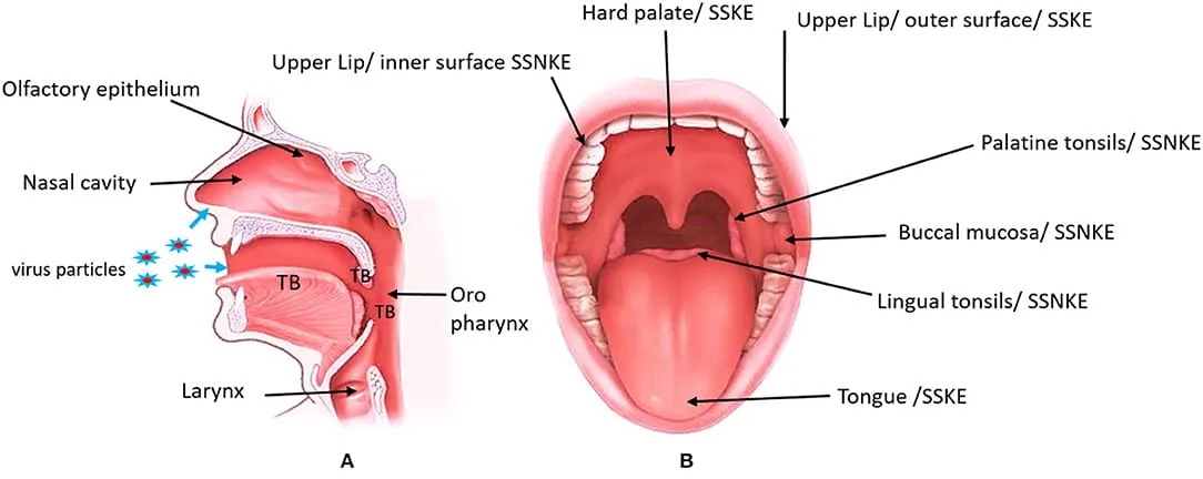
Figure 1: Mucosal Region of Mouth.
The oral cavity is bounded by the lips, cheeks, floor of the mouth, hard palate, and soft palate. The oral mucosa covers a surface area of approximately 170 cm² and consists of two main layers: the superficial stratified squamous epithelium and the underlying lamina propria. The lamina propria is a connective tissue layer containing blood vessels, nerves, supporting fibres, and immune cells. In the buccal region, the oral mucosa also contains a submucosa composed of adipose and glandular tissue, which contains blood vessels, nerve fibres, and minor salivary glands.
The oral mucosa plays a vital protective role by safeguarding the underlying tissues against mechanical trauma, acting as a barrier to microbial invasion, and limiting the penetration of foreign substances, including pharmaceutical agents, into the systemic circulation.8
Oral Mucosa as a Drug Delivery Route
The oral cavity is an easily accessible and highly vascularized site for drug administration. The rich blood supply of the oral mucosa facilitates drug absorption and makes it suitable for both local and systemic drug delivery. Buccal administration can bypass hepatic first-pass metabolism and may reduce gastrointestinal degradation, thereby improving the bioavailability of suitable drugs.
The oral epithelium is a stratified squamous epithelial layer that forms the primary protective barrier of the oral cavity. Its cells are connected by tight junctions, desmosomes, and gap junctions, which maintain tissue integrity and regulate permeability. The buccal mucosa has a cell turnover time of approximately 25 days.
Depending on the location, the epithelium may be keratinized or non-keratinized. Keratinized epithelium is present in the hard palate, gingiva, and dorsal surface of the tongue, providing protection, whereas non-keratinized epithelium lines the buccal mucosa, lips, soft palate, ventral tongue, and floor of the mouth, offering greater flexibility for chewing and speaking.
The thickness of the oral epithelium varies in different regions, with the buccal mucosa measuring approximately 500–600 μm, making it one of the thickest regions. The superficial epithelial cells contain membrane-coating granules that produce lipids, which influence the permeability of drugs across the mucosa. Drug delivery through the oral mucosa is mainly achieved via the buccal and sublingual regions, which together account for nearly 60% of the oral mucosal surface and possess a rich blood supply. These characteristics make them suitable sites for both local and systemic drug delivery, including the treatment of oral ulcers, fungal infections, and periodontal diseases.
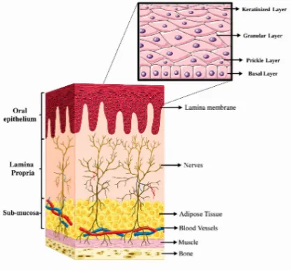
Figure 2: Layers of the Oral Mucosa.
The oral mucosa provides several sites for drug administration, including the sublingual and buccal regions. Among these, the buccal mucosa is particularly suitable for localized drug delivery because a dosage form can be placed directly against the mucosal surface and maintained at the site of application. Therefore, buccal drug delivery is a promising approach for prolonged local treatment of oral ulcerative lesions.9
Buccal drug delivery system:
Buccal drug delivery systems utilize the oral cavity as an effective site for drug administration. This route bypasses gastrointestinal degradation and hepatic first-pass metabolism, improving drug bioavailability. The mucin layer of the buccal mucosa promotes mucoadhesion, allowing prolonged drug residence and enhanced absorption. Proper formulation design also helps maintain drug stability and improves drug permeation across the buccal mucosa.10
Absorption via buccal mucosa:
Drug permeation across the oral mucosa mainly occurs through transcellular and paracellular pathways. Lipophilic drugs generally favour the transcellular route through cell membranes, whereas hydrophilic drugs mainly diffuse through the intercellular spaces via the paracellular route. Both pathways may contribute to absorption, with the route offering the least resistance becoming predominant.11
Factors Affecting Buccal Absorption:
The oral cavity presents a complex environment for drug delivery, as several physiological and anatomical factors can influence drug absorption and reduce the amount of drug available at the absorption site.
1. Membrane Factors
Membrane factors influence buccal drug absorption. These include keratinization, surface area, membrane thickness, mucus layer, lipid content, blood supply, cell turnover and enzymatic activity.
2. Environmental Factors
Saliva: The thickness, composition and movement of saliva affect drug absorption through the buccal mucosa.
Salivary Glands: Minor salivary glands secrete mucus, which helps retain mucoadhesive formulations but may reduce drug permeation.
Movement of Oral Tissues: The buccal region has less movement, making it suitable for mucoadhesive drug delivery. Mucoadhesive polymers help maintain adhesion during speaking, eating and swallowing.12
Advantages of Buccal Drug Delivery System
MECHANISM OF MUCOADHESION
Mucoadhesion is the process by which a polymeric dosage form adheres to the mucus layer or mucosal surface through interfacial interactions between the polymer and mucin. It involves various forces such as hydrogen bonding, electrostatic interactions, van der Waals forces, and polymer chain interpenetration. In buccal films, mucoadhesion helps prolong the residence time of the formulation at the buccal mucosa, thereby supporting localized and sustained drug release.
Mucoadhesion occurs in two stages depending on the nature of the dosage form and its delivery:
Stage I: Contact Stage
During the contact stage, the mucoadhesive film comes in contact with the mucosal surface, absorbs moisture, swells and spreads, resulting in close contact and initial adhesion. Mechanical pressure and oral movements may further improve contact between the film and mucosa.
Stage II: Consolidation Stage
During the consolidation stage, the polymer becomes hydrated and its chains interpenetrate with the mucus layer, forming strong adhesion through hydrogen bonding, van der Waals forces, electrostatic and hydrophobic interactions.
This stage is explained by two theories:
- Diffusion Theory: Polymer chains penetrate and intermix with mucus glycoproteins, forming secondary bonds that provide strong adhesion.
-Dehydration Theory: The polymer absorbs moisture from the mucus until osmotic equilibrium is reached, promoting adhesion. This theory is not applicable to solid or pre-hydrated formulations.14
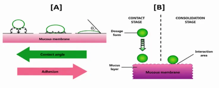
Figure 3: Structural Representation of Various Theories of Mucoadhesion;
[A] Wetting, [B] Adsorption
ORAL ULCER
Oral ulcers are painful lesions that develop on the mucous membrane of the oral cavity, commonly appearing as red, yellow or white sores on the inner cheeks and lips. They may occur due to various factors, including traumatic injuries, bacterial, viral and fungal infections, immune disorders, nutritional deficiencies, medication side effects, systemic diseases, stress, food allergies and poor oral hygiene. These ulcers can cause significant pain and discomfort during eating, drinking and speaking.15,16
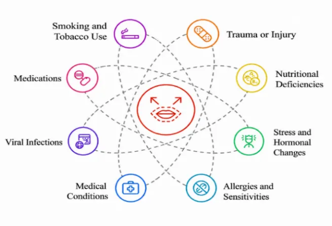
Figure 4: Causes of Mouth Ulcer
The primary symptoms of mouth ulcers are the presence of painful sores in the mouth; other symptoms may include;
Classification of Oral Ulcerative Lesions
1.Minor Aphthous Ulcers:
The most common type of recurrent aphthous stomatitis (80%), characterized by small, round or oval ulcers less than 5 mm, which usually heal within 10–14 days without scarring.
2. Major Aphthous Ulcers:
A less common and more severe type (10%), characterized by deep ulcers larger than 1 cm, which may take up to 6 weeks to heal and often leave scars.
3. Herpetiform Ulcers:
Characterized by multiple small ulcers measuring 1–3 mm, which may merge to form larger ulcers. Despite their name, they are not caused by the herpes virus.
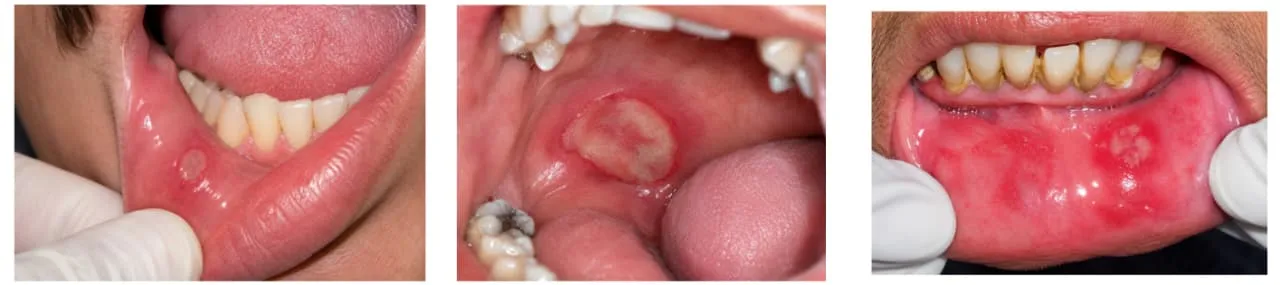
Minor ulcer Top of Form
Bottom of Form
Major ulcer Herpetiformis ulcer
Figure 5: Types of Ulcers
Oral Ulcerative Lesions Associated with Inflammatory/Immune Conditions
1. Lichenoid Lesions (Oral Lichen Planus):
A chronic immune-mediated disorder affecting the oral mucosa, causing redness, ulceration, pain and difficulty in eating and speaking.
2. Vesiculobullous Disorders:
Autoimmune conditions such as pemphigus vulgaris and mucous membrane pemphigoid that cause painful erosions and ulcers in the oral cavity.
3. Traumatic Oral Ulcers:
Caused by physical, chemical, thermal or mechanical injuries, such as cheek biting, sharp teeth, ill-fitting dental appliances and aggressive tooth brushing.18,19
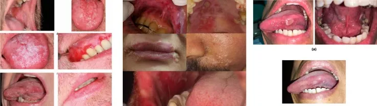
Figure 6: Types of Oral Ulcerative Lesions
Lichenoid lesions Vesiculobullous lesions Trauma-related lesions
Vitex negundo:
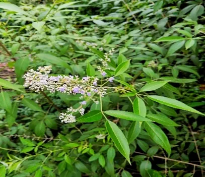
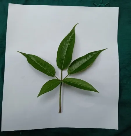
Figure 7: Vitex negundo Leave
Vitex negundo L. (Verbenaceae) is an aromatic shrub or small tree that grows up to 4.5m in height and is widely distributed throughout India.
Pharmacological activities:
It possesses diverse pharmacological properties, including anticancer, anti-inflammatory, antiseptic, antipyretic, diuretic, antihistaminic, antioxidant, antibacterial, antifungal, analgesic, hepatoprotective, insecticidal, mosquito-repellent, wound-healing, and central nervous system depressant activities.20
Vitex negundo, commonly known as the five-leaved chaste tree, is a medicinal plant widely distributed across Asia, Africa, and South America. Ayurveda, Siddha, and Unani medicine have traditionally used it to treat fever, inflammation, pain, and respiratory disorders.
Phytochemistry of Vitex negundo
Vitex negundo contains a variety of bioactive phytochemical constituents that contribute to its therapeutic properties. The major classes of constituents identified in the plant include flavonoids, iridoids, terpenoids, alkaloids, and essential oils. Researchers have extensively studied these phytochemicals and linked them to various pharmacological activities, supporting Vitex negundo's therapeutic potential.21
Role of Vitex negundoin management of oral ulcer
The plant is well known for its anti-inflammatory, analgesic, antimicrobial, antioxidant, and wound-healing properties. Its leaves contain various bioactive compounds such as flavonoids, casticin, iridoid glycosides, tannins, terpenoids, β-caryophyllene and essential oils, which contribute to its therapeutic effects. These pharmacological activities help reduce pain and inflammation, prevent microbial infections, and promote tissue repair. Therefore, Vitex negundo is considered a promising herbal candidate for the management of oral ulcers.22
BUCCAL FILM
Buccal films are promising dosage forms for drug delivery because of their ease of administration, ability to provide both local and systemic therapeutic effects, strong mucoadhesive properties that prolong drug absorption, and their thin, flexible design, which enhances patient comfort and compliance.
Mucoadhesive buccal films are thin, flexible dosage forms designed to adhere to the oral mucosa for prolonged drug release and absorption. They offer local and systemic drug delivery, improved bioavailability, ease of administration and better patient compliance. Saliva hydrates the film, allowing it to swell, adhere and release the drug in a controlled manner. Compared to gels and ointments, buccal films provide longer residence time, accurate dosing and greater patient comfort. However, they have limitations such as moisture sensitivity, limited drug-loading capacity and the need for acceptable taste and texture.
Various mucoadhesive buccal dosage forms, including tablets, gels, ointments, and films, have been developed to improve drug delivery through the oral mucosa. Among these, mucoadhesive buccal films are preferred because of their flexibility, comfort, and prolonged residence time at the site of application. Unlike gels and ointments, which can be easily removed by saliva, buccal films remain adhered to the mucosal surface for longer periods, ensuring accurate drug dosing, sustained drug release, and improved patient compliance.23
Advantages of Buccal Films
TYPES OF BUCCAL FILM
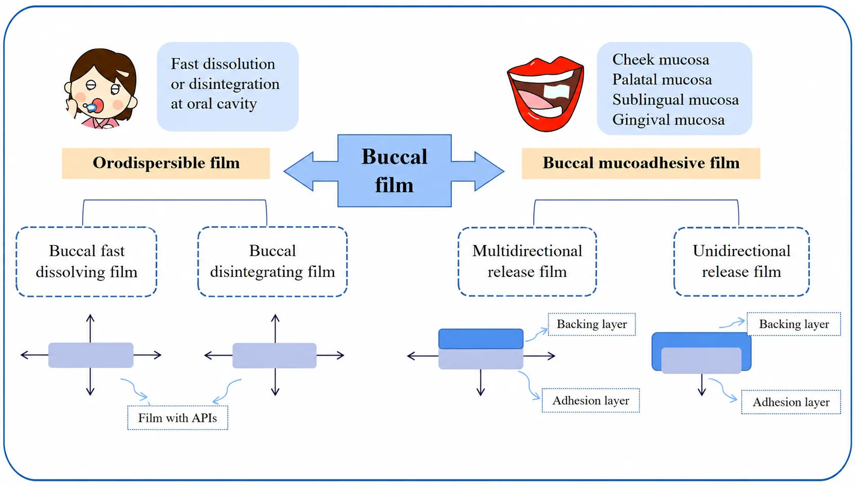
Figure 8: The Classification and Drug Release Direction of Buccal Films.
1. Fast Dissolving Buccal Films (FDDS)
Fast Dissolving Drug Delivery Systems (FDDS) are solid dosage forms that rapidly dissolve or disintegrate in the oral cavity, usually within one minute, without requiring water or chewing. Fast Dissolving Oral Films (FDOFs) are thin polymeric strips placed on the tongue, where they hydrate, dissolve and release the drug. They provide accurate dosing, rapid onset of action, portability and improved patient compliance, particularly in paediatric, geriatric, bedridden and dysphagic patients. Their formulation requires rapid dissolution, sufficient mechanical strength and effective taste masking.
2. Oral Disintegrating Films (ODFs)
Oral Disintegrating Films (ODFs), also known as fast-dissolving or orodispersible films, are thin polymeric dosage forms that dissolve within seconds when placed on the tongue without water or chewing. They form a solution or suspension in saliva and allow rapid drug release. Due to the rich blood supply of the oral mucosa, drugs may be absorbed rapidly into systemic circulation, improving bioavailability and reducing first-pass metabolism. They are formulated using film-forming polymers, plasticizers, active pharmaceutical ingredients, sweeteners, flavouring agents and other suitable excipients. ODFs are particularly useful for paediatric, geriatric and dysphagic patients.
3. Unidirectional Release Buccal Mucoadhesive Films (BMFs)
Unidirectional buccal films consist of a drug-loaded mucoadhesive layer and an impermeable backing layer, which directs drug release towards the buccal mucosa and minimizes drug loss into the oral cavity. Ethyl cellulose (EC) is commonly used as the backing layer due to its hydrophobic and water-insoluble nature. Mucoadhesive polymers such as hydroxypropyl methylcellulose (HPMC), polyvinyl alcohol (PVA) and chitosan are used in the drug-loaded layer to provide adhesion. These films improve drug absorption, prolong residence time and are suitable for both local and systemic drug delivery, particularly in the treatment of oral ulcers.
4. Multidirectional Release Buccal Mucoadhesive Films (BMFs)
Multidirectional buccal films release the drug in multiple directions within the oral cavity. They generally consist of a drug-loaded mucoadhesive layer and a backing layer. Polymers such as HPMC, sodium carboxymethyl cellulose (CMC-Na), hydroxypropyl cellulose (HPC), PVA and polyvinylpyrrolidone (PVP) are used to provide adhesion, flexibility and controlled drug release. These polymers form hydrogen bonds with the mucosal surface, increasing the residence time and improving drug absorption. The drug release profile can be modified by changing the polymer composition and viscosity. These films are suitable for both local and systemic drug delivery. This version is suitable for your Chapter 1 – Introduction while retaining the important technical details.27-30
MATERIALS AND METHODS
MATERIALS USED IN BUCCAL FILM FORMULATION:
The mucoadhesive buccal film was formulated using Vitex negundo extract as the active ingredient, which provides anti-inflammatory, antioxidant and analgesic properties. HPMC K15M was used as the film-forming polymer, while Carbopol 934P acted as a co-polymer to enhance mucoadhesion. Glycerol was incorporated as a plasticizer to improve flexibility, stevia as a sweetening agent to mask the bitter taste, and peppermint oil as a flavouring agent to improve palatability. Ethanol and distilled water were used as solvents for the preparation of the buccal film.
Table 1: Formulation Ingredients of Mucoadhesive Buccal Film and Their Uses
| INGREDIENTS | USES |
|---|---|
| Vitex negundo extract | Anti-inflammatory, antioxidant and analgesic |
| HPMC K15M | Polymers |
| Carbopol 934P | Co-Polymers |
| Glycerol | Plasticizer |
| Stevia | Sweetening Agent |
| Peppermint oil | Flavouring Agent |
| Ethanol | Solvent |
| Distilled water | Solvent |
METHODOLOGY:
1. PREPARATION OF EXTRACTS:
The leaves of Vitex negundo were collected from the local area and thoroughly washed with water to remove adhering dirt and other foreign matter. The washed leaves were shade-dried at room temperature until completely dry. The dried leaves were then coarsely powdered using a mechanical grinder and stored in an airtight container until further use. The extraction was carried out by the maceration method, in which the powdered plant material was soaked in ethanol at room temperature for approximately three days without disturbance to facilitate the extraction of phytochemicals. After maceration, the mixture was filtered to obtain the ethanolic extract. The filtrate was then concentrated under reduced pressure using a water bath maintained at 40–50°C until a semi-solid extract was obtained.31,32
2. PRE-FORMULATION STUDY:
a. Phytochemical analysis:
Qualitative tests for phytoconstituents
The ethanolic extract of Vitex negundo was subjected to preliminary phytochemical screening to identify the presence of various phytoconstituents. Qualitative tests were performed using specific chemical reagents, and the presence of flavonoids was confirmed by the formation of a yellow colour in the alkali test after adding ethanolic KOH to the extract.33,34
b. UV–Visible Spectrophotometry
UV–Visible spectrophotometry is a widely used analytical technique for determining the absorption characteristics of drug molecules and plant extracts. When exposed to ultraviolet radiation, molecules absorb light at specific wavelengths due to electronic transitions. The resulting spectrum is represented as absorbance against wavelength. In the present study, the maximum absorption wavelength (λmax) of Vitex negundo extract was determined using a UV–Visible spectrophotometer. A solution containing 10 mg of the extract in 100 mL of pH 6.8 phosphate buffer (100 µg/mL) was scanned over the wavelength range of 200–400 nm against a solvent blank to identify its characteristic absorption maximum.35,36
c. Drug Excipient Compatibility Study:
The drug-excipient compatibility studies were conducted to assess the compatibility of Vitex negundo leaf extract with the excipients used in the formulation. Fourier-transform infrared (FT-IR) spectroscopy was employed to identify potential interactions between the extract and the excipients. FT-IR spectra of Vitex negundo leaf extract and its physical mixture with the formulation excipients (peppermint oil, HPMC K15M, Carbopol 934, glycerol, stevioside, ethanol, ethyl cellulose, isopropanol, and acetone) were recorded using an FT-IR spectrometer over the spectral range of 4000–400 cm⁻¹. The spectra of the extract and the physical mixture were compared to identify any significant shifts, disappearance, or appearance of characteristic absorption peaks that could indicate potential interactions between the extract and the excipients.37
3. PREPARATION OF BACKING LAYER:
The backing layer was prepared by dissolving ethyl cellulose (5% w/v) in a mixture of isopropyl alcohol and acetone in a ratio of 3:1. The prepared solution was poured into a Petri plate and dried overnight at room temperature to obtain a thin, uniform backing layer.38
4. METHOD OF PREPARATION OF BUCCAL FILM:
The Vitex negundo buccal films were prepared by the solvent casting method. Initially, water-soluble polymers were dissolved in purified water to obtain a clear, homogeneous solution. The drug extract and other excipients were separately dissolved in a suitable solvent and mixed with the polymer solution under continuous stirring to obtain a uniform casting mixture. The prepared solution was poured into a Petri plate and spread evenly. It was then dried in a hot air oven to form a thin film. Finally, the dried film was carefully removed and cut into the required size and shape.39,40
Table 2: Formulation Chart of Mucoadhesive Buccal Film
| Ingredients | Formulation code | ||
|---|---|---|---|
| F1 | F2 | F3 | |
| Vitex negundo extract | 100 mg | 100 mg | 100 mg |
| HPMC K15M | 200 mg | 250 mg | 300 mg |
| Carbopol 934P | 50 mg | 75 mg | 100 mg |
| Glycerol | 50 mg | 65 mg | 80 mg |
| Stevia | 2.5 mg | 2.5 mg | 2.5 mg |
| Peppermint oil | 1-2 drops | 1-2 drops | 1-2 drops |
| Ethanol | q.s to 10ml | q.s to 10ml | q.s to 10ml |
| Distilled water | q. s | q. s | q. s |
5. EVALUATION OF BUCCAL FILM:
1. Physical appearance:41
Films were checked visually for colour, odour, transparency and flexibility.
2. Thickness measurement:42
The film thickness was measured using a micrometer screw gauge.
3. Weight variation test:43
The buccal films were weighed individually using a calibrated analytical balance. The weight of each film was recorded, and the average weight was calculated.
5. Folding endurance:44
Folding endurance was determined by repeatedly folding the film at the same place until it broke. The number of times the film could be folded in the same place without breaking gives the value of folding endurance.
6. Tack test:45
To evaluate tackiness, the strip was pressed between layers of aluminum foil. The observations were recorded as tack-free, slightly tacky, tacky, or very tacky based on the degree of adhesion of the strip to the aluminum foil.
7. Surface pH of the film:46
The films were allowed to swell by keeping them in contact with 1 ml of distilled water for 1hour at room temperature, and the pH is noted down by bringing the electrode in contact with the surface of the film, allowing it to equilibrate for 1min.
8. Drug Content uniformity:47
Buccal film was dissolved in 100 ml of pH 6.8 buffer separately, and the mixture was suitably diluted. The amount of drug in the film was measured spectrophotometrically. The average drug content was calculated.
9. Moisture loss:
This is used to check the integrity of films. The film was weighed and placed in a desiccator containing fused anhydrous calcium chloride. After 72 hours, it was reweighed, and the percentage moisture will be calculated.
Percent moisture loss 𝐗 100
10. Percentage moisture uptake:48
Films were kept in desiccators containing saturated Ammonium chloride solution and reweighed after 3 days.
Percent moisture uptake 𝐗 100
11. Swelling index:49
The initial weight of the film was determined using a digital balance (W0). Then the films were allowed to swell using phosphate buffer pH 6.8 solution on the surface of the Petri plate and kept in an incubator maintained at 37 °C. The weight of the swollen film was determined (Wt) at predetermined time intervals for 5 min.
S. I
Were,
Wt is the weight of the swollen film after time t
W0 is the initial weight of the film at t = 0
12. In vitro disintegration time:
It was determined visually in a Petri plate containing 2 ml of distilled water, with swirling every 10 seconds. The time at which the film started to break or disintegrate was recorded as the in vitro disintegration time.
13. In vitro diffusion study:50
The in vitro drug release study was carried out using a cellophane membrane. The buccal film was placed in the donor compartment, while the receptor compartment contained phosphate buffer (pH 6.8) maintained at 37 ± 0.5°C under continuous stirring. Samples were withdrawn at predetermined time intervals and replaced with fresh buffer. The samples were analyzed using a UV-Visible spectrophotometer, and the cumulative percentage of drug released was calculated.
14. Anti- inflammatory study:51
Protein denaturation method: In this method, 1% egg albumin solution was prepared by the albumin from the egg and diluting it with distilled water.
The test samples were prepared at concentrations of 10, 20, 50, 100µg/ml. For each sample, the reaction mixture contained 0.2 ml of 1% egg albumin, 2.8 ml of phosphate-buffered saline (PBS, pH 6.4), and 2 ml of the test sample.
The mixtures were incubated at 37°C for 15 minutes and were heated at 70°C for 5 minutes. After cooling, the absorbance is measured at 660 nm using a UV-visible spectrophotometer.
Diclofenac sodium was used as the positive control, while PBS was used as the negative control. A higher inhibition of albumin denaturation indicates greater anti-inflammatory activity.
% Inhibition
RESULTS:
1. PREPARATION OF HERBAL EXTRACTS
The herbal extracts were prepared by the maceration method, employing an ethanol: water solvent system in a 1:10 ratio.
2. Pre-formulation Studies of Herbal Extract:
a. Phytochemical analysis of herbal extract
The phytochemical analysis was performed using standard chemical tests. It shows the presence of flavonoids, tannins, saponins, terpenoids, sterols, carbohydrates, and alkaloids.
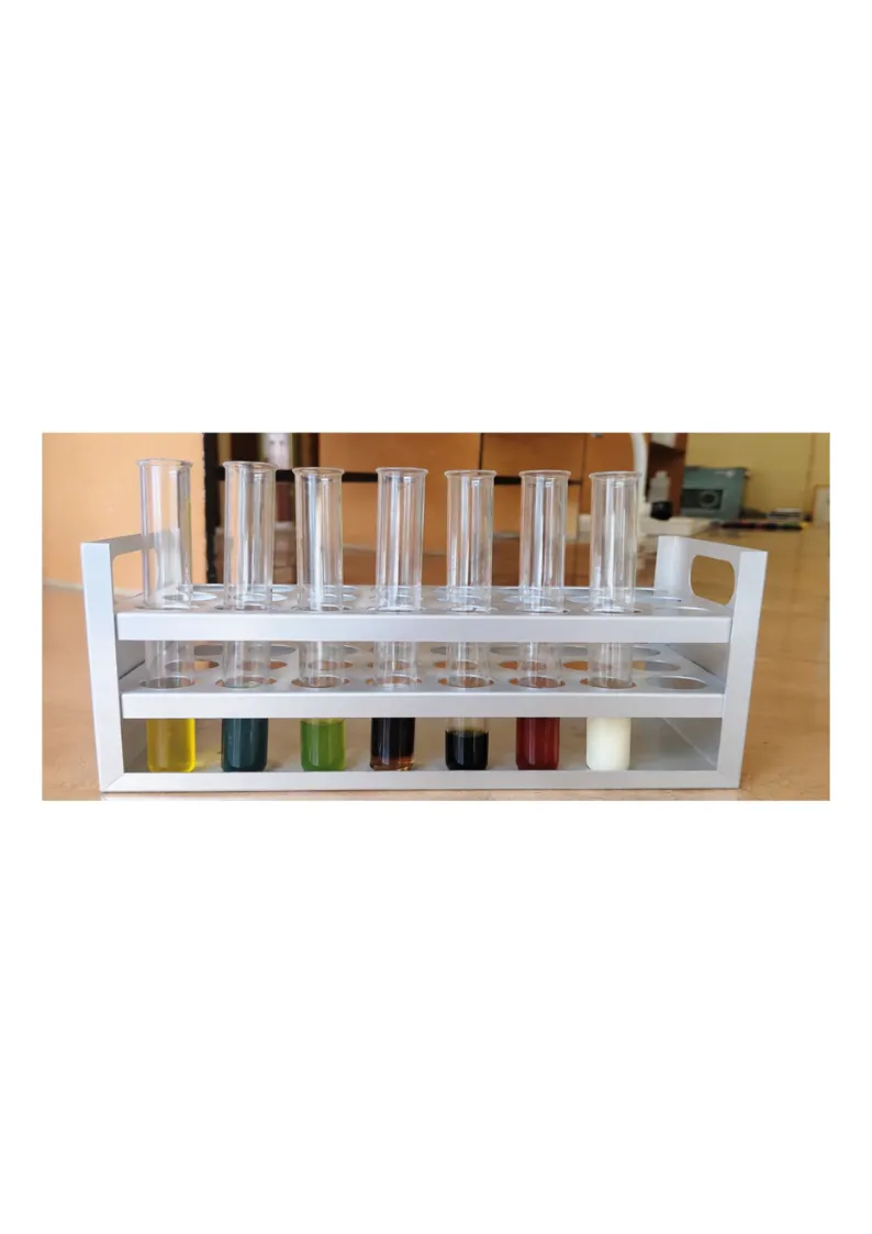
Figure 9: Phytochemical Screening of Vitex negundo Leaf Extract
b. Determination of absorbance maximum (λmax) of Vitex negundo extract:
The absorption spectra of Vitex negundo extract in phosphate buffer pH 6.8 showed the maximum absorption at 206 nm.
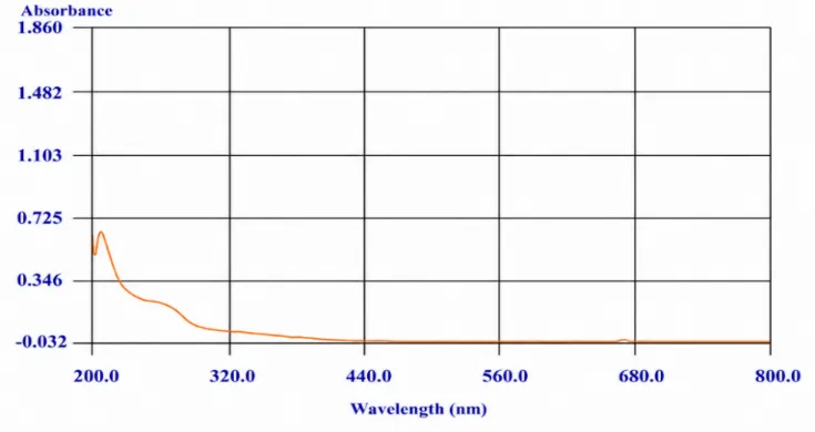
Figure 10: Λmax of Vitex negundo Extract in Phosphate Buffer pH
Table 3: Calibration Data of Vitex negundo Extract
| Concentration(μg/ml) | Absorbance |
|---|---|
| 0 | 0 |
| 2 | 0.173 |
| 4 | 0.367 |
| 6 | 0.564 |
| 8 | 0.763 |
| 9 | 0.869 |
| 10 | 0.981 |
[Figure - see the formatted PDF]
Figure 11: Standard Calibration Curve of Vitex negundo Extract
A standard calibration curve of Vitex negundo was prepared in phosphate buffer (pH 6.8) over a concentration range of 2–10 µg/ml and measured at 206 nm. The curve showed a slope of 0.1003 and an R² value of 0.886.
c. Compatibility Study:
The FT-IR analysis of the extract and formulation excipients is typically performed to detect potential interactions.
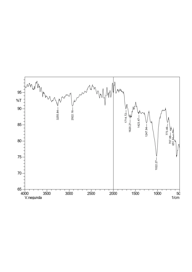
Figure 12: FT-IR Spectrum of Pure Vitex negundo
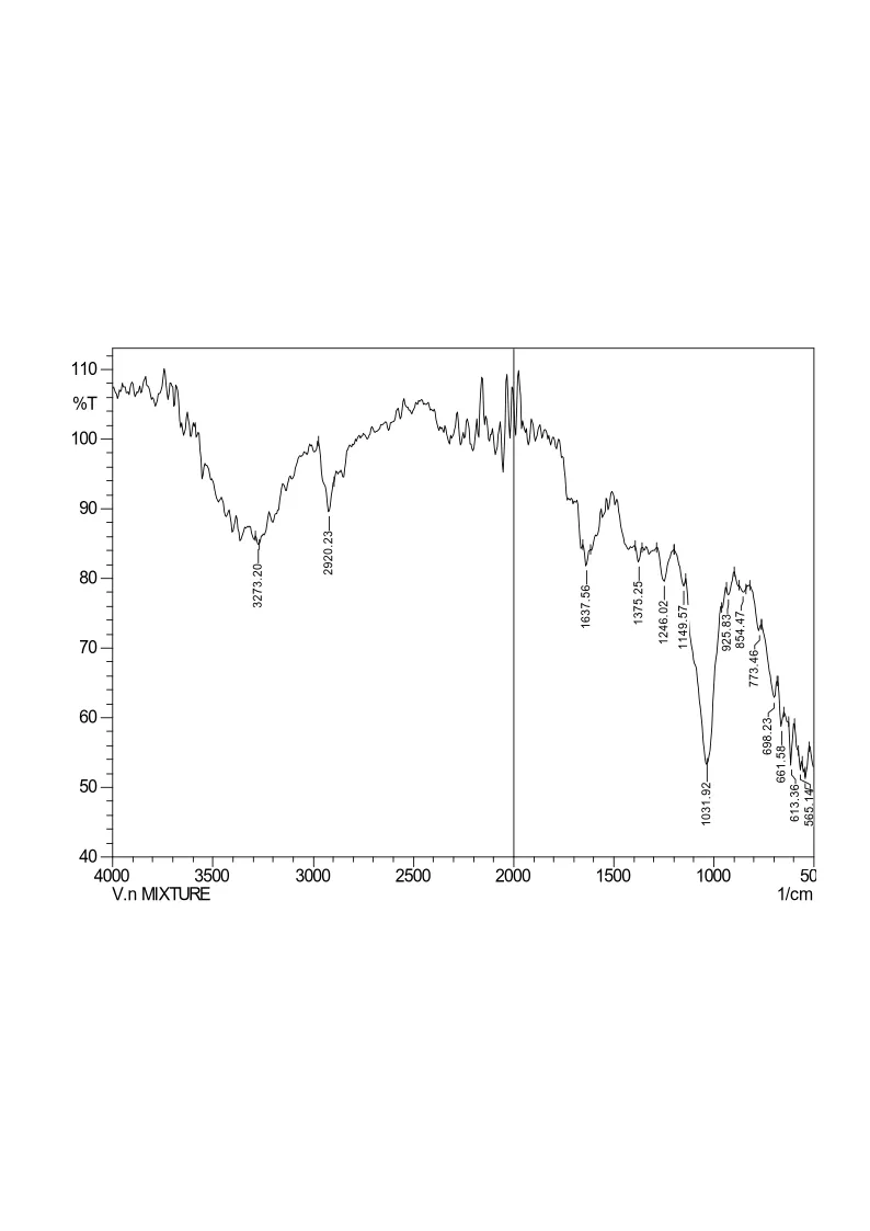
Figure 13: FT-IR Spectrum of the Physical Mixture of the Drug with Excipients
Table 4: Interpretation of FT-IR Spectra of the Drug and Physical Mixture
| Characteristic band |
Characteristic frequency (cm-1) |
Observed frequencies of compounds (cm-1) | |
|---|---|---|---|
| Vitex negundo | Physical mixture | ||
| O-H stretching | 3200 - 3600 | 3256.84 | 3273.20 |
| C-H bending | 2850 – 3000 | 2922.16 | 2920.23 |
| C=O stretching | 1640 - 1742 | 1620.21 | 1637.56 |
| C=C aromatic stretching |
1400 - 1600 | 1423.47 | - |
| N-O stretching | 1080 - 1380 | - | 1375.25 |
| C-O stretching | 1000 - 1300 | 1247.94 | 1246.02 |
| C-O stretching | 1000 - 1100 | 1022.27 | 1031.92 |
| C-H stretching | 919 - 930 | - | 925.83 |
| C-H stretching | 700 - 900 | 773.46 | 773.47 |
The compatibility of Vitex negundo leaf extract with formulation excipients was evaluated using FT-IR spectroscopy. The extract showed characteristic peaks at 3256.84, 2922.16, 1620.21, 1423.47, 1247.94, 1022.27, and 773.46 cm⁻¹. The physical mixture retained most of these peaks with minor shifts and additional bands at 1375.25 and 613.36 cm⁻¹. These findings indicate no major changes in the functional groups, suggesting possible compatibility between the extract and excipients. The observed peaks and assignments are presented in Table 4.
3. PREPARATION OF BACKING LAYER:
Three batches of backing layer were prepared by dissolving the selected water-impermeable polymer in a suitable solvent, followed by the addition of a suitable plasticizer.
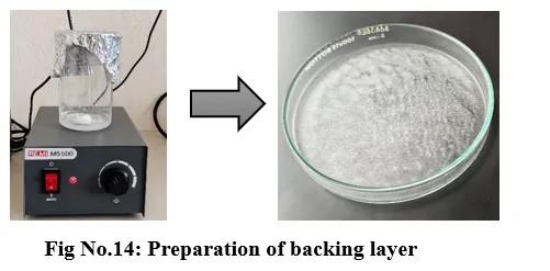
Figure 14: Preparation of Backing Layer
4. FORMULATION OF BUCCAL FILM:
Buccal films containing Vitex negundo leaf extract were prepared by the solvent casting method. The selected polymers were dissolved in a suitable solvent, followed by the addition of the plant extract, plasticizer, and other excipients.
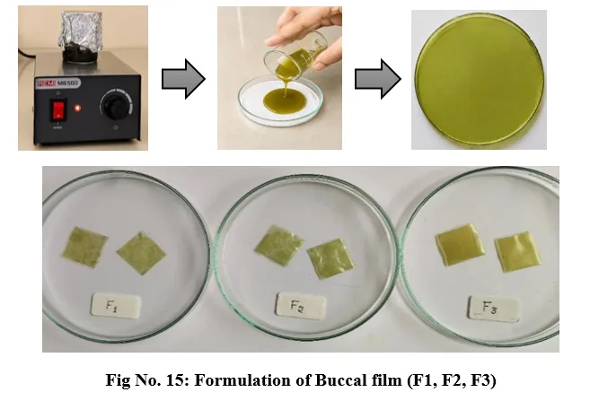
5. EVALUATION OF BUCCAL FILM:
Physical appearance:
Table 5: Physical Appearance of Buccal Film
| Formulation code | Colour | Odour | Transparency | Flexibility |
|---|---|---|---|---|
| F1 | Pale green colour | Characteristic | Transparent | Flexible |
| F2 | Pale green colour | Characteristic | Transparent | Flexible |
| F3 | Pale green colour | Characteristic | Transparent | Flexible |
Table 6: Evaluation of Buccal Films for Thickness, Folding Endurance, Weight Variation, Tack Test, and Swelling Index (%)
| Formulation Code |
Thickness (mm) | Folding Endurance | Weight variation (mg) | Tenacity | Swelling property (%) |
|---|---|---|---|---|---|
| F1 | 0.153 ± 0.005 | 145.3 ±5.03 | 97.7 ±1.51 | ++ | 99.92 ± 0.03 |
| F2 | 0.186 ± 0.013 | 208.3 ±7.64 | 130.6 ±1.49 | +++ | 99.91 ± 0.09 |
| F3 | 0.235 ± 0.011 | 342 ±7.21 | 141.2 ±0.702 | ++ | 84.2 ± 1.12 |
Table 7: Evaluation of Buccal Films for pH, Drug Content (%), Moisture Loss (%), Moisture Uptake (%), and Disintegration Time (min)
| Formulation Code |
pH | Drug Content (%) | Moisture Loss (%) | Moisture Uptake (%) | Disintegration time (min) |
|---|---|---|---|---|---|
| F1 | 6.58 ± 0.08 | 86.8 ± 0.17 | 1.22 ± 0.06 | 6.71 ± 0.04 | 4.50 ± 0.50 |
| F2 | 6.70 ± 0.05 | 84.3 ± 0.18 | 1.68 ± 0.09 | 3.79± 0.04 | 5.53 ± 0.35 |
| F3 | 6.80 ± 0.10 | 97.6 ± 0.16 | 2.74 ± 0.08 | 4.25 ± 0.03 | 6.30 ± 0.30 |
In vitro diffusion study:
The in vitro diffusion test of three buccal film formulations (F1, F2, F3) was performed and found to be in the range of 88.0 – 92.22%.
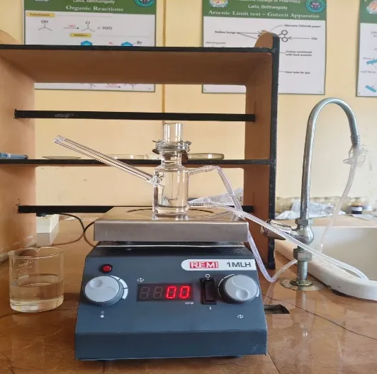
Figure 16: Determination of In vitro Diffusion Stud
Table 8: In vitro Diffusion Profiles of Buccal Film
| Time (min) |
F1 | F2 | F3 |
|---|---|---|---|
| % CDR | % CDR | % CDR | |
| 0 | 0 | 0 | 0 |
| 15 | 4.53 ± 0.03 | 5.73±0.06 | 4.93± 0.12 |
| 30 | 11.02 ± 0.12 | 12.49±0.05 | 11.96± 0.13 |
| 60 | 22.04 ± 0.08 | 23.98±0.06 | 23.93± 0.15 |
| 120 | 35.01 ± 0.15 | 37.5±0.07 | 38± 0.17 |
| 180 | 47.96 ± 0.22 | 50.95±0.08 | 51.95± 0.15 |
| 240 | 61.96 ± 0.23 | 65.46±0.09 | 66.96± 0.14 |
| 300 | 75.01 ± 0.19 | 78.49±0.10 | 79.98± 0.16 |
| 360 | 88.00 ± 0.26 | 90.2±0.12 | 92.22± 0.18 |
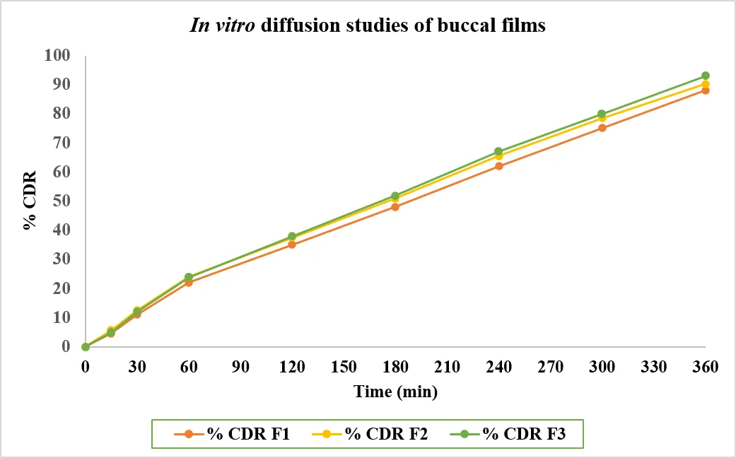
Figure 17: In vitro Diffusion Test of Three Buccal Films
Anti-inflammatory study:
Protein denaturation assay:
The Vitex negundo leaf extract was evaluated at concentrations of 10, 20, 50, and 100 µg/ml. The extract showed concentration-dependent inhibition of protein denaturation, with maximum inhibition observed at 100 µg/ml, indicating promising in vitro anti-inflammatory activity.
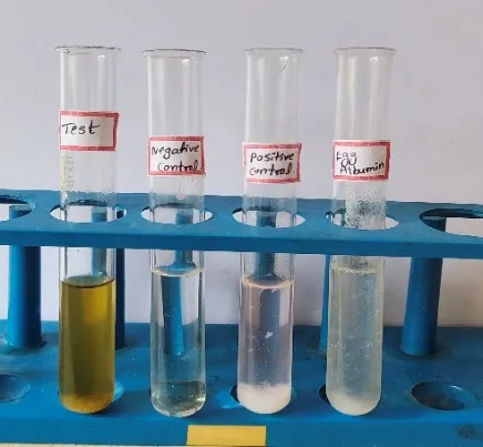
Figure 18: Inhibition of Protein Denaturation
Table 9: Percentage Inhibition of Protein Denaturation Inhibition Assay
| Concentration | % Inhibition |
|---|---|
| 10 | 25.64 |
| 20 | 40.32 |
| 50 | 51.31 |
| 100 | 68.42 |
| Control | 15.84 |
| Diclofenac sodium | 75.65 |
[Figure - see the formatted PDF]
Fig. No.19: Represents the result of the protein denaturation assay
CONCLUSION
The present study was undertaken to formulate and evaluate a mucoadhesive buccal film containing Vitex negundo leaf extract for the management of oral ulcers. The herbal extract was prepared successfully, and preliminary phytochemical screening confirmed the presence of flavonoids, tannins, phenols, saponins, terpenoids, sterols, carbohydrates, and alkaloids. Three formulations (F1, F2, and F3) were prepared using the solvent casting technique and subjected to various physicochemical and in vitro evaluations.
The prepared films showed satisfactory physical characteristics, including a pale green colour, transparency, and flexibility. The in vitro drug release study showed cumulative release values of 88.0%, 91.14%, and 92.22% for F1, F2, and F3, respectively, after 360 minutes. Among the three formulations, F3 exhibited the highest drug release and folding endurance. Furthermore, the protein denaturation assay demonstrated concentration-dependent anti-inflammatory activity of the extract, with 68.42% inhibition at 100 µg/ml.
Based on these findings, Vitex negundo leaf extract appears to be a promising herbal ingredient for the development of mucoadhesive buccal films. This dosage form may offer the advantage of prolonged contact with the oral mucosa and localized delivery of the herbal extract. However, further investigations, including stability studies, safety evaluation, and in vivo and clinical studies, are necessary to confirm its suitability and therapeutic effectiveness in the management of oral ulcers.
REFERENCES






Jalajakshi, Jayashree. B., Chandana. S., Pratibha, Jeevitha. P., Vidyashree S. P., Dr. M. Mallikarjuna Gowda, Formulation and Evaluation of Mucoadhesive Buccal Film Containing Vitex Negundo LEAF Extract for the Management of ORAL ULCERS, Int. J. of Pharm. Sci., 2026, Vol 4, Issue 11, 2749-2767. https://doi.org/10.5281/zenodo.24331694
 10.5281/zenodo.24331694