

We use cookies to ensure our website works properly and to personalise your experience. Cookies policy
1Department of Pharmaceutics,S.S.P Shiskhan Sanstha’s Siddhi College of Pharmacy, Chikhali, Pune-411062
2Department of Pharmaceutical Chemistry,S.S.P Shiskhan Sanstha’s Siddhi College of Pharmacy, Chikhali, Pune-411062
The present research work was aimed at formulation, optimisation and evaluation of Tacrolimus loaded niosomal gel for the management of rheumatoid arthritis. Method: Niosomal gel was successfully prepared by lipid thin film hydration process and optimised by using 22 full factorial designs with two independent variables (Span 60 concentration, cholesterol concentration) and three dependent variables (vesicle size,entrapment efficiency and in-vitrodrug permeation). Response surface methodology was used to evaluate the effect of all variables. Results: Evaluation of the prepared formulations were conducted to analyze particle size, in-vitro dissolution studies, encapsulation efficiency, zeta potential, viscosity and spreadability. F4 formulation turned out to be the best formulation based on response surface methodology with drug entrapment efficiency of 91.5±1.1%, in-vitro drug release of 96.2% in 12 hrs, and vesicle size 183.6±1.6(nm). Stability studies revealed that all evaluated batches were stable as no significant drug content change was observed with respect to time. Conclusion: The experiment proved that the development of Tacrolimus-loaded niosomal-gel was successful, and they had enhanced the permeability; consistent composition and duration of action prolongation was observed. Therefore, it is concluded that the niosomal gel can be a good treatment for rheumatoid arthritis.
Tacrolimus is a potent macrolide lactone immunosuppressant produced by Streptomyces tsukubaensis. It acts as a calcineurin inhibitor and possesses strong immunomodulatory and anti-inflammatory properties, making it useful in autoimmune and inflammatory disorders.Compared to cyclosporine, Tacrolimus exhibits great immunosuppressive potency at lower doses.
Systematic therapy with application of traditional formulations may increase health and financial costs by reducing the proportion of therapeutic effectiveness to side effects. Perhaps a different way of giving the drug will make it work better. [1]
The topical route of administration is the least invasive method of administering drugs since it enters the body via the skin, has a high drug retention rate, and increases patient compliance by lowering dosage frequency while maintaining excellent safety and efficacy.The invention, optimisation, and assessment of Tacrolimus-loaded niosomal-gel for dermal distribution served as the foundation for the current study.[2,3] Niosomes are a non-ionic vesicular delivery mechanism that can capture lipophilic and hydrophilic substances.
RHEUMATOID ARTHRITIS:
Persistent synovial inflammation, cartilage degradation, bone erosion, and increasing joint deformity are the hallmarks of rheumatoid arthritis (RA), a chronic autoimmune inflammatory disease. It frequently causes discomfort, oedema, stiffness, and decreased mobility in the tiny joints of the hands, wrists, feet, and knees. Pannus development, which invades osseous tissue and chondrix and causes irreparable joint degeneration, is encouraged by chronic inflammation. Figure 1 shows the structural variations between a normal joint and a joint affected by rheumatoid arthritis.[4]
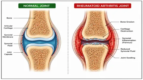
Genetic, environmental, hormonal, and immunological variables interact intricately in the RA pathophysiology. Pro-inflammatory cytokines, such as TNF-α, IL-1, and IL-6, are released when T cells, B cells, macrophages, and synovial fibroblasts are activated. These cytokines cause inflammation and joint degradation. The lungs, skin, eyes, and cardiovascular system are examples of extra-articular organs that RA may impact, exacerbating the disease's severity and morbidity. Despite the adverse consequences and restricted site-specific delivery of traditional treatments such NSAIDs, corticosteroids, and DMARDs. For the enhancement of therapeutic results, sophisticated formulations are being investigated. The potential of niosomes to improve drug stability, permeability, and sustained drug release has drawn a lot of attention.[5]
Although tacrolimus is a powerful calcineurin inhibitor with anti-inflammatory and immunosuppressive qualities, its poor aqueous solubility and systemic side effects limit its clinical use in the treatment of autoimmune diseases. Therefore, by increasing drug retention at the inflammatory site, lowering systemic exposure, and boosting therapeutic efficacy, tacrolimus-loaded niosomal gel represents a viable localised treatment approach for RA. [6]
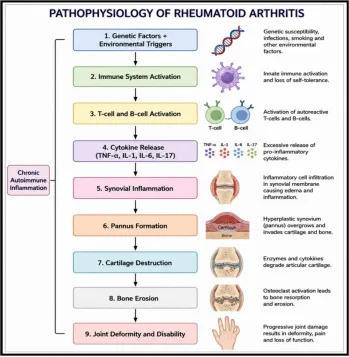
Figure 2: Pathophysiology of Rheumatoid Arthritis [7,8]
NIOSOMES:
Novel drug delivery systems (NDDS) are developed to overcome the limitations of conventional dosage forms by improving drug stability, therapeutic efficacy, controlled release, and patient compliance.
Among NDDS, vesicular systems such as liposomes, transferosomes, ethosomes, and niosomes are widely used for enhancing drug permeation and targeted delivery.[9] These systems can encapsulate both lipophilic and hydrophilic medications, improving bioavailability and therapeutic performance.
Niosomes are non-ionic surfactant-based vesicles formed by self-assembly that can entrap drugs in both the aqueous core and lipid bilayer, making them efficient drug carriers. [10,11,12]
Compared to liposomes, niosomes are more economical, stable, and less prone to oxidative degradation, resulting in better shelf life and pharmaceutical applicability. Surfactants like Span, Tween, and Brij along with cholesterol significantly influence vesicle characteristics like size, stability, and drug release behavior.[13]
Niosomes enhance skin permeation, drug retention, and provide controlled and sustained release, making them appropriate for topical and transdermal delivery. They also protect drugs from degradation and improve stability and bioavailability.[14]
In RA (Rheumatoid Arthritis), noisome help in localising drugs at inflamed sites, thereby reducing systemic exposure and side effects.Tacrolimus-loaded niosomes further enhance solubility, permeation, and retention, and incorporation into a gel system improves application and patient compliance.[15]
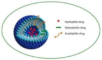
Fig 3: Structure of Niosome [16]
Advantages of Niosomes in Topical Medication Administration:
Because dermal drug delivery (topical drug delivery) techniques eliminate first-pass metabolism, avoid gastrointestinal degradation, and promote patient compliance, they are frequently chosen. However, there are drawbacks to traditional topical dose forms such as creams, ointments, lotions, and gels, such as poor skin penetration, insufficient drug retention, low bioavailability, and corneum instability.[17]
Noisomes are vesicular formulations that can contain both lipophilic and hydrophilic medications. They are made of sterol and non-ionic surfactants. These systems' capacity to increase drug penetration, improve drug retention, and offer regulated and sustained release has drawn a lot of interest for topical and transdermal delivery.[18]
As demonstrated, noisome improve medication penetration by interacting with stratum corneum lipid components, enhancing membrane fluidity, and enabling greater penetration of encapsulated medicines into dermal and epidermal layers. Additionally, niosomes serve as drug reservoirs, releasing the medication gradually over time to maintain therapeutic levels and lower the frequency of doses.[19]
Table 1: Advantages of Niosomal-Drug Delivery System
|
Advantage |
Therapeutic Benefit |
|
Enhanced Permeation |
Improved skin penetration |
|
Sustained Release |
Prolonged drug action |
|
Drug Protection |
Increased stability |
|
Targeted Delivery |
Better localization |
|
Reduced Toxicity |
Fewer systemic effects |
MATERIAL AND METHODS:
Materials:
The chemicals and the medication utilised in this study, drug used in the present study, namely tacrolimus purchased from Majisa Pharmaceuticals 102, ram rahim colony,punayata road pali Pali Rajasthan-306401, cholesterol, and span 60, cholesterol,chloroform, methanol,PBS(Phosphate Buffer Saline), Carbopol 934, Triethanolamine,Glycerin and distilled water were used. All reagents were of the highest analytical grade.
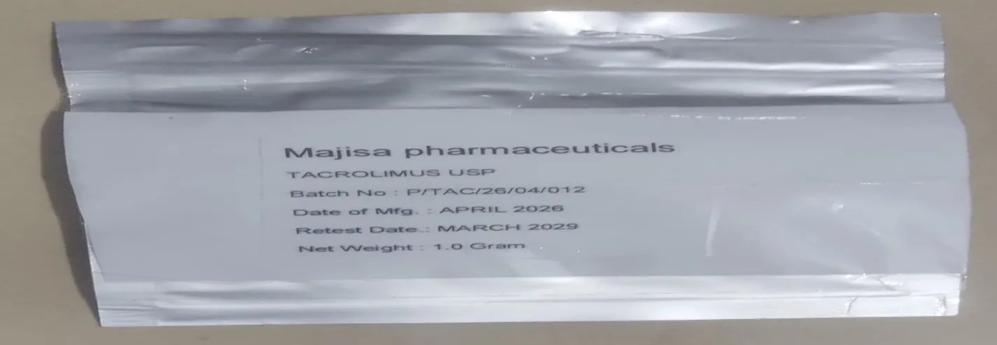
Fig 4: Tacrolimus drug
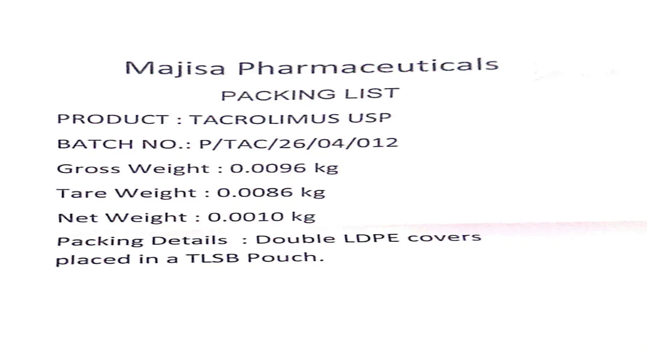
Table 2: List of materials used [20]
|
Sr.No. |
Material |
Category/use |
|
1 |
Tacrolimus |
Active pharmaceutical ingredient |
|
2 |
Span 60 |
Non-ionic surfactant |
|
3 |
Cholesterol |
Vesicle stabilizer |
|
4 |
Chloroform |
Organic solvent |
|
5 |
Methanol |
Organic solvent |
|
6 |
PBS (Phosphate Buffer Saline) |
Hydration and dissolution medium |
|
7 |
Carbopol 934 |
Gelling agent |
|
8 |
Triethanolamine |
PH adjustment agent |
|
9 |
Glycerin |
Humectant and stabilizer |
|
10 |
Distilled Water |
Vehicle |
Preformulation Study:
Active pharmaceutical ingredient (API) Tacrolimus was identified by analysis of absorption maxima by UV spectrophotometer in methanol and scanned Shimadzu-180. Spectrophotometer UV.Organoleptic characterization of Tacrolimus was completed by visual and sensory examination to determine its physical appearance, color, texture, and odor.[21,22]The sample of the drug was compared with reported standard characteristics to confirm its identity, purity, and suitability for formulation development.Solubility studies of Tacrolimus were performed in different solvents such as water, methanol,and allowed to reach equilibrium under controlled conditions.The point of fusion of Tacrolimus was determined to confirm its identity and assess purity. A small amount of Tacrolimus was filled into a capillary tube and subjected to gradual heating using a melting point apparatus. Drug compatibility study was taken and interactions between Tacrolimus and formulation excipients of niosomal gel were carried out.The compatibility was assessed using Fourier Transform Infrared Spectroscopy (FTIR) and Differential Scanning Calorimetry (DSC). The obtained spectra and thermograms were analyzed in order to identify any noteworthy alterations in functional groups or thermal behavior.
Fig 5: Niosome Formation I
Fig 5: Niosome Formation II
Methods of Preparation for Niosomes:
A Bangham method (lipid thin film hydration method) was accustomed to preparing niosomes. Briefly, in a round bottom flask required amount of was put into 5 ml of methanol. In a separate beaker, span 60 and cholesterol were dissolved in 5 ml of chloroform. Both organic solutions were mixed, and the organic solvent was evaporated until a complete dry film was obtained under reduced pressure using a rotary evaporator. Then this dry film was moisturized using a phosphate buffer with a pH value of 7.4.[23]
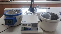
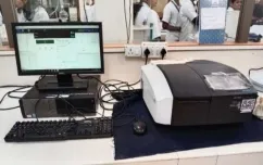
Figure 7: UV–Visible spectrophotometer [25]
The compositions of all four formulations are mentioned in Table 3.
Table 3: Composition of Tacrolimus Niosomal Formulation
|
Ingredient |
Batch 1(F1) For 10g |
Batch 2(F2) For 10g |
Batch3(F3) For 10g |
Batch4(F4) For10g |
|
Tacrolimus (Drug) |
10mg |
10mg |
10mg |
10mg |
|
Span 60 |
50mg |
50mg |
50mg |
50mg |
|
Cholesterol |
50mg |
50mg |
50mg |
50mg |
|
Chloroform: Methanol |
q.s |
q.s |
q.s |
q.s |
|
PBS (Hydration medium) |
q.s |
q.s |
q.s |
q.s |
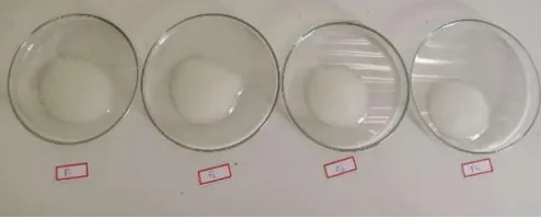
Figure 8: Formulation of Tacrolimus Niosomal gel batches
Optimization of Various Parameters of Niosomal Gels by Factorial Design:
Response surface methodology served to evaluate the effect of various independent factors on formulation parameters. The four formulations of niosomal gels loaded with tacrolimus were developed using Factorial 22. The experiment's design software was utilized for the calculation of the response. As shown in Table 4, two independent factors—Span 60 concentration (X1) and cholesterol concentration (X2)—were chosen. In relation to these, three dependent variables were chosen: Y?: Vesicle Size (nm), Y?: % of Entrapment Efficiency and Y3: In-vitro Drug Permeation (%). These responses were selected because they directly influence drug delivery
efficiency, stability, and therapeutic performance of the non-ionic surfactant vesicular gel.Low and high (-1, +1) were the two levels at which the two independent variables were chosen. [26,27]
Table 4: The Independent Variables
|
Independent Variable |
Symbol |
Low Level (-1) |
High Level (+1) |
|
Span 60 Concentration |
X? |
Low |
High |
|
Cholesterol Concentration |
X? |
Low |
High |
Experimental Design Matrix:
The four experimental runs generated using the 2² factorial design are shown below:
Table 5: Experimental Design Four Experimental Runs [28]
|
Formulation |
X? |
X? |
|
F1 |
-1 |
-1 |
|
F2 |
+1 |
-1 |
|
F3 |
-1 |
+1 |
|
F4* |
+1 |
+1 |
Polynomial Equation
The following first-order polynomial equation represented the connection involving the independent variables and response variables:
???? = ????0 + ????1????1 + ????2????2 + ????12 ????1 ????2
where:
Explanation of Regression Coefficients
runs. [29,30,31]
b??: Represents the interaction between Span 60 and cholesterol, showing whether the combined effect differs from their individual effects. An indicator of a positive coefficient is a synergistic effect, whereas a coefficient that is less than zero indicates an antagonistic effect on the response.
Statistical Analysis:
The experimental data were statistically analyzed using ANOVA (Analysis of Variance) to assess the significance of the model and individual formulation The following statistical parameters were considered:
A p-value less than 0.05 was considered statistically significant
Response Surface and Optimisation
Three-dimensional response surface plots and two-dimensional contour plots were generated
to visualise the effect of formulation variables on the selected responses.
Numerical optimisation was executed using the desirability function approach to get the optimised formulation with the desired pharmaceutical characteristics.
Optimization Criteria
The optimised formulation was selected on the basis of the following criteria:
High desirability value.[32]
Advantages of 2² Factorial Design
The use of factorial design offers several advantages:
variability.[33]
The use of the 2² factorial design enabled efficient optimisation of the Tacrolimus-loaded niosomal formulation by establishing a quantitative relationship between formulation variables and decisive quality attributes, thereby ensuring the development of a stable and effective dermal drug delivery system.[34]
Characterisation of Noisome:
The prepared Tacrolimus-loaded niosomal formulations were systematically evaluated to determine their physicochemical properties, vesicular characteristics, stability behaviour, as well as drug release performance. Comprehensive evaluation is essential to verify the suitability of the developed system for effective drug delivery, as these parameters directly influence therapeutic efficiency, skin permeation, stability, and overall formulation performance. The evaluation studies help in selecting the optimised formulation by correlating formulation variables with decisive quality attributes such as particle size, surface charge, drug encapsulation, and release kinetics. [35,36]
Particle Size Analysis
Analysis of particle size was performed using a DLS (Dynamic Light Scattering) technique-based particle size analyser. Before analysis, the niosomal dispersion was appropriately diluted with distilled water for avoiding the multiple scattering effects. The diluted sample was then filled into a clean cuvette and analysed at a fixed scattering angle under controlled temperature conditions. DLS measures Brownian motion of particles and converts it into a distribution of particle size using the Stokes–Einstein equation.
PDI (Polydispersity Index)
The PDI (Polydispersity Index) was determined simultaneously during analysis of particle size using the same DLS instrument. PDI is a numerical value that describes the width distribution of particle size in a formulation. [37,38,39
Table 6: PDI (Polydispersity Index) Values
|
PDI Value |
Interpretation |
|
0.0 – 0.3 |
Highly uniform, monodisperse system |
|
0.3 – 0.5 |
Moderately uniform system |
|
> 0.5 |
Broad distribution, heterogeneous and unstable system |
|
PDI Value |
Interpretation |
Zeta Potential:
The zeta potential of the prepared Tacrolimus-loaded niosomes was determined using a Zeta Potential Analyzer on the basis of Electrophoretic Light Scattering (ELS)principle. Before analysis, the optimized niosomal dispersion was appropriately diluted with double-distilled water to avoid multiple scattering effects and obtain accurate measurements. The specimen sample was transferred into a disposable folded capillary cell, and the analysis was carried out at 25 ± 2°C. The electrophoretic mobility of the vesicles was measured and automatically converted into zeta potential values (mV) using the Smoluchowski equation.[40]
Table 7: Zeta Potential Value
|
Zeta Potential |
Stability Level |
|
0 to ±10 |
Highly unstable |
|
±10 to ±20 |
Incipient instability |
|
±20 to ±30 |
Moderately stable |
|
Greater than ±30 |
Highly stable |
Entrapment efficiency:
Entrapment efficiency serves as a key parameter that determines the quantity of drug successfully encapsulated within the niosomal vesicles. It reflects the capability of the formulation to keep the drug inside the bilayer or aqueous core.
Method: Centrifugation Method
The niosomal dispersion was subjected to high-speed centrifugation (typically 10,000–15,000 rpm for 30–45 minutes). This process separates:
The amount of substance in the supernatant (free drug) was analyzed using UV spectrophotometry at the predetermined λmax of Tacrolimus.
The concentration of free drug present in the supernatant was analyzed using UV spectrophotometry at the predetermined λmax of Tacrolimus.
Formula:
%???????? = (???????????????????? ????????????????−???????????????? ????????????????) ???????????????????? ????????????????× 100
Drug Content:
The consistent concentration of Tacrolimus within the formulation with niosomes were used. This parameter is essential for confirming dose accuracy, formulation reproducibility, and therapeutic reliability.
Morphological Study:
Morphological evaluation of Tacrolimus-loaded niosomes was carried out to examine the shape, surface characteristics, structural integrity, and vesicle formation behavior. This study provides visual confirmation of successful vesicle formation and helps correlate structural features with formulation performance.[41]
Techniques Used:
In-vitro Permeation Examination
To assess the drug's capacity to pass across a dialysis membrane under physiologically realistic conditions, an investigation of in vitro penetration study of niosomal gel filled with tacrolimus was conducted. The study offers details on the medication transport properties, diffusion behavior, and penetration profile of the ready niosomal gel.[42]
Methodology: Sac Dialysis Method Using Franz Diffusion Cell or VDC (Vertical Diffusion Cell)
Using a Franz diffusion cell or VDC (Vertical Diffusion Cell) and the sac dialysis method, the permeation research was conducted. The permeation barrier was a pre-soaked dialysis membrane (MWCO 12,000–14,000 Da). The dialysis sac was put in the donor compartment of the VDC or Franz diffusion cell, firmly knotted, and filled with a predetermined amount of Tacrolimus niosomal gel as well as the experiment, the receptor compartment was filled with PBS (Phosphate Buffer Saline) with pH 7.4 and stored at 37 ± 0.5°C with constant magnetic stirring.
Sampling Procedure:
Aliquots of the receptor medium were removed at predefined intervals (0, 1, 2, 4, 6, 8, 10, and 12 hours) and promptly replaced with an equivalent volume of new phosphate buffer to maintain a constant volume. Using UV-visible spectrophotometry at the preset wavelength, the Tacrolimus content of the removed samples was assessed, and the cumulative % drug permeation was computed. [43,44]
Characterization of Tacrolimus NSV or niosomal gel:
The developed Tacrolimus-loaded NSV was systematically evaluated to assess its physicochemical properties, rheological behavior, drug distribution, and performance characteristics. These evaluations are essential to confirm the suitability of the topical application formulation, ensure patient compliance, and verify its effectiveness as a controlled medication. The parameters of evaluation also help in understanding the relationship between formulation composition and its performance in biological conditions.
Physical Characteristics and Homogeneity
The prepared niosomal gel or nsv was examined visually under normal daylight and against a black and white background to evaluate its appearance and uniformity. The composition was assessed for colour, clarity, consistency, and the presence of NSV was considered. A well-formulated gel should exhibit a smooth, elegant, and homogeneous appearance without grittiness or aggregation. Homogeneity ensures uniform distribution of niosomal vesicles within the gel matrix, which is critical for consistent drug delivery. The absence of phase separation or precipitation during storage further indicates good physicochemical stability and compatibility between niosomes and gel base components.
pH Determination
The niosomal gel's pH was measured using a calibrated pH meter that is digital.Before measurement, the electrode was carefully immersed in the gel sample after suitable dispersion in distilled water to ensure accurate reading.[45] The pH value is an important factor for topical formulations because it directly affects:
The gel formulation was adjusted using triethanolamine to maintain pH within the physiological skin range of 5.5–6.5, which is considered ideal for minimizing irritation and maintaining the natural acid mantle of the skin. A pH within this range also ensures better patient acceptability and long-term stability of Tacrolimus within the formulation.
Viscosity
The niosomal gel's viscosity was measured using a Brookfield rotational viscometer equipped with an appropriate spindle. The measurements were carried out at controlled rotational speed and temperature to ensure reproducibility. Viscosity is a key rheological property that influences:
Higher viscosity formulations generally provide prolonged contact time with the skin, which enhances drug absorption and therapeutic effect. However, excessively high balance between viscosity and spreadability is required for effective topical delivery.[46]
The flow behaviour of the niosomal gel also indicates whether the system exhibits Newtonian or non-Newtonian (pseudoplastic) characteristics, which is common in Carbopol-based gels.
Spreadability
Spreadability is a significant parameter that determines the ease with which the gel can be utilised over the skin surface in a uniform layer. It directly affects patient compliance and dosing accuracy.
The spreadability was ascertained utilising the glass slide method, in which a fixed amount of gel was placed between two glass slides and subjected to a known weight.[47]
The time required for the upper slide to move a specific distance was recorded.
Formula:
???? =???? × ?????T
Where:
Drug Content
Drug content uniformity was evaluated to ensure consistent distribution of Tacrolimus throughout the gel formulation. A known quantity of gel was accurately weighed and subjected to complete disruption using a suitable solvent system such as methanol or phosphate buffer containing organic modifiers. The mixture was sonicated to ensure complete extraction of Tacrolimus from niosomal vesicles and gel matrix. The solution was then filtered and analyzed using UV spectrophotometry at the predetermined wavelength (λmax). [48,49]
Extrudability
Extrudability refers to the ease with which the gel can be expelled from a collapsible tube when pressure is applied. It is an important parameter that reflects the usability and patient convenience of the formulation.
The test was carried out by filling the gel into aluminum or plastic collapsible tubes and applying a standardized force. The quantity of gel extruded in a given time was measured and documented.
Evaluation Criteria:
Smooth and uniform extrusion → good extrudability
Excessive force required → poor extrudability
In Vitro Drug Release by using (VDC) Vertical diffusion cell or Franz diffusion Cell (FDC)
The study of in vitro drug release of Tacrolimus-loaded nsv gel was performed by using a (Franz diffusion cell) apparatus, which is widely used to simulate drug permeation through egg membranes (semi-permeable membrane). A dialysis membrane (MWCO 12,000–14,000 Da) was mounted between receptor and donor compartments to act as a synthetic barrier mimicking skin diffusion. [50,51]
Experimental Conditions:
Temperature: 37 ± 0.5°C
Continuous stirring: to maintain uniform diffusion
A known amount of gel was placed in the donor compartment. At certain intervals of time, samples were withdrawn from the receptor compartment followed by immediate replacement with a fresh buffer to maintain constant volume. The collected samples were analysed spectrophotometrically for Tacrolimus concentration.[52]
Stability Study
Studies on the stability of Tacrolimus-loaded niosomal gel were performed to assess the physicochemical integrity, formulation robustness, and drug retention capability under different environmental conditions. These investigations are essential to make certain that the developed formulation maintains its quality, safety, and effectiveness over the course of its shelf life.The research was carried out in accordance with ICH (International Council for Harmonisation) guidelines, which provide standardized conditions for stability testing of medicinal compounds.[53]
Storage Conditions:
The formulated niosomal gels were kept in storage under the following conditions:
Refrigerated conditions: 2°±8°C
Room temperature conditions: 25C ± 2°C %RH
Accelerated conditions: 40 ± 2°C / 75% RH
Each condition was selected to simulate different storage environments and to predict long-term stability behaviour.[54]
RESULTS AND DISCUSSION:
Preformulation study:
Tacrolimus was observed as a white to off-white crystalline powder with no characteristic odour. The drug exhibited poor aqueous solubility, which justifies the need for a vesicular formulation such as niosomes to enhance its permeability. The melting point or point of fusion of Tacrolimus was discovered to be within the reported range, confirming its purity.
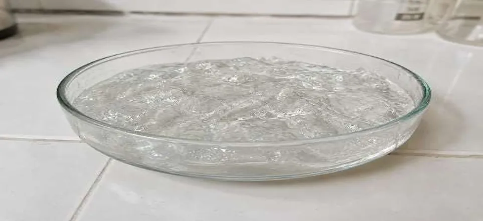
Figure 9: Blank niosomes
CHARACTERISATION OF NIOSOMES:
Particle size:
The size of the particles in the prepared Tacrolimus-loaded niosomes was identified utilising the dynamic light scattering technique. The findings showed that the vesicles were present in the nanometre range, which is favourable for dermal drug delivery. The optimised formulation showed a size of the particles of 183.6 nm, indicating the development of nanosized vesicles. [55]
Polydispersity index (PDI):
The polydispersity index (PDI) was discovered to be 0.266, which indicates uniform distribution of vesicle size. A low PDI value (<0.5) confirms homogeneity and steadiness of the formulation.[56
Table 8: Polydispersity (PDI) Batch values
|
Formulation Code |
PDI (Polydispersity index) Value |
|
F1 |
0.051 |
|
F2 |
0.120 |
|
F3 |
0.235 |
|
F4* |
0.266 |
Morphological Study:
The surface morphology of the niosomes was investigated by the TEM and SEM methods. Niosomes had a spherical shape, smooth, vesicular in nature, and were morphologically similar without agglomerations. [57,58]
Zeta potential:
The Electrokinetic potential, or Zeta Potential, of the niosomal formulation was measured to evaluate the consistency of the vesicular system. The optimised batch showed an electrokinetic potential value of -32. 4mV.The obtained zeta potential value indicates sufficient vesicle surface charge, which prevents aggregation due to electrostatic repulsion between particles. This contributes to the stability of the physical the niosomal system.
Table 9: Zeta Potential Batches Values
|
Formulation Code |
Zeta potential |
|
F1 |
?25.9mV |
|
F2 |
?29.7mV |
|
F3 |
?30.5mV |
|
F4* |
?32.4mV |
EE% (Entrapment efficiency%):
Entrapment efficiency is a key parameter that determines the quantity of medication successfully encapsulated within the niosomal vesicles. It reflects the capability of the mixture to keep the drug inside the bilayer or aqueous core.[59]
Table 10: EE% (Entrapment Efficiency %) Batches Values
|
Formulation Code |
EE% (Entrapment Efficiency%) |
|
F1 |
71.4 ± 1.2 |
|
F2 |
78.6 ± 1.5 |
|
F3 |
84.2 ± 1.3 |
|
F4* |
91.5 ± 1.1 |
In vitro Permeation study:
Tacrolimus niosomal gel compositions (F1–F4) were subjected to an in-vitro permeation research utilizing a VDC or (Franz diffusion cell) and the sac dialysis method. The gel formulation was included in the dialysis membrane after it had been hydrated in a PBS [(phosphate buffer (pH 7.4)] and placed within the diffusion cell. With constant magnetic stirring, the compartment of receptors containing PBS (phosphate buffer, pH 7.4)} was kept at 37 ± 0.5°C. The total % of medication penetration was computed using a UV-visible spectrophotometer after samples were removed at predefined intervals (0–12 h) and swapped out for a new buffer.[60]
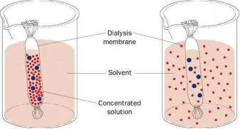
Figure 10: Sac Dialysis Method
Table 11: Permeation Study
|
Time (hrs) |
F1(%) |
F2(%) |
F3(%) |
F4(%) |
|
0 |
0 |
0 |
0 |
0 |
|
1 |
10.2 |
13.8 |
16.5 |
18.9 |
|
2 |
18.6 |
23.4 |
27.8 |
31.6 |
|
4 |
30.5 |
37.9 |
43.6 |
49.8 |
|
6 |
42.3 |
50.8 |
57.4 |
64.9 |
|
8 |
52.7 |
60.9 |
68.2 |
76.5 |
|
10 |
60.4 |
69.2 |
76.8 |
85.3 |
|
12 |
66.8 |
75.6 |
83.5 |
92.1 |
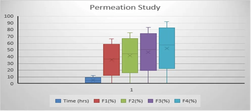
Figure 11: Permeation studies graph
Drug release investigation in vitro:
The In-Vitro drug release investigation study of Tacrolimus niosomal gel compositions (F1–F4) was completed using a membrane for dialysis in a suitable diffusion medium. Samples were removed at prearranged intervals and analysed spectrophotometrically. [61,62,63]
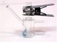
Figure 12: Vertical Diffusion Cell or (Franz diffusion cell)
Table 12: In vitro drug release study [64]
|
Time (hrs) |
F1(%) |
F2(%) |
F3(%) |
F4(%) Optimize batch |
|
0 |
0 |
0 |
0 |
0 |
|
1 |
12.4 |
15.2 |
18.6 |
20.3 |
|
2 |
21.8 |
26.5 |
30.9 |
34.7 |
|
4 |
35.6 |
42.3 |
48.5 |
55.2 |
|
6 |
48.2 |
55.7 |
63.4 |
70.6 |
|
8 |
58.9 |
66.8 |
74.2 |
82.5 |
|
10 |
66.5 |
74.9 |
82.6 |
90..8 |
|
12 |
72.3 |
80.6 |
88.4 |
96.2 |
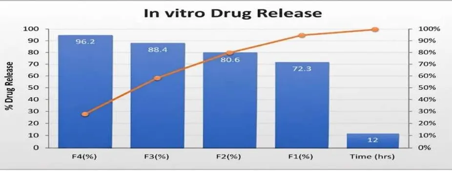
Figure 13: In-vitro drug release graph
CHARACTERISATION OF TOPICAL GEL (NIOSOMAL GEL):
Parameters Evaluated:
The stored samples were assessed for the following parameters:
Physical Appearance
The formulation remained smooth, homogeneous, and free from phase separation, grittiness, syneresis, or colour change during the research period under both storage conditions. No visible microbial growth or precipitation was observed, indicating good physical stability. [65]
pH
The formulation's pH ranged between 5.6 and 6.7 during the study period. Only minor variations were observed, which were within the acceptable range for topical preparations. The stable pH indicates the lack of significant chemical degradation of Tacrolimus or incompatibility between formulation components.
Spreadability
The spreadability of the drug-loaded niosomal gel was discovered to be 19.15 gm cm/sec±0.1, which is in an acceptable range.
Viscosity
The viscosity showed only negligible changes during storage, demonstrating that the Carbopol 934 gel network remained stable. The formulation retained its consistency and exhibited no evidence of polymer degradation or structural breakdown with a viscosity of 10 Pa. [66]
Drug Content
The initial amount of drug content was 98.4%, slightly decreased to 96.2% following three months of rapid storage. However, the amount of drug content remained within the acceptable pharmacopeial limits (90–110%), indicating good stability of the chemical with minimal degradation.
Overall Stability Assessment
Statistical analysis {p > 0.05} revealed no significant variations in the evaluated parameters during the storage period. The optimised formulation exhibited excellent stability under both long-term and accelerated storage conditions. [67,68]
The improved stability may be credited to:
· Cholesterol, which increased the inflexibility of the rigidity of the niosomal bilayer and minimised drug leakage.
· Non-ionic surfactants that maintained vesicle integrity.
· Carbopol gel matrix, which reduced vesicle fusion and aggregation while improving storage stability.
· Appropriate storage conditions that protected the formulation from environmental stress.
The stability analysis confirms that the optimised Tacrolimus-loaded niosomal gel possesses adequate shelf stability and is appropriate for topical use. [69]
Factorial Design
Table 13: Factorial Design [70]
|
Formulation Code |
Cholesterol (mg) (A) |
Span 60 (mg) (B) |
Vesicle Size (nm) |
EE%{Entrapment Efficiency (%)} |
|
F1 |
50 |
100 |
245.8 ± 2.1 |
71.4 ± 1.2 |
|
F2 |
100 |
100 |
221.6 ± 1.8 |
78.6 ± 1.5 |
|
F3 |
50 |
200 |
198.4 ± 2.4 |
84.2 ± 1.3 |
|
F4* |
100 |
200 |
183.6 ± 1.6 |
91.5 ± 1.1 |
Stability studies
The stability analysis of the optimised Tacrolimus-loaded niosomal gel formulation was completed in accordance with the International Council for Harmonisation (ICH) Guideline Q1A(R2): Stability Testing of New Drug Substances and Products to evaluate the physical, chemical, and pharmaceutical stability of the formulation under different storage conditions.[71] The optimised formulation was packed in a tightly closed container and stored under the following conditions:
· Refrigerated conditions: 2°±8°C
· Long-term storage: 25 ± 2°C / 60 ± 5% RH
· Accelerated storage: 40 ± 2°C / 75 ± 5% RH
The study was conducted for 3 months, and samples were withdrawn at 0, 1, 2, and 3 months for evaluation.[72]
CONCLUSION:
Tacrolimus-loaded topical gel with better penetration, superior homogeneity, and increased length of action was successfully developed, In line with the study. Particle size, entrapment efficiency, in-vitro drug release, zeta potential, tests for stability, spreadability, and viscosity were used to evaluate the developed nano gel formulations. The F4 formulation is thought to be the best formulation with the highest entrapment effectiveness and drug release rate based on the response surface methodology results. The formulation was stable at the normal room temperature with very little drug loss, according to stability study data. Therefore, it might be said that the created gel may be a useful remedy for rheumatoid arthritis.
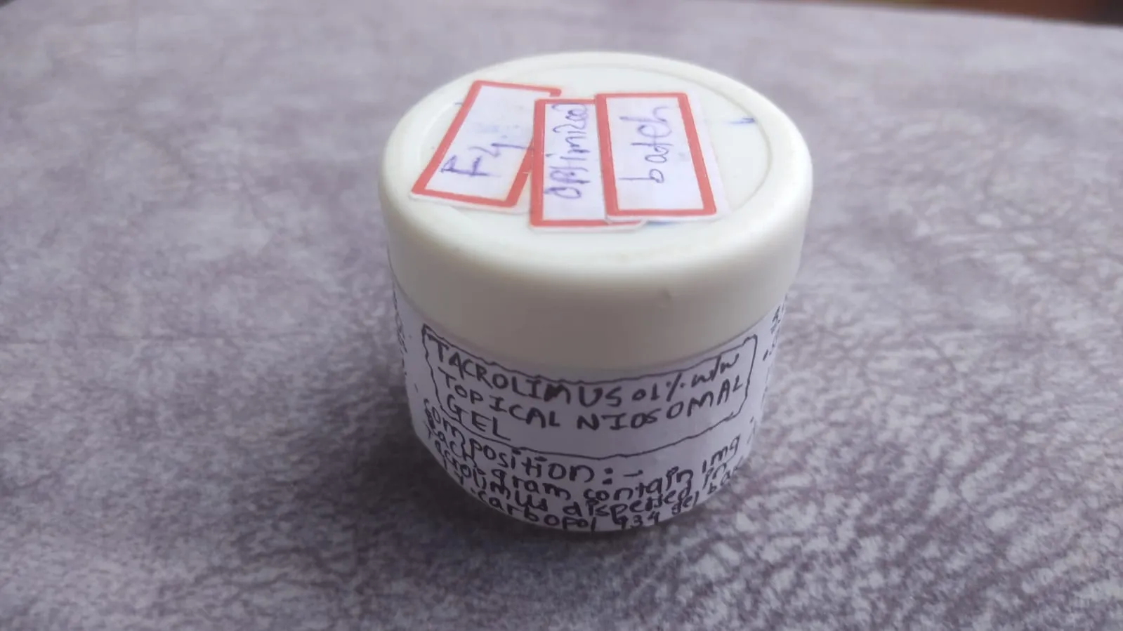
Figure 14: Tacrolimus-loaded niosomal topical gel I(F4 optimised batch)
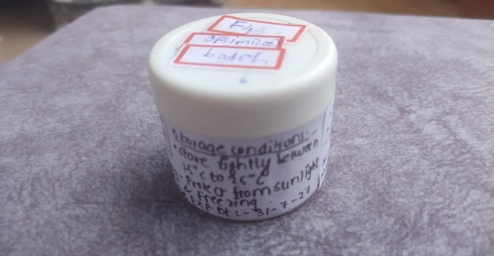
Figure 14: Tacrolimus-loaded niosomal topical gel II (F4 optimised batch)
CONFLICTS OF INTEREST: None
ACKNOWLEDGEMENT:
I want to sincerely thank everyone who gave me the chance and encouragement to do this research project on "Formulation and Evaluation of Niosomal Gel Using Tacrolimus Drug for Rheumatoid Arthritis. “First and first of all, I want to convey my sincere gratitude to my co-author and project guide, Dr. Pravinkumar Sable, for their essential advice, steadfast assistance and continual supervision during this work. Overcoming the difficulties encountered throughout the formulation and evaluation stages was made possible by their technical know-how and perceptive recommendations.
I have an obligation of appreciation to the management and administration of S.S.P. Shikshan Sanstha's Siddhi College of Pharmacy, Chikhali, Pune-411062 for providing the chemicals, well-equipped laboratories, and infrastructure needed to carry out this research project successfully. My profound gratitude is extended to the faculty and lab assistants of the Pharmaceutics and Pharmaceutical Chemistry departments for their prompt assistance, technical cooperation, and assistance during the laboratory evaluation procedures. Finally, I would want to convey my heartfelt appreciation to my family and peers for their steadfast encouragement, tolerance and moral assistance, all of which enabled me to complete this project.
REFERENCES
Formulation of Nanospanlastics as a Promising Approach for Improving the Topical Delivery of a Natural Leukotriene Inhibitor (3-Acetyl-11-Keto-β-Boswellic Acid): Statistical Optimization, in vitro Characterization, and ex vivo Permeation Study
Drug Design Development and Therapy [Internet]. 2020 Sep 1; Volume 14:3697–721. Available from: https://doi.org/10.2147/dddt.s265167
Formulation of Nanospanlastics as a Promising Approach for Improving the Topical Delivery of a Natural Leukotriene Inhibitor (3-Acetyl-11-Keto-β-Boswellic Acid): Statistical Optimization, in vitro Characterization, and ex vivo Permeation Study
Drug Design Development and Therapy [Internet]. 2020 Sep 1; Volume 14:3697–721. Available from: https://doi.org/10.2147/dddt.s265167

Revati Nampalle*, Dr.Pravinkumar Sable, Formulation And Evaluation Of Niosomal Gel Using Tacrolimus Drug For Rheumatoid Arthritis, Int. J. of Pharm. Sci., 2026, Vol 4, Issue 8, 1082-1110. https://doi.org/10.5281/zenodo.21835717
 10.5281/zenodo.21835717