

We use cookies to ensure our website works properly and to personalise your experience. Cookies policy
Bachelor of Pharmacy, Dhanalakshmi Srinivasan College of Pharmacy.
Onychomycosis is a common fungal infection of the nail caused mainly by dermatophytes, yeasts, and non-dermatophyte moulds. Conventional topical treatment is often limited by the dense keratinized structure of the nail plate, which restricts drug penetration. The present study was aimed at formulating and evaluating a fluconazole-loaded medicated nail lacquer for enhanced trans ungual drug delivery. Fluconazole was incorporated using ethyl cellulose as a film-forming polymer, triacetin as a plasticizer, polyethylene glycol 400 as a co-plasticizer, propylene glycol as a penetration enhancer, and ethanol as a solvent. Two formulations were prepared and evaluated for smoothness of flow, drying time, glossiness, non-volatile content, water resistance, UV-Visible spectral characteristics, and antifungal activity. Both formulations showed good smoothness and glossiness, while formulation F1 exhibited a drying time of 63 seconds and non-volatile content of 35%. Antifungal activity was demonstrated against Candida albicans and Aspergillus niger. Overall, the developed fluconazole nail lacquer showed promising characteristics for localized treatment of onychomycosis
NAIL: A tough, horn-like protective layer that covers the upper surface of the tips of fingers and toes in humans, primates, and some other animals. Composition: Primarily composed of alpha-keratin, a resilient structural protein found in hair and hooves as well. Nails mainly consist of hard keratin, which is a fibrous protein. They also contain small amounts of water, lipids (fats), calcium, and other minerals. Keratin imparts strength, hardness, and protection to the nail. The nail plate is made up of around 10–30% water1. 1
A nail is a hard, protective plate covering the tips of the fingers and toes. It is mainly composed of hard keratin, which provides strength, hardness, and protection. It also contains small amounts of water, lipids, calcium, and other minerals.
Main components: Nail plate, nail bed, nail matrix, nail fold, cuticle, lunula, and hyponychium.
Functions:
Nail Anatomy & Structure –The nail apparatus is a complex structure made up of several important parts: Nail Plate: The visible, hard part of the nail, mainly made of keratin. It protects the fingertips and toes, supports grasping, and acts as a major barrier to transungual drug delivery. Nail Matrix: The growth-producing part located beneath the proximal nail fold. It produces keratinized cells that form the nail plate.2 Nail Bed: The tissue beneath the nail plate that supports and anchors it. Its blood vessels contribute to the nail’s pink appearance. Lunula: The whitish, crescent-shaped area at the base of the nail, representing the visible part of the distal nail matrix. Cuticle: A thin protective tissue between the nail fold and nail plate that prevents microorganisms from entering. Proximal & Lateral Nail Folds: Skin folds surrounding the nail that protect the growing nail, provide support, and prevent entry of foreign materials. Hyponychium: Thickened skin beneath the free edge of the nail that forms a protective barrier against the external environment.3
Onychomycosis is a common fungal infection of the nails, affecting toenails more frequently than fingernails. It causes nail discoloration, thickening, brittleness, deformation, and separation from the nail bed. Dermatophytes, particularly Trichophyton rubrum and Trichophyton mentagrophytes, are the major causative organisms, while yeasts and non-dermatophyte moulds may also cause infection. Risk factors include increasing age, humidity, tight footwear, repeated nail trauma, diabetes, poor circulation, and weakened immunity.4
The thick keratin structure of the nail makes drug penetration difficult, reducing the effectiveness of conventional topical treatments. Medicated nail lacquer provides an effective approach by forming a thin film on the nail after application. This film acts as a drug reservoir and allows sustained release and improved penetration of the antifungal agent.5
Fluconazole is a broad-spectrum antifungal drug commonly used against dermatophytes and yeasts. Formulating fluconazole as a topical nail lacquer may provide localized and prolonged drug delivery while reducing the limitations associated with systemic therapy. The development of an effective formulation requires suitable film-forming polymers, solvents, plasticizers, penetration enhancers, and antifungal agents. Therefore, the project focuses on developing and evaluating a stable, fast-drying fluconazole nail lacquer with suitable drug release, penetration, and antifungal activity for the treatment of onychomycosis.6
Common symptoms: Thickened nails, Yellow, white, or brown discoloration, Brittle, crumbly, or ragged nails, Distorted nail shape, Nail lifting away from the nail bed (onycholysis), Sometimes a foul odour
Treatment: Topical antifungals (best for mild infections affecting a small portion of the nail): Ciclopirox nail lacquer, Fluconazole nail lacquer7
NAIL LACQUER
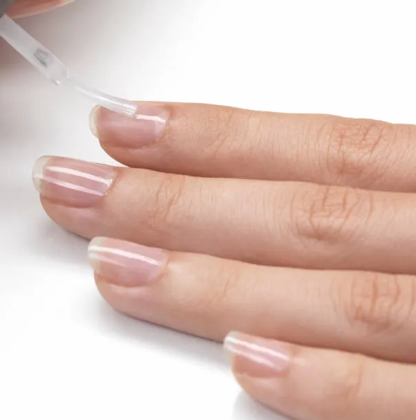
Nail lacquer is a liquid preparation applied to the nail plate that dries to form a thin, adherent film. It may be cosmetic or medicated.8
COLLECTION OF FLUCONAZOLE:
Fluconazole was received as a gift sample from aarti drugs ltd, Mumbai, Maharashtra, India. Ethyl cellulose, PEG 400, Triacetin are received from my senior gifted this to me. Ethanol is buying in Phoenix scientific supplies, Trichy, Tamil Nadu. All other chemicals and reagents were of analytical grade
PREPARATION OF NAIL LACQUER:
Dissolve 0.10 g fluconazole in 2.0 ml ethanol.
↓
Dissolve 0.40g ethyl cellulose in 6.0 ml ethanol with continuous stirring until a clear solution is obtained.
↓
Slowly add the drug solution (2.0ml) to the polymer solution (6.0ml) while stirring.
↓
Add 0.20ml triacetin and mix well.
↓
Add 0.20 ml PEG 400 and stir until the formulation is homogeneous.
↓
Add ethanol q.s. to make the final volume exactly 10ml (the exact amount depends on the volume occupied by the dissolved solids, so add it gradually while checking the final volume).9
|
S.NO |
INGREDIENTS |
FORMULA 1 |
FORMULA 2 |
|
1 |
FLUCONAZOLE |
0.10g |
0.10g |
|
2 |
ETHYL CELLULOSE |
0.40g |
0.40g |
|
3 |
PEG 400 |
0.20ml |
0.15ml |
|
5 |
ETHANOL |
9.1ml |
9.1ml |
|
6 |
TRIACETIN |
0.20ml |
0.25ml |
INGREDIENTS:
|
S.NO |
INGREDIENT |
ROLE OF INGREDIENT |
|
1 |
Fluconazole |
Active pharmaceutical ingredient act as anti-fungal activity |
|
2 |
Ethyl cellulose |
It acts on polymer. |
|
3 |
Triacetin |
It acts on plasticizer |
|
4 |
Polyethylene glycol-400 |
It acts on co-plasticizer. |
|
5 |
Propylene glycol |
It acts on permeation enhancers |
|
6 |
Ethanol |
It acts on solvents. |
EVALUATION TEST
ANTIFUNGAL ASSAY:
The antifungal activity of bark extracts was assayed by agar well diffusion method similar to antibacterial assay, except that Rose Bengal Agar medium was used instead of Nutrient Agar medium. The sterilized Rose Bengal agar medium was aseptically poured (20ml) into the sterile petriplates and allowed to solidify. The A. niger fungal broth culture was swabbed on petriplate using a sterile bud. Then wells were made by well cutter. The organic and aqueous extracts of bark (100µl) were added to each well aseptically. Amphotericin B disc was used as a positive control. The petriplates were incubated at 37˚C for 48 hours. The antifungal activity was assayed by measuring the diameter of the inhibition zone formed around the well (NCCLS, 1993). The same procedure was repeated for C. albicans also. The experiment was repeated three times and mean values calculated for conclusion.
SMOOTHNESS OF FLOW
The sample was poured from a height of 1.5 inches into a glass plate and spread on a glass plate. Made to rise vertically and visually observed for smoothness of flow.
GLOSSINESS
The glossiness of the prepared formulation was determined by visual inspection.
NON-VOLATILE CONTENT
First taken initial weight of petri dish (M1) and then Place 1gm (M) of each sample was taken into Petri dishes and spread evenly. Then weight of each Petri dish with sample was recorded. The Petri dish was kept in the hot air oven for 1 hr at 105 ± 2 ◦C. After 1 hour the Petri dish was cooled down and weighed again (M2). The percent non-volatile content was calculated by determining the weight difference.
DRYING TIME
A layer of the sample was administered onto a petri dish using a brush. The duration requires for the formulation of a film that was dry to the touch was observed using stopwatch.\
WATER RESISTANCE TEST
To determine the resistance of the formulation into water the water resistance test is performed. The test was carried out by spreading required amount of nail lacquer was spread on a uniform area of a glass plate and was dried. The sample containing glass was weighed and then placed in a beaker filled with distilled water. After 24 hr, the plate was dried with the use of filter paper and was weighed again. Water resistance was determined by initial and final weight difference, and the results were expressed in weight loss (%).
UV-VIS SPECTRA ANALYSIS
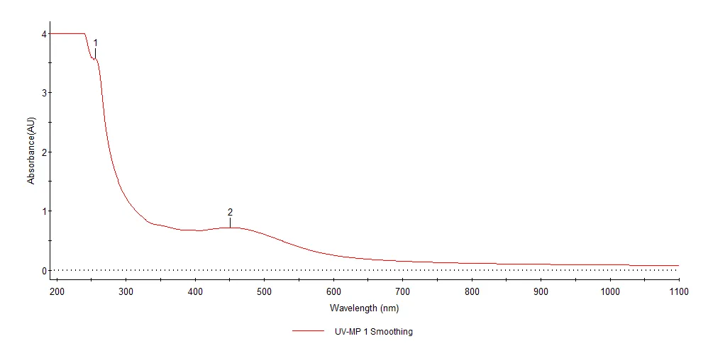
The bio reduction of Ag+ ions in solutions was monitored by measuring the UV-VIS spectrum of the reaction medium. The UV-VIS spectral analysis of the sample was done by using U-3200 Hitachi spectrophotometer at room temperature operated at a resolution of 1 nm between 200 and 800 nm ranges.
RESULT AND DISCUSSION:
SMOOTHNESS OF FLOW
|
S.No |
Formulation I |
Formulation Ii |
|
1. |
GOOD |
GOOD |
DRYING TIME
|
S.No |
Formulation I |
Formulation Ii |
|
1. |
63s |
67s |
GLOSSINESS
|
S.No |
Formulation I |
Formulation Ii |
|
1. |
GOOD |
GOOD |
NON-VOLATILE CONTENT
In order to have adequate coverage on nail plate, non-volatile content should be 20% or more. Non-volatile content depends upon the concentration of polymers used and was found to be directly proportional to the concentration of polymer. The non-volatile content of different formulation F1 & F2 was calculated.
|
S.No |
Formulation I |
Formulation Ii |
|
1. |
35% |
25% |
WATER RESISTANCE TEST
Higher was the increase in weight, lower was the water resistance of nail lacquer. Formulation F1 showed very high permeation as compared to F2.
|
S.No |
Formulation I |
Formulation Ii |
|
1. |
98% |
92% |
ANTI-FUNGAL ASSAY
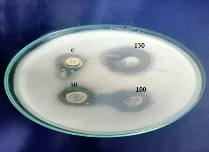
Candida albicans,
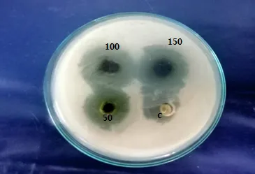
Aspergillus niger
|
Organism |
Zone of inhibition (mm/ml) |
|||
|
50 µl |
100 µl |
150 µl |
Control- Fluconazole-B |
|
|
Aspergillus niger |
06 |
12 |
18 |
20 |
|
Candida albicans, |
05 |
10 |
17 |
18 |
UV-VIS SPECTRA ANALYSIS
|
S.NO |
WAVELENGTH (nm) |
ABSORBANCE |
|
1. |
256.3 |
3.575 |
|
2. |
450.8 |
0.721 |
CONCLUSION
Fluconazole-loaded medicated nail lacquer was formulated as a promising topical drug delivery system for the treatment of onychomycosis. The optimized formulation is expected to provide satisfactory film formation, sustained drug release, and enhanced transungual permeation of fluconazole. Therefore, the developed nail lacquer may offer an effective and patient-friendly alternative for delivering antifungal drugs directly to the site of infection.
REFERENCES

S. Iyswarya, S. Mohamed Halith, P. Dharani, N. Dharanitharan, K. Dheva Dharshini, H. Fahima Almas, M. Faruk Basha, Formulation And Evaluation of Transungual Permeation of Fluconazole Nail Lacquer for Onychomycosis, Int. J. of Pharm. Sci., 2026, Vol 4, Issue 9, 1069-1075, https://doi.org/10.5281/zenodo.22688841
 10.5281/zenodo.22688841