

We use cookies to ensure our website works properly and to personalise your experience. Cookies policy
1Research Scholar, Pacific Academy of Higher Education and Research, Udaipur, Rajasthan – 313024.
2Professor, Pacific College of Pharmacy, Pacific Academy of Higher Education and Research University, Udaipur, Rajasthan – 313024
3HOD and Professor, Department of Pharmaceutics, Indrayani Vidya Mandir Krishnarao Bhegade Institute of Pharmaceutical Education and Research, Talegaon Dabhade, Pune, Maharashtra – 410507,
4Professor and Additional Principal, Geetanjali Institute of Pharmacy, Geetanjali University, Udaipur, Rajasthan
This study evaluated the antifungal effects of Miconazole Nitrate combined with Carica papaya seed oil and Nigella seed oil in microemulsion form, and investigated potential synergistic activity. Microemulsions were prepared by titrating various oil-to-Smix (surfactant and co-surfactant) ratios with water, and the resulting regions were identified using a pseudoternary phase diagram. Formulations were characterised for viscosity, pH, drug content, globule size, zeta potential, and stability. Optimised formulations (CPM3 and NOM3) were incorporated into 1% w/w Carbopol gel to create CPM3-G1 and NOM3-G2, which were then evaluated for physicochemical properties, drug release, and antifungal activity. In vitro antifungal studies confirmed that combining Miconazole Nitrate with essential oils in a microemulsion produces a synergistic antifungal effect compared to using the drug or oils alone. Miconazole nitrate microemulsions using Carica papaya seed oil and Nigella seed oil were developed for topical use. The CPM3 and NOM3 formulations were optimised due to their high transmittance, low viscosity, high drug content, suitable pH, enhanced in vitro drug release, and stability. Antifungal activity was evaluated in vitro against the Candida albicans strain. The microemulsion system improved the therapeutic effect of topical drugs, and combining the drug with essential oils in this form demonstrated synergistic activity for enhanced efficacy.
Topical preparations are administered to localised areas of the body. This term refers to formulations intended to act exclusively at the site of application while minimising systemic drug absorption. Conventional topical drug delivery methods typically involve either disrupting the stratum corneum at the molecular level or modifying the skin's barrier function, as seen with topical antibiotics, antibacterials, emollients, and sunscreen agents, to facilitate drug delivery to the viable epidermal and dermal tissues without employing oral, systemic, or alternative therapies. 1
Microemulsions are effective for oral delivery of poorly water-soluble drugs because they enhance drug solubilisation. Higher thermodynamic activity in the vehicle increases drug absorption rates.2
Microemulsions are widely used in topical drug delivery because they are optically isotropic, thermodynamically stable mixtures of water, oil, surfactant, and co-surfactant. They enhance the solubility of poorly water-soluble drugs, improve topical and systemic availability, and promote rapid, effective skin penetration.3
This makes microemulsions useful for applying medications to the skin. Microemulsions are clear, uniform, and stable mixtures of two liquids that do not usually mix. They are created with a soap-like substance called a surfactant, often helped by a co-surfactant.4 The surfactant not only helps dissolve the drug, improving its stability and availability, but also helps the drug spread better through the skin. Because microemulsions are very small, they are an effective way to deliver drugs. For these reasons, microemulsions show great promise for delivering drugs through the skin.5
Miconazole nitrate, a synthetic imidazole derivative, exhibits broad-spectrum antibacterial activity and is utilised for the treatment of fungal infections both locally and systemically. It is particularly effective against Microsporum, Trichophyton, Epidermophyton, and Candida species, and demonstrates some efficacy against gram-positive bacteria. The primary site of action is the cell membrane. Studies on Candida albicans indicate that miconazole selectively inhibits the uptake of mucopolysaccharides (such as glutamine) and RNA and DNA precursors (purines) at low concentrations by primarily targeting the yeast cell membrane.6, 7
Therefore, the goal of the current study was to test how well Miconazole Nitrate works with essential oils like Carica Papaya seed oil and Nigella seed oil to make a microemulsion gel and see if this creates a stronger antifungal effect.
MATERIALS AND METHODS
Materials
The study utilised the following chemicals and reagents: Miconazole Nitrate (Pharma grade, Mahrshee Laboratories, Gujarat) as the active antifungal agent; Carica Papaya seed oil and Nigella Seed oil (LR grade, RV Essential, New Delhi) as natural antifungal agents; Tween 20 and Tween 80 (LR grade, Thomas Baker Pvt. Ltd., Mumbai) as non-ionic surfactants for microemulsion stabilisation; Propylene glycol and PEG 400 (LR grade, S D Fine Chem. Ltd., Mumbai) as co-surfactants and penetration enhancers; Carbopol 934 (LR grade, CDH Pvt. Ltd., New Delhi) as a gelling agent; Methanol (LR grade, S D Fine Chem. Ltd., Mumbai) as a solvent; and Potassium dihydrogen orthophosphate (Thermo Fisher Scientific, Mumbai) and Sodium hydroxide (S D Fine Chem. Ltd., Mumbai) for buffer preparation and pH adjustment. All chemicals were of analytical or laboratory reagent grade.
Method of Preparation of Microemulsion Gel
The drug was initially dissolved in the selected oil, after which a fixed ratio of surfactant and co-surfactant was added. The mixture was vortexed continuously for 15 minutes to ensure thorough mixing. Demineralised water was then added dropwise with constant stirring until a clear and transparent liquid formed, indicating successful microemulsion formation. This microemulsion was subsequently incorporated into a 1% w/w Carbopol 934 gel base to produce the final microemulsion gel formulation for topical application.9


3:1
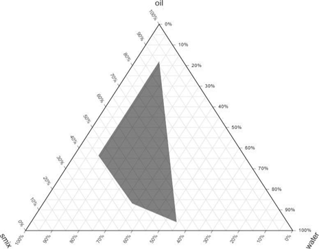
Figure 1: Pseudoternary phase diagram of Carica papaya seed oil, Tween 80, and Propylene glycol with different Smix ratios (1:1, 2:1, and 3:1).
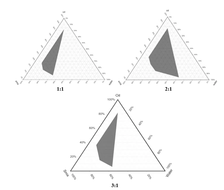
Figure.2: Pseudoternary phase diagram of Nigella seed oil, Tween 80, and Propylene glycol containing different Smix ratios (1:1, 2:1 and 3:1)
Table 1: Formulation development based on Pseudo-ternary phase
|
Formulation Code |
Smix ratios |
Co-surfactant |
Surfactant |
Oils |
Percentage w/w component in formulation |
|||
|
Oil % |
Smix % |
Water % |
Drug % |
|||||
|
CPM1 |
1:1 |
Propylene glycol |
Tween80 |
Carica Papaya seed oil |
16 |
64 |
20 |
1 |
|
CPM2 |
2:1 |
30 |
54 |
15 |
1 |
|||
|
CPM3 |
3:1 |
37 |
50 |
13 |
1 |
|||
|
NOM1 |
1:1 |
Nigella seed oil |
15 |
55 |
30 |
1 |
||
|
NOM2 |
2:1 |
20 |
50 |
30 |
1 |
|||
|
NOM3 |
3:1 |
35 |
50 |
15 |
1 |
|||
EVALUATION OF MICROEMULSION
Percentage transmittance: The transparency of the microemulsion was determined by measuring the percentage transmittance at 272 nm against distilled water as a blank by using a UV spectrophotometer.10
Percent Transmittance= -Log 10(2-Absorbance)

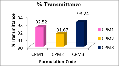
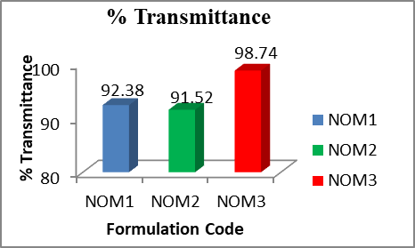
Fig.3: % Transmittance of CPM1-CPM3 Fig.4: % Transmittance of NOM1-NOM3
Viscosity measurements: The Rheological behaviour of the microemulsion formulation was evaluated using an Ostwald viscometer at room temperature.
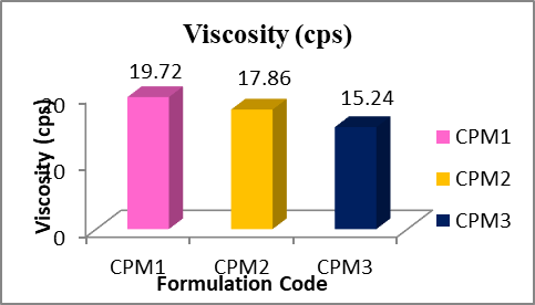
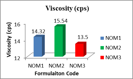
Fig.5: Viscosity of COM1-COM3 Fig.6: Viscosity of NOM1-NOM3
Measurement of pH: The pH of Miconazole Nitrate microemulsion formulations was determined by using a digital pH meter. The pH of each formulation was measured in triplicate, and the average values were calculated.11
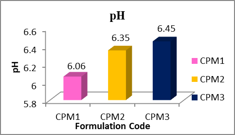
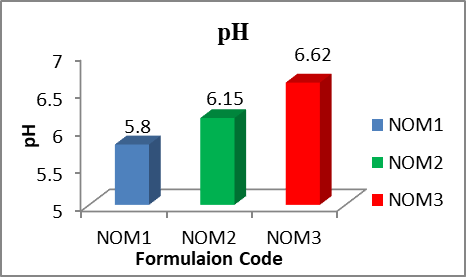
Fig.7: pH of COM1- COM3 Fig.8: pH of NOM1- NOM3
Percentage Drug content: For the determination of drug content, about 1 mL of each microemulsion formulation was transferred to a 10 mL volumetric flask and dissolved in methanol. It was diluted appropriately and analysed by spectrophotometry at 272 nm.
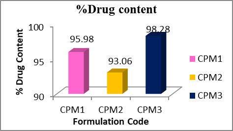
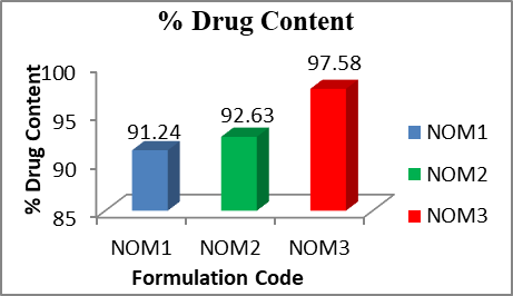
Fig.9: % Drug content of COM1-COM3 Fig.10: % Drug content of NOM1-NOM3
Measurement of globule size and zeta potential: The average globule size and zeta potential of the optimised microemulsions were measured using a Malvern ZetaSizer instrument at a temperature of 25°C.12
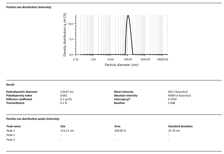
Fig.11: Globular size of CPM3
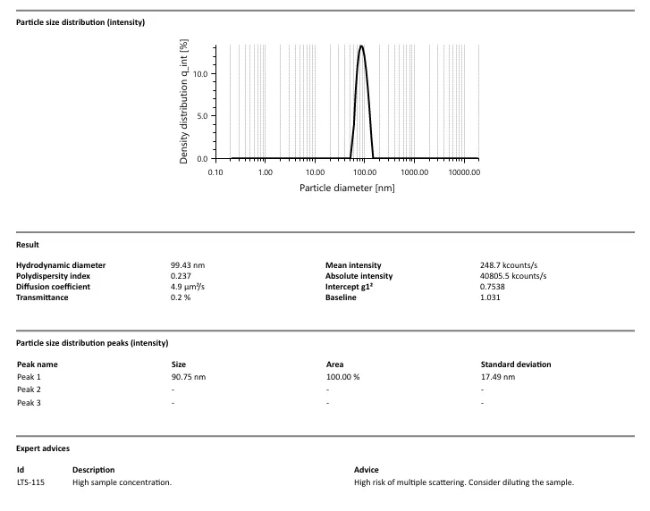
Fig.12: Globular size of NOM3
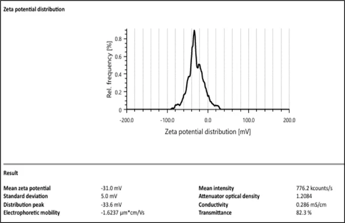
Fig.13: Zeta potential of CPM3
Fig.14: Zeta potential of NOM3
Surface morphology: Surface morphology of the optimised microemulsion formulations CPM3 and NOM3 was determined by using a scanning electron microscope (SEM).13
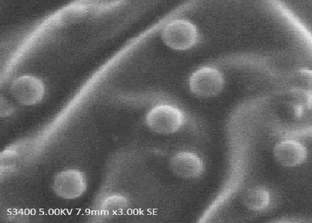
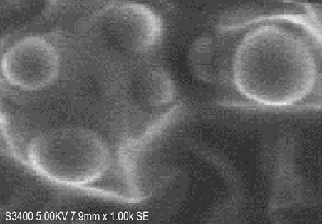
Fig.15: SEM of CPM3 and NOM3 Microemulsion
In vitro diffusion study:
Phosphate buffer at pH 6.8 was used as the medium. The diffusion cell, designed according to specified dimensions, had an effective diffusion area of 3.14 cm2. For in vitro permeation studies, the egg membrane was carefully mounted to prevent air bubble entrapment and was clamped tightly to ensure close contact with the receptor fluid..14
Diffusion cells were placed in the receptor compartment with a magnetic stirrer. One gram of microemulsion was added to the donor compartment, and 200 mL of phosphate buffer (pH 7.4) was added to the receptor compartment. Stirrer speed and temperature remained constant throughout the experiment. At 60-minute intervals from 0 to 12 hours, 1 mL samples were withdrawn from the receptor compartment using a pipette and replaced with an equal volume of receptor medium to maintain sink conditions. Samples were diluted as needed, and absorbance was measured at 272 nm using a UV spectrophotometer.15
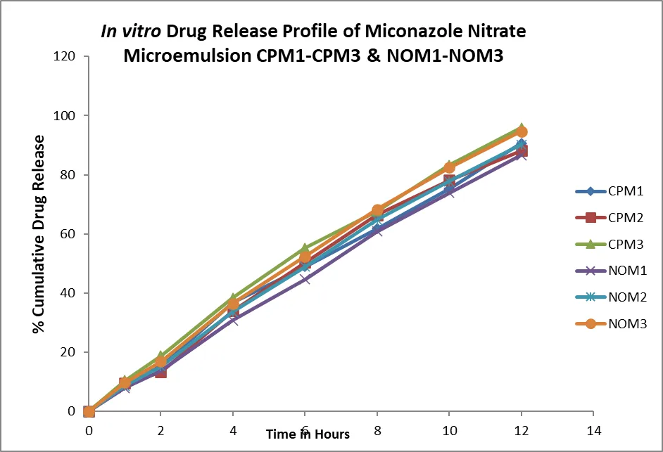
Fig.16: Comparison of Percentage Cumulative Drug Release of CPM1-CPM3 and NOM1-NOM3
EVALUATION OF MICONAZOLE NITRATE MICROEMULSION GEL
Viscosity and Rheological studies: A Brookfield digital viscometer (Model LVDV–E, USA) was used to determine the viscosity and rheological properties of the microemulsion-based gel using spindle no. 6. 10 g of sample was placed in a small sample holder, and the viscosity of the gel was measured at 25°C.16
Determination of pH: The apparent pH of the gel was determined by a pH meter in triplicate at 25±1ºC.
Determination of Percentage drug content: For the determination of drug content, 1 g of the gel formulation was weighed into a 10 mL volumetric flask and dissolved in methanol. It was diluted appropriately and analysed by spectrophotometry at 272 nm. 17
In vitro release studies: An in vitro drug release study was performed using a diffusion cell. The egg membrane was placed between the receptor and donor compartments. Microemulsion gel equivalent to 0.2 g was placed in the donor compartment, and the receptor compartment was filled with phosphate buffer pH 6.8. The diffusion cells were maintained at 37 ± 0.5℃ with stirring at 100 rpm throughout the experiment. At fixed time intervals, 5ml of sample was withdrawn at 1, 2, 4, 6, 8, 10, and 12 hrs, and the same volume was replaced with receptor fluid solution to maintain sink conditions. The collected samples were analysed using a UV spectrophotometer at λ max 272nm.18
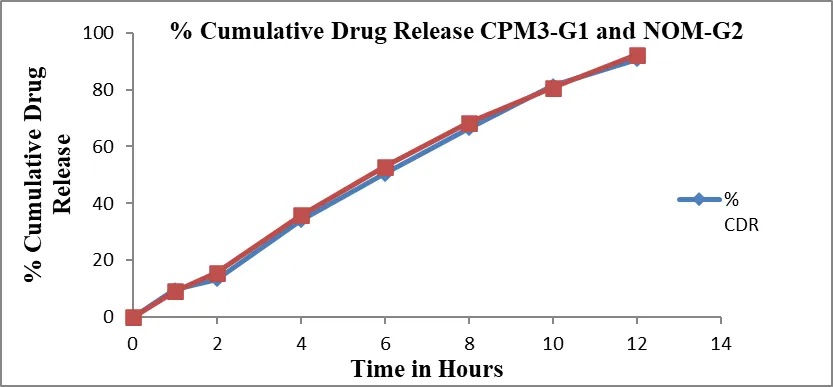
Fig.17: Percentage Cumulative Drug Release of CPM3-G1 and NOM-G2
In vitro antifungal studies: Sterile Sabourd Dextrose Agar plates were prepared by pouring the sterile agar into sterile Petri dishes under aseptic conditions. 0.1 ml of the test organism (Candida Albicans) was spread on agar plates. 5 mm diameter holes were made in the agar plates using a sterile bore. 500µg/ml drug, 30µl of formulations (CPM3 & NOM3) 20µl of essential oils (CPS & NS) and 60mg of gels (CPM3-G1 & NOM3-G2) were added into each hole separately. The plates were maintained at +4˚C for 4 hours to allow the solution to diffuse into the agar medium. All plate cultures containing Candida albicans were incubated at 28˚C for 48 hours. Zones of microbial growth inhibition around the well were measured and recorded after incubation.19
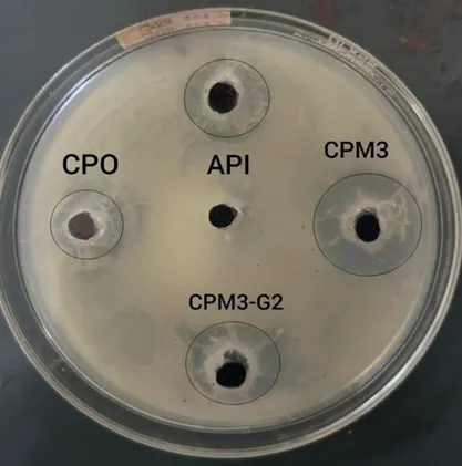
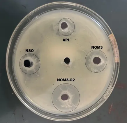
Fig. 18: Antifungal activity against Candida albicans using the agar well diffusion method.
Ex vivo skin permeation studies: An Ex vivo drug permeation study was conducted using a Franz diffusion cell containing 150 ml of phosphate buffer (pH 6.8) using an excised goat skin. The goat's skin was obtained from a local slaughterhouse within 15 minutes of the goat's sacrifice. After removing the hairs, the skin was stored on ice-cold phosphate buffer (pH 6.8). The skin was immediately immersed in Ringer's solution. The freshly excised skin was mounted on the diffusion cell, and a 20 mg microemulsion gel containing an equivalent dose was placed on it. Throughout the study, the buffer solution in the chamber was maintained at 37±1°. At predetermined time intervals (1, 2, 4, 6, 8, 10, 12 hours), 1 mL of the sample was withdrawn and replaced with an equal volume of phosphate buffer. The samples were appropriately diluted and filtered, and absorbance was measured spectrophotometrically at 272 nm using a UV/Vis Spectrophotometer, taking phosphate buffer (pH 6.8) as the blank.20
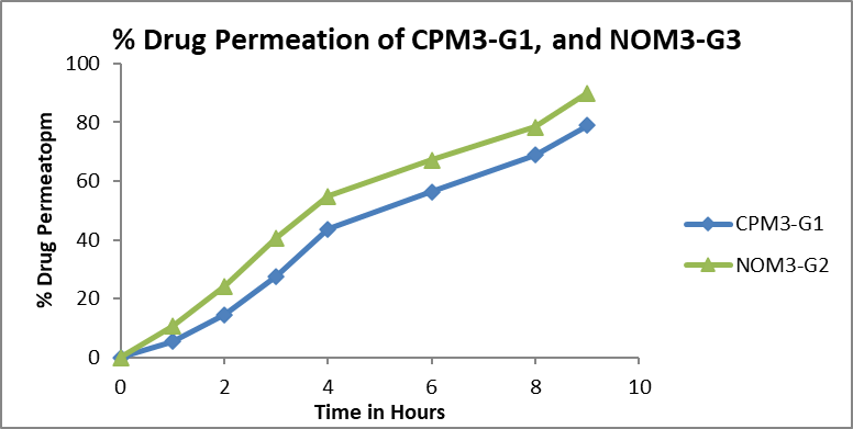
Fig.19: Ex vivo drug permeation profile of CPM3-G1 and NOM3-G2.
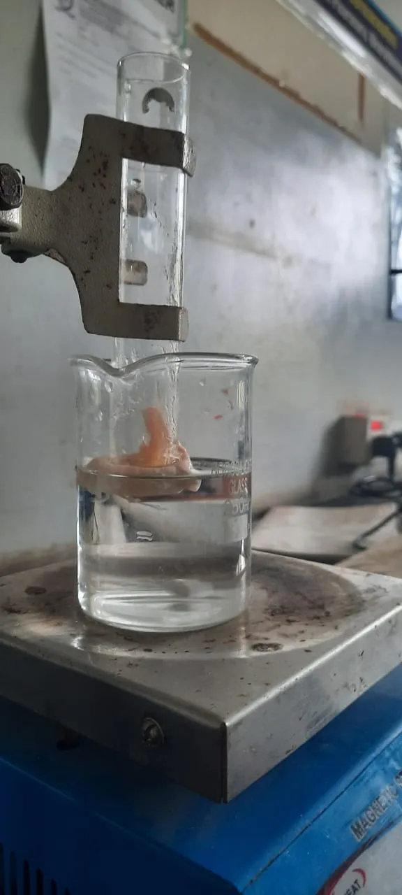
Fig. 20: Ex vivo skin permeation studies
Spreadability: Spreadability was performed using two 7.5 cm glass slides. 350 mg of Microemulgel was weighed accurately and placed on one slide. Another glass slide was placed above it from a height of 5 cm. A weight of 5 g was placed on the upper slide, and after 1 minute, the diameter of the circle formed was measured in cm. The observed diameter indicates the type of gel. 21
RESULTS AND DISCUSSION
For formulation development, a pseudoternary phase diagram was constructed using Tween 80 and propylene glycol at Smix ratios (1:1, 2:1, and 3:1) to identify the microemulsion region. Increasing the surfactant ratio expanded the microemulsion zone. Based on this, suitable proportions of oil, Smix, and water were selected for formulation. The prepared microemulsions were clear, transparent, and stable, with % transmittance above 90%, confirming the nanometric droplet size. The viscosity ranged from 13–17 cps, showing Newtonian flow, while pH values (5.8–6.6) were within the skin-compatible range.
The drug content was uniform (91–98%), and zeta potential values indicated stability without aggregation. The globule size (around 90 nm) and SEM images confirmed the presence of smooth, spherical droplets. No phase separation was observed after centrifugation, confirming good physical stability, and in vitro drug release studies revealed sustained release for 12 hours, with 93.86% release from CPM3 and 94.98% from NOM3. Nigella seed oil-based formulation showed slightly higher release than the Carica Papaya seed oil-based formulation.
Stability studies showed no significant changes, confirming the formulation's stability. Optimised microemulsions (CPM3 and NOM3) were converted into 1% Carbopol 934-based gels (CPM3-G1 and NOM3-G2). These gels showed good spreadability, a suitable viscosity, and a pH (6.2–6.6) compatible with the skin. The optimised formulations CPM3, NOM3, CPM3-G1, and NOM3-G2 showed higher antifungal activity with inhibition zones of 24 mm, 21 mm, 21 mm, and 25 mm, respectively, compared to 19 mm for the pure drug, 13 mm for Carica Papaya seed oil, and 17 mm for Nigella seed oil. It indicates a strong synergistic effect that enhances antifungal efficacy. Ex vivo permeation showed sustained drug release, with cumulative percentage release of 81.98% for CPM3-G1 and 80.89% for NOM3-G2, confirming prolonged retention and controlled release. The drug content was above 93%, and the gels exhibited sustained release over 12 hours.
All formulations exhibited high cumulative drug release (86–95%). The kinetic models revealed that among the formulations (CPM- CPM3), CPM3 provided the best fit to the zero-order model (R² = 0.9972), indicating concentration-independent drug release. The Peppas model also showed a high correlation (R² = 0.9968), suggesting controlled release behaviour. The ‘n’ value (0.901) indicates a Non–Fickian transport mechanism, and among the formulations (NOM1-NOM3), NOM3 showed strong linearity in zero-order kinetics (R² = 0.9962) along with a high Peppas correlation (R² = 0.9977). The ‘n’ value (0.9237) further confirms non-Fickian transport, indicating a combination of diffusion and polymer relaxation mechanisms. Among the gel-based formulations, CPM3-G1 and NOM3-G2 also demonstrated zero-order kinetics with high correlation coefficients (R2 ≈ 0.9930-0.9939). The Peppas model indicated ‘n’ values of 0.9715 and 0.9649, respectively, suggesting super case II transport mechanisms. Among all formulations, NOM3 showed the highest drug release (94.73%), suggesting it as the optimised formulation for controlled drug delivery. Stability studies showed no major changes during storage.
Table No. 2: Data for different kinetic models resulting from the model fitting.
|
Formulation code |
%CDR |
Zero order |
First order |
Higuchi |
Peppas |
‘n’ values |
|
CPM1 |
85.042 |
0.9831 |
0.9685 |
0.9286 |
0.9891 |
1.0077 |
|
CPM2 |
87.709 |
0.9652 |
0.9615 |
0.9383 |
0.9857 |
0.9576 |
|
CPM3 |
93.985 |
0.9972 |
0.9047 |
0.9392 |
0.9968 |
0.9237 |
|
NOM1 |
86.570 |
0.9986 |
0.9456 |
0.9290 |
0.9976 |
0.9862 |
|
NOM2 |
90.140 |
0.9964 |
0.9385 |
0.9374 |
0.9976 |
0.9672 |
|
NOM3 |
94.730 |
0.9962 |
0.9024 |
0.9424 |
0.9977 |
0.9394 |
|
CPM3-G1 |
90.690 |
0.9939 |
0.9463 |
0.9332 |
0.9851 |
0.9715 |
|
NOM3-G2 |
92.180 |
0.9930 |
0.9382 |
0.9431 |
0.9951 |
0.9649 |
Table no. 3: Intermediate stability studies at 30ºC±2ºC and 65±5% RH
|
Parameter |
Duration in months |
|||||
|
0 |
3 |
6 |
||||
|
CPM3 |
NOM3 |
CPM3 |
NOM3 |
CPM3 |
NOM3 |
|
|
Drug content |
97.86 |
97.58 |
97.24 |
96.57 |
93.46 |
95.84 |
|
%CDR |
95.68 |
93.89 |
94.20 |
92.86 |
93.12 |
91.65 |
Table No 4: Report of Antifungal Activity against Candida albicans
|
Sl. No. |
Samples |
Quantity Used |
Zone of Inhibition in mm |
Sensitivity |
|
1. |
Miconazole Nitrate |
500µg/ml |
19 |
sensitive |
|
2. |
CPM-3 |
30µl |
24 |
sensitive |
|
3. |
NOM-3 |
30µl |
21 |
sensitive |
|
4. |
CPM3- G1GEL |
60mg |
21 |
sensitive |
|
5. |
NOM-3 GEL |
60mg |
25 |
sensitive |
|
6. |
Carica papaya seed oil |
20µl |
13 |
sensitive |
|
7. |
Nigella seed oil |
20µl |
17 |
sensitive |
CONCLUSION
Pseudo-ternary phase diagrams were used to guide microemulsion preparation by varying water and oil-to-Smix (surfactant and co-surfactant mixture) ratios. CPM3 and NOM3 formulations were chosen for their high transmittance, suitable viscosity, high drug content, and pH values near 6.2, which support enhanced drug release. After stability testing, these formulations were incorporated into gels containing 1% w/w Carbopol 934. The resulting gels exhibited good spreadability, appropriate pH, high drug content, and acceptable viscosity. In vitro studies demonstrated that CPM3-G1 and NOM3-G2 provided sustained drug release. Both optimised microemulsion and gel formulations showed stronger antifungal activity against Candida albicans than the pure drug or oils alone, indicating a synergistic effect. Larger inhibition zones reflected improved drug penetration and efficacy. Ex vivo permeation studies further confirmed sustained drug release and prolonged retention, supporting effective topical delivery and the potential to replace synthetic antifungal agents.
CONFLICT OF INTEREST
The authors declare no conflict of interest.
REFERENCES


Mohd. Ali, Dr. Jayesh Dwivedi, Dr. Amol Rakte, Dr. Udichi Kataria, Formulation and Optimization of Miconazole Nitrate-Loaded Topical Microemulsion Gel Using Carica Papaya Seed Oil and Nigella Sativa Seed Oil, Int. J. of Pharm. Sci., 2026, Vol 4, Issue 8, 264-276, https://doi.org/10.5281/zenodo.21772465
 10.5281/zenodo.21772465