

We use cookies to ensure our website works properly and to personalise your experience. Cookies policy
Chhatrapati Shivaji College of Pharmacy, Deori, Gondia, Maharashtra, India.
Whilst the placenta provides a partially selective barrier to drug transfer between maternal and fetal circulations, many drugs can still be transferred to the fetus to some degree. Whether a drug reaches the fetus will depend on drug properties, passive diffusion and active transport processes via placental transporters. This review provides an overview of how placental transporters are regulated throughout gestation and by pathology. This will impact on drug exposure in the mother and fetus. There are a number of transporter superfamilies including ATP-binding cassette transporters (ABC). The ABC transporters that have been identified within the placenta include P-glycoprotein (P-gp/ABCB1), breast cancer resistance protein (BCRP/ABCG2) and multidrug resistance associated proteins (MRPs). Transporters from the solute carrier family (SLC) have also been identified including organic anion transporters, organic anion transporting polypeptides and organic cation transporters. Some drugs may not reach the fetus due to P-gp and BCRP acting as barriers to transfer by transporting selected substrates back into the maternal circulation. On the other hand SLC transporters play a role in drug uptake and exchange. There may be changes in transporter expression throughout gestation. Infection, inflammation, metabolic disturbances and other complications can affect placental transporter regulation. Molecular factors have been shown to play a role including inflammation via cytokines, oxidative stress, hypoxia, hormone signalling and nuclear receptors. Genetic polymorphism and epigenetic mechanisms can also affect transporter expression. This may alter drug exposure to the fetus. However, it cannot be determined from placental transporter expression if a drug will transfer or if a drug will result in toxicity.
One of the critical aspects that necessitates attention is placental transfer and fetal exposure to drugs taken by the mother. The placenta acts as a barrier between the maternal and fetal circulations but is not impermeable to drugs. Certain drugs can readily cross the placenta, exposing the fetus to potential risks such as teratogenicity, developmental abnormalities, or other adverse effects.[1]. Therefore, studying placental transfer and fetal exposure is essential for assessing the safety of drug therapy during pregnancy and minimizing harm to the developing fetus.
Statistics indicate that approximately 80% of pregnant women take at least one medication throughout their pregnancy for treatment of a variety of complications, such as viral infection (e.g., human immunodeficiency virus [HIV]), smoking cessation, or pregnancy-induced condi tions like hypertension, depression, preeclampsia, nausea, and gestational diabetes. Some drugs are prescribed to pregnant women to treat both the mother and fetus (e.g., antiretroviral drugs to treat HIV-positive mother and prevent perinatal transmission of HIV) or just the fetus (e.g., digoxin and amiodarone to treat fetal tachyar rhythmia). When pregnant women receive drugs, their fetuses are also exposed to thedrugs. However, mostdrugs are administered to pregnant women off-label without in formation about the dose, maternal-fetal pharmacoki netics, safety, or efficacy of the drugs. A major concern of drug use by pregnant women is the transfer of drugs across the placental barrier, leading to fetal drug exposure and potential toxicity to the developing fetus. Indeed, 5%–10% of pregnant women receive drugs which are potentially teratogenic, and 50% of drugs are used in the first trimester when the fetus is most vulnerable to devel opmental toxicity. While most drugs can cross the placenta to a certain extent by passive diffusion, the placenta is richly endowed with a variety of adenosine triphosphate (ATP)-binding cassette (ABC) efflux trans porters and solute carrier (SLC) uptake transporters. Drug transporters in the placenta play crucial roles in fetal drug exposure. The localization and activity of transporters as well as changes of their expression in the placenta over gestation have a profound impact on fetal drug exposure and hence efficacy or toxicity of the drugs to the fetus. In this review, first, we describe human placenta physiology and transporter expression in the placenta [2].
The maternal-fetal interface in the placenta consists of a layer of syncytiotrophoblasts and cytotrophoblasts that separate the maternal from the fetal circulation. This cellular layer constitutes the blood-placenta-barrier and various transporters may be expressed in the apical or basolateral membrane of these cells. Transporters may additionally be expressed in the endothelial membrane lining the interior surface of the fetal blood vessels and separating the fetal blood from the stroma and trophoblasts [3].
The human placenta is unique in structure and can only be represented by placentas of certain primate species. The human placental barrier consists of the singular epithelium called the syncytiotrophoblast arising from cytotrophoblast, whose apical membrane is in direct contact with the maternal blood and basal membrane is close to the fetal circulation through the umbilical vessels into the placental villi. The syncytiotrophoblast represents the rate-limiting barrier for trans-placental transfer of most substances. As pregnancy advances, the thickness of the human placental barrier diminishes, primarily due to the partial disappearance of the cytotrophoblast . The function of the placenta varies during the gestational period. In early pregnancy, the main function of the placenta is to mediate implantation of the embryo into the uterus and produce hormones that prevent the end of the ovarian cycles [4].
2. Placental Drug Transporters
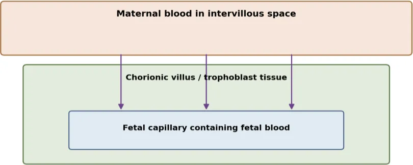
Figure 1: Simplified Placental Exchange Interface Showing Maternal Blood, Trophoblast Tissue and Fetal Capillary
The maternal-fetal interface in the placenta consists of a layer of syncytiotrophoblasts and cytotrophoblasts that separate the maternal from the fetal circulation. This cellular layer constitutes the blood-placenta-barrier and various transporters may be expressed in the apical or basolateral membrane of these cells. Transporters may additionally be expressed in the endothelial membrane lining the interior surface of the fetal blood vessels and separating the fetal blood from the stroma and trophoblasts.[5].
Placental transfer can be considered using a series of steps which include; maternal plasma concentration, transfer to the maternal aspect of the placenta, crossing of the trophoblast, binding/metabolism within the cell, transfer across the fetal aspect of the placenta and distribution/excretion within the fetus. Some drugs may pass quickly via passive diffusion but can be affected by active transport. Other drugs may accumulate within the placental tissue but not produce an equivalent concentration in the fetus. This can be further affected by binding to proteins in the maternal plasma and changes in volume of distribution. As such it is important to consider placental permeability, transporter activity and overall pharmacokinetics [6].

Figure 2: Simplified Pathway of Maternal–Placental–Fetal Drug Transfer.
2.1 Structure and function of the placental barrier.
The human placenta acts as a barrier between maternal blood in the intervillous space and fetal blood in the capillaries of the chorionic villi. An important interface for exchange is formed by trophoblast cells, particularly the syncytiotrophoblast. The placenta is not an absolute barrier to transfer and allows oxygen, nutrients and waste products to pass through. However, drug transfer will depend on the drug and circumstances. Transporters are an integral part of the placental cell membrane and help to create this selective barrier.[7].
2.2 Passive and transporter-mediated transfer.
Passive diffusion occurs along the concentration gradient. It is affected by the molecular size, lipid solubility, ionization, protein binding and surface area. Some drugs may also be transported across via aqueous channels or other specific mechanisms. Transporter mediated transport involves the membrane protein recognizing the substrate. This can lead to facilitative transport. This can cause a saturation effect and be very specific. Active transport uses the energy of the cell either directly or indirectly. Two drugs with similar plasma concentrations may have very different placental transfer due to differences in transporter interaction.[8].
3. Major Placental Drug Transporters
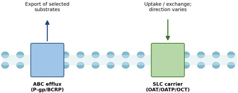
Figure 3: Conceptual Representation of ABC Efflux and SLC Carrier Transport at a Membrane.
Table 1: Major Placental Transporter Families and Their General Roles.
| Transporter group | Examples | General role | Interpretation for fetal exposure |
|---|---|---|---|
| ABC efflux | P-gp (ABCB1), BCRP (ABCG2), MRPs |
ATP-dependent export of selected substrates | May limit fetal passage for some substrates; depends on location and drug |
| SLC carriers | OATs, OATPs, OCTs | Uptake, exchange or carrier-mediated movement |
Net effect depends on localization, direction and substrate |
The strongest evidence is available for a limited number of transporters. P-gp and BCRP are generally described as protective efflux transporters. However, the actual effect will depend on where the transporter is located and whether the drug is a transported substrate at clinically relevant concentrations. MRPs show variable substrate specificities. They may play a role in efflux of organic anions, conjugated metabolites etc. However, there is not necessarily a quantitatively significant amount of protein expressed if only messenger RNA is detected [9].
3.1 ABC efflux transporters: P-gp, BCRP and MRPs
ATP-binding cassette (ABC) transporters utilise ATP hydrolysis to facilitate the transport of substrates across cell membranes. P-glycoprotein (P-gp/ABCB1) and BCRP (ABCG2) are two well known efflux transporters that have been discussed in relation to placental drug disposition. These transporters are expressed on the maternal-facing trophoblast membrane and may be responsible for transporting certain substrates back into the maternal circulation. This can reduce the availability of certain substrates to reach the fetal side. However, this protective function depends on the substrate present [10]. P-gp has been studied to understand its expression and function within the placenta. It has been seen to vary depending on the gestational age. BCRP is also an important transporter and has been seen to transport a variety of pharmaceutical compounds at the maternal-fetal interface.[11]
3.2 SLC uptake transporters: OATs, OATPs and OCTs
Transporters from the solute carrier (SLC) superfamily play a role in the uptake or exchange of endogenous molecules and drugs. Organic anion transporters (OAT), organic anion transporting polypeptides (OATPs) and organic cation transporters (OCTs) have all been considered in placental transport studies. Uptake transporters may play a role in aiding placental cell entry, inter-compartmental movement and endogenous substrate processing. Their overall impact on fetal exposure will depend on the location, directionality and process of the transporter [8].
3.3 Localization, function and representative substrates
Transporter localization determines the direction in which a compound is moved relative to maternal blood, trophoblast cells and fetal circulation. A transporter on the maternal-facing membrane may have a different effect from one on the fetal-facing membrane. Substrate lists should be interpreted cautiously because transporter recognition may vary across experimental systems and concentrations. For any specific medicine, evidence of transporter interaction should ideally be combined with data on placental expression, functional transport and maternal–fetal pharmacokinetics [10].
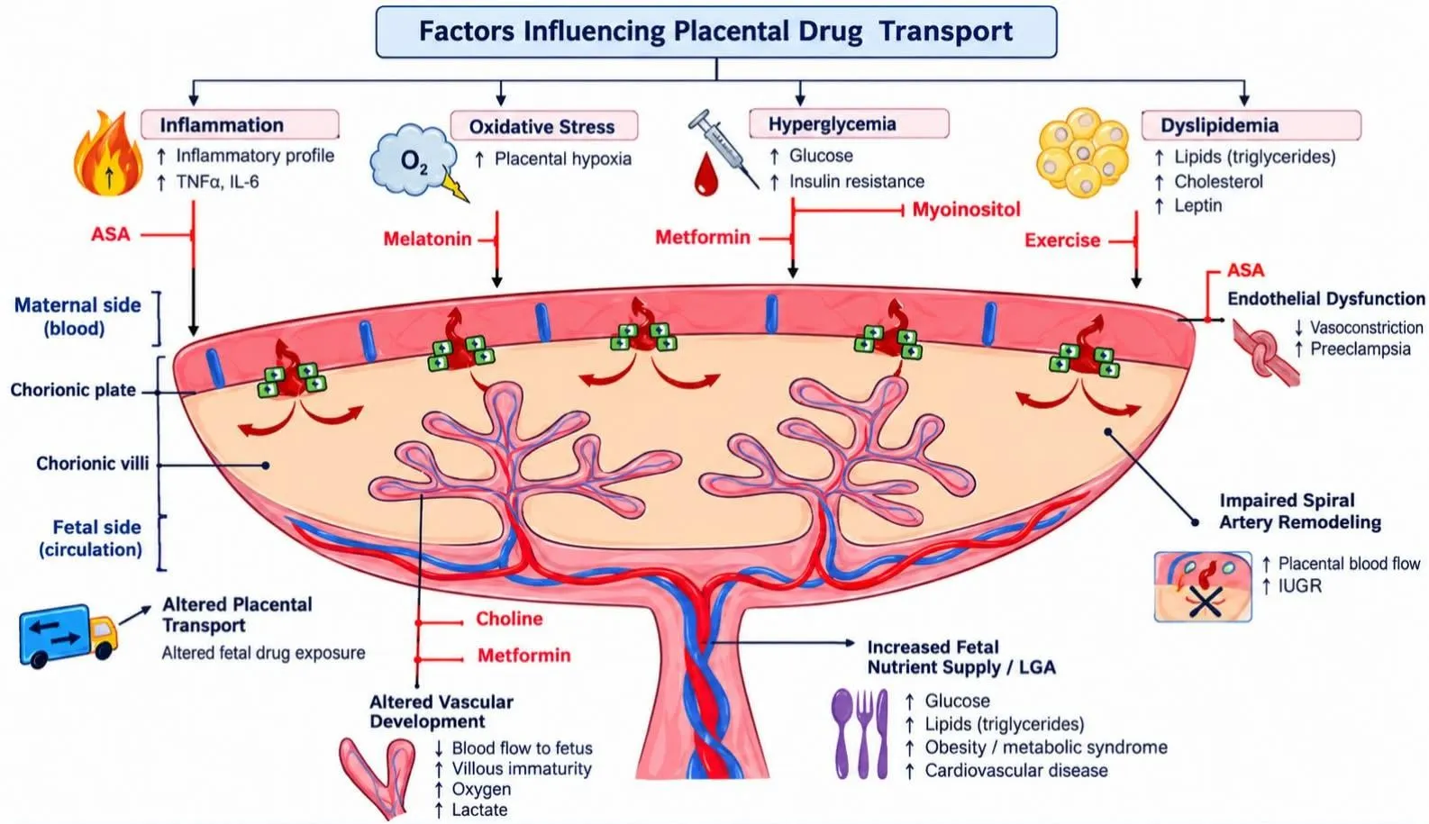
Figure 4: Key Factors Affecting Placental Drug Transport & Potential Therapeutic Interventions.[21]
4. Gestational Regulation of Placental Drug Transporters.
Table 2: Gestational Stage: Review Considerations Based on the Supplied Outline.
| Pregnancy stage |
Review focus | Key interpretation |
|---|---|---|
| First trimester | Early development and organogenesis | Early-gestation human transporter data are limited; avoid assuming term patterns apply |
| Second trimester | Placental maturation and ongoing fetal growth | Expression and activity may differ from early pregnancy |
| Third trimester / term | Late gestation and delivery tissue studies | Term placenta data are useful but may not represent earlier stages |
The placenta undergoes considerable changes during pregnancy with transporter expression being affected by growth, differentiation, vascular changes and endocrine/metabolic changes. Human placental tissues have demonstrated gestational age-dependent changes in expression of P-gp, BCRP and other ABC transporters. Quantitative data from Mao and Chen indicates that transporter expression is not constant throughout gestation. P-gp protein decreased by approximately 55%, BCRP decreased by approximately 69% and OCT3 and OAT4 increased by approximately 2-fold between first trimester and term. OATP2B1 decreased by approximately 32% between first and second trimester. However, NET and SERT did not show significant changes with gestation. This data is useful to demonstrate trends for individual transporters but should not be used universally to correct fetal exposure. Variations in cohort, sample, normalisation and platform can cause changes in expression estimates [12].
5. Pathological Regulation of Placental Drug Transporters
Table 3: Pregnancy Complications and Issues to Consider When Evaluating Transporter Regulation.
| Condition | Potential placental influence | Evidence note |
|---|---|---|
| Infection / inflammation |
May alter transporter expression and signaling | References in the supplied list address bacterial infection and multidrug-resistance transporters [14,15,17] |
| Preeclampsia | Placental dysfunction and altered transport processes | The listed review addresses transport of ions and small molecules; drug-specific conclusions need verification [16] |
| Gestational diabetes / obesity | Metabolic, hormonal and inflammatory changes may affect placental function |
Disease- and transporter-specific evidence should be assessed before drawing conclusions |
| Fetal growth restriction / preterm birth | May coexist with altered placental physiology | Direction and clinical significance are not uniform across drugs |
Complications during pregnancy can cause changes to placental physiology and therefore transporter expression/activity. Evidence varies depending on the complication, transporter family and drug, therefore disease specific changes should be mentioned [5]. A key theme in the literature is that complications during pregnancy can cause changes to the placental barrier and transporter expression/function. Kozlosky, Barrett and Aleksunes looked at the evidence for placental efflux transporters and how they are affected by inflammation/infection, chorioamnionitis, hypertensive disorders, metabolic disorders and fetal growth restriction [13].
5.1 Infection and inflammation
Maternal infection and placental inflammation can affect trophoblast signaling pathways and drug transporter expression. Bacterial infection can affect transporter expression in the human placenta and infection may cause disruption to multidrug resistance transporters. Changes associated with inflammation could affect placental disposition of certain drugs. The clinical implications will depend on the transporter and drug.[14]
5.2 Gestational diabetes, maternal obesity and other complications
Metabolic disorders, including gestational diabetes and maternal obesity, can cause changes in the maternal-placental environment, including hormonal, inflammatory, metabolic and oxidative changes. These are plausible candidates for regulating transporters, however, evidence may vary depending on the transporter and study design. Placental dysfunction can occur alongside fetal growth restriction and preterm birth. Further disease specific research is needed before general conclusions can be drawn about individual drugs [15].
5.4 Autoimmune and inflammatory conditions
Autoimmune and chronic inflammatory diseases may affect placental function via cytokine production, immune cell activity and drug exposure. Disease and drug treatments may both be relevant. Careful consideration is required to determine whether changes to placental transporters are due to the disease present in the mother, or the medication itself [15].
6. Molecular Mechanisms of Transporter Dysregulation.
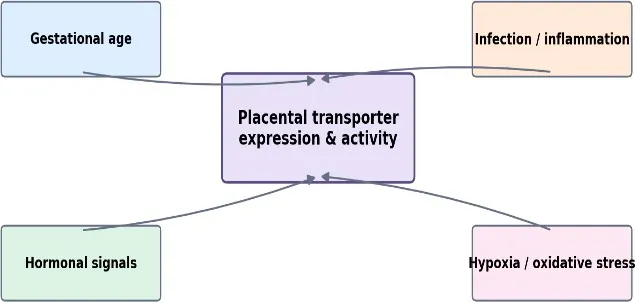
Figure 5: Gestational and Pathological Factors That May Regulate Transporter Expression and Activity.
Regulation of transporters can occur at many levels, including gene regulation of transporter expression, regulation of the mRNA translation, protein degradation, targeting of the protein to the correct membrane and direct regulation of transporter function. Inflammation can activate cytokine mediated pathways which will impact gene regulation and protein turnover. Oxidative stress and hypoxia can also impact mitochondrial function, membrane integrity and signalling pathways. This can have an impact on ATP dependent efflux transporters. Different responses can be seen from the same stimulus in different trophoblast models and therefore it is important to state the model used, exposure time, concentration and endpoint measured [13].
Nuclear receptors and other transcription factors can regulate transporter expression following hormonal or xenobiotic stimulation. Hormonal changes that occur during pregnancy can influence the regulation of transporter expression. Genetic polymorphisms can also have an impact on substrate recognition and function. Epigenetic regulation such as DNA methylation, histone modifications and microRNA regulation can also impact transporter expression. There is literature surrounding chorioamnionitis showing that a specific placental ABC-transporter signature has been associated with increased miR-331-5p in preterm placenta [15].
6.1 Inflammatory cytokines
Inflammatory mediators can alter gene transcription, cell signaling and protein turnover. In the placenta, infection and inflammation has been associated with changes in ABC transporter profiles indicating that these molecules are influenced by the local inflammatory environment [15].
6.2 Oxidative stress and hypoxia
Placental dysfunction can result in oxidative stress and hypoxia. These can affect signal transduction pathways, cellular energy status and membrane protein stability. Transporter function is dependent on the availability of the protein and the cellular environment. Therefore, changes in oxidative/hypoxia may lead to altered transporter function. However, the extent and direction of effect will depend on transporter specific functional data.[17]
6.3 Hormonal regulation and nuclear receptors
Pregnancy is associated with fluctuating levels of steroid and peptide hormones. This allows hormones and their nuclear receptors to control genes involved in xenobiotic sensing, metabolism and transport. This could provide a mechanism by which transporter expression is controlled throughout gestation. However, the precise effect will depend upon the transporter, stage of gestation and physiological/pathological state.[18]
6.4 Genetic, epigenetic and microRNA mechanisms
Genetic differences may contribute to variation in transporter expression and function between individuals. Epigenetic control can alter gene availability and expression. miRNAs can also control target mRNAs. This reference describes how chorioamnionitis can cause an altered ABC-transporter signature in the placenta with increased levels of miR-331-5p in human preterm placenta. Another review has looked at the effect of infection and inflammation on placental amino acid transporters [16].
7. Consequences for Maternal–Fetal Drug Exposure and Developmental Safety
Altered activity of transporters at the placenta can impact fetal drug exposure, with decreased efflux increasing fetal exposure to certain substrates. However, increased efflux can reduce transfer to the fetus. Uptake transporter changes can have more complex effects. As substrates can vary in affinity, passive permeability, protein binding and elimination, transporter changes for one drug may be clinically significant but have no effect for another [9].
Fetal drug concentration does not always equate to fetal toxicity. The development of the fetus depends on several factors including the dose, duration of exposure, distribution and clearance from the fetus, as well as the stage of development. It is important to consider when the drug is being administered. If there is a change in concentration during organogenesis this may be different from later in pregnancy. However, transporter expression alone will not determine the outcome of development and evidence of a clinical effect must be considered [12].
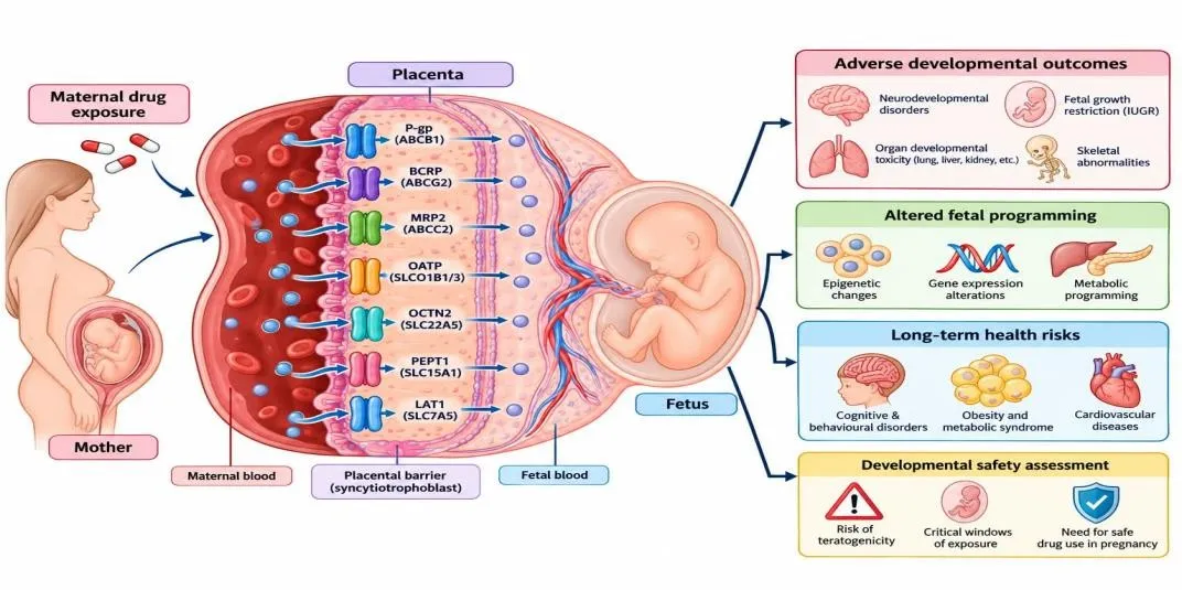
Figure 6: Consequences of Maternal–Fetal Drug Exposure and Implications for Developmental Safety.[2]
Practical considerations should be taken into account including the indication for the drug in the mother, existing pregnancy specific data, placental transfer characteristics, transporter interactions, gestational age and maternal disease. If there is uncertainty about the evidence then individualised decisions should be made with appropriate clinical oversight. Understanding the mechanism behind transporter changes may help to understand why there is variability in fetal exposure and help to determine when closer monitoring or alternative treatment may be required. Transporter changes can have at least three different impacts on the drug. This should not be confused. Decreased efflux transporters may increase fetal exposure to the transported substrate but does not mean all drugs will have increased transfer or fetal toxicity. Placental retention may increase which can impact placental cells even if fetal blood concentrations are relatively low [5].
The safety of development depends on several factors including the compound, dose, duration, timing, target organ in the fetus, maternal disease and benefit of treatment. A robust risk assessment should be performed to understand the maternal disease without treatment, known and unknown risks of treatment, using the best available pregnancy specific data. Transporter data should not be interpreted in isolation. If there is limited data then this should be clearly stated. Mechanistic studies can help to understand what factors may impact risk. Clinical outcome data and validated exposure models are required to understand if these will meaningfully impact outcomes in human pregnancy [2].
8. Experimental and Emerging Approaches.
Table 4: Experimental Approaches for Studying Placental Drug Transport.
| Approach | What it can contribute | Important limitation |
|---|---|---|
| Trophoblast cell models | Controlled study of transporter expression and substrate movement | May not fully reproduce intact placental tissue |
| Ex-vivo placental perfusion | Direct assessment of transfer across collected placenta | Usually reflects tissue obtained at delivery, not early gestation |
| Placental organoids | Study of trophoblast biology and selected disease processes | Needs validation for transporter localization and function |
Depending on the question being asked, different experimental models can be used. Trophoblast cell lines are relatively easy to access and can be used to screen for substrates or inflammatory responses. However, they may not fully recapitulate the polarised and differentiated nature of trophoblast. Primary cells and placental tissue can be used to provide a greater biological relevance however there are limitations to the availability of placental tissue, variability from different donors and the short time window. Ex-vivo perfusion can be used to directly measure transfer between maternal and fetal circulations in a functioning term placenta. However, great care must be taken in maintaining flow, tissue viability, mass balance and non-specific binding to ensure reliable data [2].
8.1 In-vitro trophoblast models
Trophoblast cell models are useful tools to study transporter expression, substrate transport and responses to inflammatory/hormonal stimulation. They allow comparison of experimental conditions and can provide insight into mechanisms. However, they do not fully represent whole placental tissue and can have issues with cell differentiation and transporter expression.[20].
8.2 Ex-vivo human placental perfusion
Ex-vivo perfusion of human placental tissue can be used to study compound transfer across the placenta between maternal and fetal circulations. This can provide direct placental transfer data which can be used alongside cellular models. However, the tissue is usually obtained post-delivery and so cannot fully represent early gestation. The length of the experiment and viability of the tissue can limit interpretation [8].
8.3 Placental organoids/advanced cellular models
Placental organoids/advanced cellular models may provide a better opportunity to study trophoblast differentiation, cell-cell interactions and disease changes. However, how well the model represents placental structure and transporter distribution/function will depend on how well the model recapitulates the in-vivo placenta. Validation using human tissue and functional transport data will be important.[22].
8.4 Physiologically based pharmacokinetic modelling
Physiologically based pharmacokinetic (PBPK) models can be used to predict exposures by incorporating maternal drug kinetics, placental transfer, transporter parameters and fetal physiology. This may be used to support dose and risk assessments when direct fetal data is not available. However, predictions will depend on the quality of the data inputs, particularly transporter activity at specific gestations and disease changes [10].
8.5 Multi-omics and computational modelling
Using transcriptomics, proteomics, metabolomics and microRNA can provide insight into how placental transporters respond to gestational development and disease. Combining this data with functional transport studies and PBPK models can provide insight into trends which may not be picked up by individual measurements. Computational modelling should be validated using independent experimental/clinical data. Current Knowledge Gaps and Future Perspectives
There are several limitations to predicting fetal drug exposure. Firstly there is limited data available in humans in early gestation. Secondly, transporter mRNA/protein expression do not always correlate with activity. Thirdly, whilst major ABC efflux transporters have been well characterised, SLC uptake transporters may not be as well understood. Finally, transporter systems may be impacted differently by complications of pregnancy, meaning disease and trimester specific investigations are required [5]. Future studies should investigate transporter localisation and activity, using relevant drug concentrations and multiple gestational stages if possible. It should be clear whether any associations observed are causal and confounding factors such as maternal disease severity, drug use and placental pathology should be considered. Correlating transporter data with maternal and fetal pharmacokinetics will help translate findings into a clinical context. Ultimately the aim will be to improve personalized prediction of fetal exposure. This will involve creating valid models which take into account maternal physiology, gestational age, placental function, drug properties, transporter genotype/ regulation and pathological conditions. This may help provide safer and more personalized pharmacotherapy during pregnancy. However, maternal therapy and disease management will remain an important aspect of pregnancy.[19]
Table 5: Summary of Key Review Themes
| Theme | Main point | Caution when interpreting evidence |
|---|---|---|
| Transporter families | ABC proteins commonly mediate efflux; SLC systems Support carrier-mediated movement. |
Net effect depends on substrate, membrane location and activity. |
| Gestational change | Transporter expression may vary with gestational age. | Term tissue cannot automatically represent early pregnancy. |
| Pregnancy complications | Infection/inflammation and other disorders may affect placental transport. |
Evidence differs by disease, transporter and drug. |
| Developmental safety | Fetal exposure is one factor in risk assessment. | Transporter expression alone does not prove toxicity. |
| Emerging methods | Cell models, perfusion, organoids and PBPK models complement one another. |
Models require validation with human and functional data. |
Quantitative data from early and mid-gestation is needed. The placenta can easily be obtained at term however this does not reflect all stages of development. The abundance of transporter proteins should be measured, with localization and function. Transporter specific effects should be distinguished from overall placental changes in perfusion, metabolism, inflammation and maternal pharmacokinetics. Clinically relevant substrate concentrations should be used along with well-characterised pregnancy complications so that results can be more carefully translated into clinical practice [13].
CONCLUSION
The placenta has an important role in controlling the transport of drugs from the maternal circulation to the fetus. This transport is influenced by many factors including drug physiochemical properties, passive diffusion, transporter expression/activation and maternal/fetal pharmacokinetics. Some drugs may have limited fetal exposure due to ABC efflux transporter expression including P-glycoprotein (P-gp/ABCB1) and breast cancer resistance protein (BCRP/ABCG2). However, solute carrier (SLC) transporters are involved in drug uptake and transport. Throughout pregnancy, expression and function of placental transporters can change and can also be affected by different pathological conditions including infection, inflammation and other complications. This can affect maternal-fetal transport of drugs. However, this will depend on the transporter and drug as it cannot be reliably predicted based solely on transporter expression.
Understanding how placental drug transporters are regulated during gestation and under pathological conditions is key to improving the assessment of medication safety during pregnancy. Future studies should investigate how placental transporters are localised and function during different stages of gestation, how complications affect transporters and how these findings can be applied to human pharmacokinetics. Using advanced techniques such as placental models, ex-vivo perfusion, organoids and PBPK modelling can help improve the prediction of fetal drug exposure. Ultimately by combining transporter knowledge with clinical evidence and individualised risk assessment, this can aid safe and effective pharmacotherapy during pregnancy.
REFERENCES





Revati Donode, Yogini Kawale, Priyanka Lilhare, Kunal Zode, Upadesh Lade, Gestational and Pathological Regulation of Placental Drug Transporters: Implications for Maternal–Fetal Drug Exposure and Developmental Safety, Int. J. of Pharm. Sci., 2026, Vol 4, Issue 11, 3964-3975. https://doi.org/10.5281/zenodo.24415432
 10.5281/zenodo.24415432