

We use cookies to ensure our website works properly and to personalise your experience. Cookies policy
P.R. Pote Patil College of Pharmacy, Amravati, Maharashtra, India.
Green pharmacy is an emerging approach that aims to make pharmaceutical processes simpler, safer and environmentally sustainable. The development of biodegradable polymeric implants is an important area of modern drug delivery that can be integrated with the principles of green pharmacy. These implants are designed to provide controlled and prolonged drug release and to degrade gradually into safer products within the body, which reduces the need for surgical removal after treatment. Natural biodegradable polymers such as collagen, chitosan, alginate, gelatin, starch and cellulose are often preferred because of their biocompatibility, biodegradability and renewable origin, while synthetic biodegradable polymers such as polylactic acid, polyglycolic acid, poly (lactic-co-glycolic acid) and polycaprolactone are also widely investigated for implant development. A green pharmacy approach decreases the use of hazardous chemicals and solvents, reduces pharmaceutical waste and energy consumption, and encourages environmentally friendly manufacturing methods. Biodegradable implants also improve patient compliance. Green pharmacy principles combined with biodegradable polymer technology therefore support the development of simple, sustainable and effective drug-delivery systems that promote pharmaceutical care while minimizing environmental impact. This review discusses the evolution, classification, drug-release mechanisms, natural polymers, advantages and manufacturing methods of biodegradable polymeric implants from a green pharmacy perspective.
1.1 Green Pharmacy and Pharmaceutical Implants
Green pharmacy is an approach to the design, development, manufacturing, use, and disposal of pharmaceutical products that applies the principles of green chemistry and sustainability to minimize the use and generation of hazardous substances, reduce waste and energy consumption, promote the use of safer and renewable materials, and minimize environmental impacts throughout the pharmaceutical product life cycle.1,2 Pharmaceutical implants are specialized drug-delivery systems designed to be placed within the body to provide prolonged, controlled, or site-specific release of a therapeutic agent. Unlike conventional dosage forms such as tablets, capsules, and injections, implantable drug-delivery systems can maintain drug concentrations for extended periods and may reduce dosing frequency, improve patient adherence, and provide drug delivery directly to a desired anatomical site.3
Polymeric implants provide a long-acting drug-eluting device that ensures patient compliance and in many cases aids with drug targeting when implanted at the required site of action.3 There are currently several US Food and Drug Administration (FDA)–approved polymeric implants. Implantable drug-delivery systems include a broad range of technologies, including polymeric matrices, drug-containing reservoirs, implantable pumps, biodegradable implants and drug-eluting devices.4
1.2 Historical Evolution of Polymeric Implants
The development of polymeric implants is closely associated with the evolution of controlled drug-delivery systems. Early implantable drug-delivery approaches were developed to provide prolonged exposure to therapeutic agents and overcome the limitations of conventional dosage forms.5 A major development occurred during the 1960s with the introduction of silicone rubber as a polymeric carrier for prolonged drug delivery. In 1964, Folkman and Long reported the use of silicone rubber (Silastic) capsules containing therapeutic agents. The drug was incorporated into an implanted silicone capsule and released gradually through the polymer. This work is considered an important milestone in the development of modern polymer-based controlled-release implants.6
Another important development was the application of polymeric implants to long-acting contraception. The development of levonorgestrel-releasing silicone implants eventually led to the introduction of Norplant, which was approved by the US FDA in 1990. Norplant consisted of six silicone capsules containing levonorgestrel and provided long-term contraceptive drug release. This represented one of the major clinical applications of polymer-based implantable drug delivery.7 The major milestones in the evolution of polymeric implants are summarised in Table 1.
Table 1: Evolution of Polymeric Implants
| Period | Major development in polymeric implants | Ref. |
|---|---|---|
| 1930s | Early hormone-containing implants | 8 |
| 1940s–1950s | Development and use of prolonged-release hormone implants | 8 |
| 1960s | Silicone/Silastic-based controlled-release implants | 6 |
| 1970s | Development of polymeric systems for sustained release of proteins and macromolecules | 9 |
| 1980s | Expansion of biodegradable polymer systems | 8 |
| 1990s | Clinical development and application of biodegradable polymeric implants | 3,8 |
| 2000s | Increased use of PLA, PLGA and PCL for controlled drug release | 10 |
| 2010s | Advanced manufacturing approaches and development of 3D-printed implants | 3 |
| 2020s–Present | Research on biodegradable, sustainable, personalized and advanced implant systems | 3 |
1.3 Need for Pharmaceutical Implants
The development of pharmaceutical implants has been driven by the limitations of conventional drug delivery routes. Oral administration is convenient and widely accepted, but many drugs exhibit poor absorption, degradation in the gastrointestinal tract, first-pass metabolism, or short biological half-lives.11 Patients receiving long-term treatment may have difficulty maintaining adherence to frequent dosing schedules. Implantable drug-delivery systems can address some of these limitations by providing prolonged drug release from a single administration.12 They may therefore be particularly useful for chronic diseases, hormone therapy, cancer treatment, pain management, contraception, and other conditions requiring sustained pharmacological activity.13
2. Classification of Implantable Drug Delivery Systems
2.1 Biodegradable Implants
Biodegradable implants were developed to overcome the drawbacks of non-biodegradable implants. These devices are made using polymers or block copolymers that can be broken down into smaller fragments that will be subsequently excreted or absorbed by the body.14 Normally they are made using polymers such as polycaprolactone, polylactic acid, polyglycolic acid and natural biodegradable polymers such as collagen, cellulose, dextran.15
2.2 Non-Biodegradable Implants
Non-biodegradable implants are commonly prepared using polymers such as silicones, poly (urethanes), poly (acrylates) or copolymers such as poly (ethylene vinyl acetate).16,17 This type of device can be monolithic or reservoir type implant, as shown in Figure 1. Monolithic type implants are made from a polymer matrix in which the drug is homogeneously dispersed.15 On the other hand, reservoir-type implants contain a compact drug core covered by a permeable non-biodegradable membrane. The membrane thickness and the permeability of the drug through the membrane will govern the release kinetics. Non-biodegradable implantable drug delivery systems have been extensively used for contraceptive delivery.16
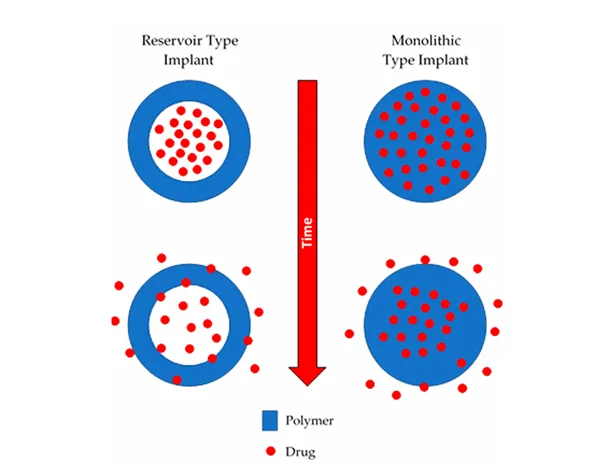
Figure 1: Schematic Illustration of Reservoir-Type and Monolithic-Type Implants.3
3. Mechanisms of Drug Release
3.1 Implantable Polymeric Drug Delivery Systems
Mechanisms of drug release from implantable systems are mainly classified into four groups: matrix degradation; controlled swelling; osmotic pumping; and passive diffusion.18 For systems based on controlled swelling, solvent penetration into the matrix of the device controls the rate of release. This is usually much slower than diffusion of the drugs, and will, therefore, lead to a lower release rate. Although the diffusion from swollen matrices is mainly responsible for the drug release, matrix degradation could also contribute in the effectiveness of these systems.19 On the other hand, osmotic pumping and passive diffusion mechanisms of drug delivery are the most promising for linear delivery of drugs. Osmosis is the overall movement of water from a dilute solution to a more concentrated solution through a partially permeable membrane, and it causes a hydrostatic pressure difference between the two compartments.20
Diffusion is a process by which molecules transfer spontaneously from one region to another to equilibrate chemical potential or thermodynamic activity. In this mechanism, migrating molecules are usually known as the diffusants or permeants, and the membrane or matrix in which the diffusant migrates is called the diffusional barrier. Additionally, the external phase is termed medium. The driving force of this drug release mechanism is the concentration gradient or profile of the diffusant within the diffusional barrier.21
3.2 Biodegradable Implants
Implantable drug delivery devices generally consist of a drug reservoir surrounded by a polymer or a drug polymer mixture. When inserted into the desired area of the body, the drug will be released at a pre-determined rate as the polymer degrades. Drug release from a reservoir system is controlled by the rate of polymer degradation or drug dissolution into, and then diffusion through, the polymer wall, or a combination of both. Drug release from a drug polymer mixture is controlled by diffusion, swelling or erosion. The release of drug from the system will be dependent on: the solubility and permeability of the drug in the polymer; the drug load; and the in vivo degradation rate of the polymer.22 Degradation of the polymer and subsequent drug release may occur through one, or a combination of processes including: hydrolysis, in which bonds (e.g., ester bonds) in the polymer backbone are broken down; enzymatic degradation, in which hydrolytically susceptible bonds (e.g., amide bonds) degrade in the presence of a catalyst; oxidation; and physical degradation, in which bonds are broken as a result of physical forces such as swelling or mechanical loading.23
The degradation time of polymers can vary extensively depending on features such as polymer molecular weight and surface properties. This will, in turn, affect the release of any drug contained within a formulation. In addition, degradation will also be dependent on in vivo factors such as: pH; and temperature. Therefore, the in vivo degradation time of any polymer needs to be fully characterised.24
4. Biodegradable Polymers Used in Implants
4.1 Overview and Classification
Biodegradable polymers used in implantable drug-delivery systems are broadly classified as natural or synthetic.25 The natural polymers discussed in this review are cellulose, chitosan, gelatin, alginate, chitin, dextran, pectin, collagen, starch and hyaluronic acid. The synthetic polymers include polylactic acid (PLA), polyglycolic acid (PGA), poly (lactic-co-glycolic acid) (PLGA), polycaprolactone (PCL), polydioxanone (PDS) and polyhydroxyalkanoates (PHAs).25 Because natural polymers are obtained from renewable sources and are generally biocompatible and biodegradable, they align closely with the principles of green pharmacy; their key features are summarised in Table 2.
4.2 Natural Polymeric Implants
4.2.1 Cellulose
Cellulose is a natural, linear polysaccharide and one of the most abundant organic compounds in the world. It is produced predominantly by plants and is therefore considered an important naturally derived polymer. Cellulose consists of long polymer chains made up of repeating β-D-glucopyranose units. These glucose units are covalently linked through β-(1→4)-glycosidic bonds, forming a linear polymeric structure. Cellulose is among the natural polymers with potential applications in implantable devices, since natural polymers can provide biocompatibility, biodegradability and non-cytotoxicity.26
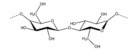
Figure 2: Chemical Structure of Cellulose.26
Cellulose is mostly produced by plants and represents a major naturally occurring polymeric material. Plant-derived cellulose can therefore be considered a renewable polymer source. Bacterial cellulose (BC) is another important form of cellulose used for biomedical applications.27 Biodegradability is particularly important when developing sustainable biomedical materials and implantable systems. Bacterial cellulose fibres possess a high degree of crystallinity. This structural characteristic contributes to the distinctive properties of bacterial cellulose and its potential biomedical applications. Bacterial cellulose possesses a nanostructure that makes it attractive for a range of medical applications.28 Plant-derived polymers are cost-effective to produce and straightforward to prepare for implantation. Cellulose can be converted into different forms and derivatives, including cellulose ethers/esters, micro- and nanoscale cellulose materials, bacterial cellulose, cellulose nanocrystals and cellulose nanofibrils.29
4.2.2 Chitosan
Chitosan is a cationic polysaccharide produced by deacetylation of chitin, which is found in the exoskeletons of insects, the cell walls of fungi, and certain hard structures in invertebrates and fish.30 This natural polymer is structurally composed of N-acetyl-D-glucosamine and D-glucosamine units with one amino (NH2) group and two hydroxyl (–OH) groups in each repeating glycosidic units. The structure of chitosan is shown in Figure 3. Due to its properties, it has been extensively used in a large amount of applications ranging from medical to industrial areas, including application in implantable drug delivery devices.31
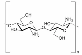
Figure 3: Chemical Structure of Chitosan.
Biodegradation plays a major role in the metabolic fate of chitosan in the body and is important for all polymers used in drug delivery systems. Both chemical and enzymatic biodegradation can provide fragments suitable for renal clearance. Chemical degradation is referred to acid catalyzed degradation such as in the stomach. Enzymatically, chitosan can be degraded by enzymes able to hydrolyse glucosamine–glucosamine, glucosamine–N acetyl-glucosamine and N-acetyl-glucosamine–N-acetyl glucosamine linkages. Chitosan is known to be degraded in vertebrates predominantly by lysozyme and by certain bacterial enzymes in the colon. Chitosan has proved to be a safe excipient in drug formulations over the last decades.32 Chitosan is considered as being a non-toxic, biologically compatible polymer. It is approved for dietary applications in Japan, Italy and Finland, and it has been approved by the FDA for use in wound dressings. Chitosan offers several advantages, and these include its ability to control the release of active agents and avoid the use of hazardous organic solvents while fabricating particles since it is soluble in aqueous acidic solution. Chitosan is extensively used in developing drug delivery systems.33
Chitosan is used in the field of tissue engineering. Tissue engineering is a highly interdisciplinary field that combines the principles and methods of life sciences and engineering to utilize structural and functional relationships in normal and pathological tissue to develop biological substitutes to restore, maintain, or improve biofunction. It involves the in vitro seeding and proliferation of relevant cells in a scaffold support. Since it is biodegradable, non-toxic and can be formulated in a variety of forms including powders, gels and films for applications, it has a wide range of potential applications in tissue engineering. In addition to its use in the formulation of controlled delivery systems, a wide range of chitosan modifications can be made to improve cell seeding. Many tissue analogs including cartilage, bone, liver, and nerve have been prepared using this engineering technology.34 Chitosan has been extensively used in bone tissue engineering, since it was shown to promotes cell growth and mineral rich matrix deposition by osteoblasts cells in culture. The biocompatibility of chitosan minimizes additional local inflammation, and it can be molded into porous structures to allow osteoconduction.35
4.2.3 Gelatin
Gelatin is a naturally occurring polymer derived from the hydrolytic breakdown of collagen, and its unique amino acid composition imparts various medicinal advantages. It is widely used in the pharmaceutical, food, cosmetic and photographic industries because of its gelling, texturizing, stabilizing and emulsifying properties.36 Its favourable attributes, such as biodegradability, biocompatibility and low toxicity, promote enhanced cell adhesion, differentiation and proliferation. Gelatin is degraded by endogenous enzymes, including metalloproteinases, without eliciting an immunogenic reaction.37
Tissue engineering is an emerging field that integrates biology, engineering, medicine, chemistry, materials science and pharmacy. Natural polymers such as gelatin, collagen and chitosan are favoured in tissue engineering because of their low antigenicity, natural degradability, biological compatibility and resemblance to the native extracellular matrix (ECM).38
4.2.4 Alginate
Alginate is a naturally occurring polysaccharide obtained mainly from brown seaweeds and also produced by certain bacteria. It is a copolymer consisting of β-D-mannuronic acid and α-L-guluronic acid residues. Alginate is considered a biocompatible, biodegradable, non-antigenic and non-toxic natural polymer. Its ability to form gels in the presence of divalent and multivalent cations, particularly calcium ions, makes it useful in pharmaceutical and biomedical applications.39 The major commercial sources of alginate are brown seaweeds belonging to the class Phaeophyceae. Important alginate-producing seaweeds include Laminaria, Macrocystis, Ascophyllum, Sargassum, Lessonia and Ecklonia species. Alginate can also be produced by microorganisms, particularly Azotobacter vinelandii and Pseudomonas species. Alginate possesses several properties that make it suitable for pharmaceutical and biomedical applications. It exhibits high viscosity and shear-thinning behaviour. Alginate can undergo ionic crosslinking with calcium ions to produce hydrogels and three-dimensional polymeric networks. These characteristics make alginate useful for controlled drug delivery, tissue engineering, wound dressing and cell encapsulation.40
Alginate degradation involves cleavage of the polymer backbone and reduction in molecular weight. The polymer can undergo chemical degradation under acidic or alkaline conditions. Enzymatic degradation can also occur through alginate lyase, which cleaves alginate chains. The degradation behavior is influenced by factors such as molecular weight, composition of mannuronic and guluronic acid residues, degree of crosslinking and environmental conditions. Alginate has extensive applications in pharmaceutical, biomedical, food and biotechnology fields. In pharmaceutical formulations, it is used for oral drug delivery, controlled-release systems, tablets, hydrogels, microspheres, microcapsules, nanoparticles, beads and floating drug-delivery systems. Biomedical applications include wound dressings, bone regeneration, neovascularization, protein delivery, cell delivery and tissue engineering. Alginate is also used for immobilization of biological materials and for the treatment of environmental contaminants.41 In the food industry, alginates are used to produce restructured foods with improved texture and structural properties and to maintain product stability at low and high temperatures; the gelation rate and gel strength are controlled by the concentrations of Ca2+ and H+ ions.42
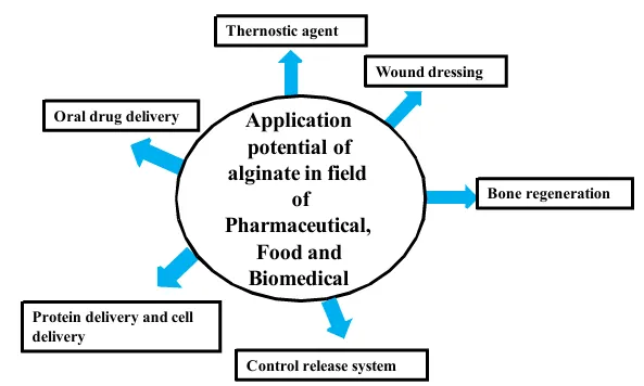
Figure 4: Application Potential of Alginate in the Pharmaceutical, Food and Biomedical Fields.42
4.2.5 Chitin
Chitin (C8H13O5N)n is the second most abundant biopolymer in the world, a long-chain polymeric polysaccharide, renewable organic resource and the supporting material of some plants and many sea creatures and is produced by a number of living organisms. It is obtained at a relatively low price from these organic resources especially shells of shellfish (mainly shrimps, lobsters, crabs and krill) and waste of seafood processing industries. This natural polymer has very good properties such as biocompatibility, biodegradability, and non-toxicity.43 Chitin has a high molecular weight and its structure consists of linear repeating units of β-1,4-linked N-acetyl D-glucosamine and chemically resembles cellulose, in which the hydroxyl group at carbon-2 in cellulose has been substituted by acetamido group. Chitin is white, hard, semi-transparent material, nitrogenous polysaccharide, and inelastic, low chemical reactivity and insoluble in most organic solvents. It is provided in forms of granules, sheets and powders. Pure chitin is inexpensive and commercially available.44 Pure chitin is inexpensive and commercially available. The word “chitin” is derived from the Greek word “chiton”, meaning a coat. It was apparently used by Braconnot in 1811 for the first time.45
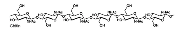
Figure 5: Chemical Structure of Chitin.45
Chitin is a natural material with a structure consisting of an assembly of microfibrils of about 20 sugar chains. These microfibrils are structural components of biocomposites which constitute the cell walls and some particular extracellular matrices. Chitin is synthesized by fungi and insects, which extremely depends on biosynthesis machinery involving several soluble enzymes. It has various applications in waste water treatment (removal of metal ions, dyes and pigments as membrane in purification processes), food industry (anti-cholesterol and fat binding, packaging material, preservative and food additive), agriculture (seed and fertilizer coating, controlled agrochemical release), pulp and paper industry (surface treatment, photographic paper), cosmetics and toiletries (moisturizer, body creams, lotions) and biomedical applications (tissue engineering, drug delivery, wound healing, cancer diagnosis).46
Chitin and its most important derivative, chitosan, show fungistatic and fungicidal activity and dissolve in organic acids, which makes them good candidates for cosmetic applications such as hair care, oral care and skin care. The biocompatibility, biodegradability and non-toxicity of chitin are the distinctive properties that make it suitable for biomedical applications.47
4.2.6 Dextran
Dextran is a naturally occurring microbial polysaccharide belonging to the class of neutral polyglucans. It is composed of D-glucose units and possesses a predominantly α-(1→6)-linked main chain, together with varying proportions of α-(1→2), α-(1→3), and α-(1→4) linkages that form branches. The proportion and arrangement of these linkages depend strongly on the bacterial strain responsible for dextran production. The α-(1→6) linkages may account for approximately 50–97% of the total glycosidic linkages. Dextran is particularly important among microbial polysaccharides because of its applications in medical, biomedical and industrial fields. Its usefulness is associated with its water solubility, biocompatibility and ability to undergo degradation under suitable biological or physical conditions. The polymer also contains several hydroxyl groups, which provide opportunities for chemical modification and preparation of derivatives with tailored physicochemical and functional properties.48
Dextran is primarily produced by microorganisms, particularly species belonging to the genera Leuconostoc and Streptococcus. Leuconostoc mesenteroides is one of the most important organisms used for dextran production. These microorganisms are capable of synthesizing dextran mainly from sucrose. Dextran-producing microorganisms have also been associated with fermented foods of plant origin. Dextran can also occur in dental plaque. Streptococcus mutans produces water-soluble glucans, including dextran-like glucans, from sucrose. These glucans contribute to the matrix that supports colonization of cariogenic bacteria on tooth surfaces. Dextranases, which hydrolyse specific glycosidic bonds, can be used to degrade these polysaccharides.49 Dextran degradation occurs mainly through cleavage of its glycosidic linkages. Dextran can be depolymerised by α-1-glycosidases, commonly referred to as dextranases. These enzymes are reported to occur in tissues including the liver, spleen, kidney and lower gastrointestinal tract. Therefore, enzymatic hydrolysis contributes to the biodegradation of dextran in biological environments. The degradation behaviour can be influenced by chemical modification of the dextran backbone. For example, ionic derivatives such as diethylaminoethyl dextran and carboxymethyl dextran are degraded more slowly by α-1-glucosidases than unmodified dextran. However, cross-linked dextran can still undergo enzymatic degradation. This characteristic is useful for developing biodegradable hydrogel systems. Controlled degradation can also be used during the preparation of dextran fractions. Partial hydrolysis using hydrochloric or sulfuric acid, followed by fractionation, allows the molecular weight of dextran to be controlled. Enzymatic degradation using dextranases is another approach for obtaining defined dextran fractions.50
Dextran and its derivatives have also been investigated for drug-delivery applications. Chemical modification of dextran can alter its solubility, hydrophilic–lipophilic balance and degradation characteristics. Amphiphilic dextran derivatives can form micelles and can therefore be investigated for encapsulation of hydrophobic drugs. Dextran-based ether derivatives can also incorporate spacers that facilitate drug attachment and influence drug release. Dextran-based hydrogels are additionally investigated as matrices for drug delivery and tissue engineering. For example, cross-linked carboxymethyl dextran hydrogels can show pH- and ionic-strength-dependent changes in swelling, porosity and protein transport.51
4.2.7 Pectin
Pectin is universally present in every organ of terrestrial plant in the meristematic tissue as well as parenchyma. In plant cell, pectin is present in cell wall and middle lamella, while the quality and quantity of pectin could vary between different plant species. Traditionally, pectin is extracted with hot water (60-100 °C) acidified under the pH range of 1.5-3 for several hours. Different acids like oxalic acid, citric acid, sulfuric acid, hydrochloric acid and nitric acid are used for pectin extraction. But, this is a time-consuming method with very low efficiency. In order to improve the quality and efficiency of pectin, different extraction techniques were established like heat refluxing extraction, solvent extraction through the heating and stirring, microwave, enzymatic, ultrasound-assisted extraction and combination of subcritical water with ultrasound-assisted extraction.52
Chemically, pectin (methylated ester of polygalacturonic acid) mainly contains α-D-galacturonic acid residues. Pectin is composed of at least 17 different kinds of monosaccharides with D-galacturonic acid as main component followed by the D-galactose and L-arabinose. The degree of esterification (DE) is an important term that influences various properties of plant pectin and could be defined as carboxyl group percentage esterified with methanol. Based on DE, there are two different classes in pectin, i.e., high methoxyl pectin (HMP) (more than 50% DE) and low methoxyl pectin (LMP) (less than 50% DE). Moreover, DE varies according to the source of pectin.53 Commercially, apple pomace and citrus peels are the main source for the production of pectin. Also, the large quantities of wastes generated from citrus fruits processing are particularly the main source for production of citrus pectin. It is estimated that 25-30% dry weight of citrus peel is composed of pectin. Moreover, sugar beet and sunflower are also sources of great interest for industries related to pectin extraction. All these sources are important because of their biomasses availability as well as the physiochemical quality presented by their pectin. Novel sources such as cocoa husks are also being explored.54
Pectin has attracted considerable attention as a natural polymeric material for pharmaceutical, food, nutraceutical, cosmetic and biomedical applications. Its gelling ability, binding capacity, emulsifying properties, biodegradability and biocompatibility make it suitable for use as a carrier and wall material for bioactive compounds. Pectin can be incorporated into nanocomplexes, nanohydrogels, nanoliposomes, nanoemulsions, nanoparticles and electrospun nanofibers for protection and controlled delivery of active compounds. In pharmaceutical applications, pectin has been investigated as a carrier for drugs and bioactive compounds such as curcumin, doxorubicin, indomethacin, itraconazole and gentamicin. Pectin-based systems can improve the stability of encapsulated compounds and provide controlled or targeted release. Pectin-based hydrogels and nanohydrogels have also been investigated for delivery of vitamin C, curcumin, indomethacin and anticancer drugs.55
Pectin is particularly useful for oral and colon-targeted drug delivery because it is poorly absorbed in the upper gastrointestinal tract and can undergo degradation by pectinolytic enzymes produced by colonic microorganisms. Therefore, pectin-based nanocarriers can provide protection of active compounds during gastrointestinal transit and facilitate their controlled release in the intestine or colon. Pectin-containing systems have been investigated for the delivery of curcumin, rutin and neohesperidin under simulated gastrointestinal conditions. Pectin can also be used as a coating material for liposomes. Pectin-coated liposomes can improve the stability and encapsulation efficiency of liposomal systems, while its mucoadhesive properties may contribute to improved interaction with biological surfaces. Pectin-coated nanoliposomes have been investigated for delivery of vitamin C and phloridzin and have potential applications in pharmaceutical and food products.56
Pectin is also applicable in biomedical delivery systems. Pectin-based electrospun nanofibers possess high porosity, large specific surface area and interconnected structures and have been investigated for drug delivery and other biomedical applications. Pectin-containing formulations have also been investigated for local delivery of gentamicin in infected wounds, where an alginate–pectin formulation provided sustained drug penetration for approximately 3–6 days. Thus, the properties of pectin make it a versatile natural polymer for the development of biodegradable delivery systems. Its ability to form gels, interact with other polymers and protect active compounds supports its potential application in controlled drug delivery, colon-targeted delivery, wound-care formulations, nutraceutical delivery and other biomedical applications.57
4.2.8 Collagen
Collagen is a naturally occurring proteinaceous polymer and is one of the major structural components of the ECM. It is highly conserved among different species and accounts for approximately one-third of the total protein present in mammalian tissues. Collagen is particularly abundant in connective and musculoskeletal tissues, where it provides structural support and contributes to the mechanical integrity of tissues such as skin, bone, tendon and cartilage. At least 27 types of collagen have been identified. Collagen molecules are organized into fibrillar and other structural arrangements, and their density, packing, orientation and degree of cross-linking determine the mechanical characteristics of individual tissues. Type I collagen is particularly important in connective and matrix-rich tissues and is extensively used in the preparation of biomaterials and tissue-engineering scaffolds. Collagen is therefore considered an important natural polymer for biomedical applications because its structure closely resembles the extracellular matrix of native tissues. Its architecture can also be processed and modified to produce three-dimensional scaffolds for tissue regeneration and repair.58
Collagen is a naturally occurring component of mammalian tissues and provides biological motifs that support interaction between cells and the extracellular matrix. Cell–collagen interactions occur primarily through integrin receptors. Collagen can be processed into hydrogels, porous scaffolds, meshes and electrospun structures. These forms allow collagen to be used for different tissue-engineering applications. The mechanical performance of collagen depends strongly on its density, packing, orientation and degree of cross-linking. This allows collagen scaffolds to be engineered for different tissue environments.59 Collagen is a biodegradable protein and can undergo degradation after implantation. The degradation behavior depends on the organization of the collagen matrix and the processing or cross-linking treatment applied to the material. During processing, collagen may be dispersed or broken down into polymers, oligomers or monomers depending on the treatment conditions. In biological environments, collagen is susceptible to degradation by collagenolytic enzymes, particularly collagenases. The degradation rate can therefore be influenced by the degree of cross-linking. For example, glutaraldehyde cross-linking makes collagen less susceptible to collagenase degradation and consequently increases its persistence in vivo.60
4.2.9 Starch
Starch is an abundant natural biopolymer and an important energy-storage material in plants. It serves as a major source of dietary carbohydrate for humans and animals and is also widely used as a raw material in non-food industries. Two major forms of starch occur in plants: storage starch and transitory starch. Transitory starch is synthesized in the chloroplasts of leaves and undergoes a daily cycle of synthesis and degradation, whereas storage starch is produced mainly in storage organs such as seeds and tubers and remains stored until it is required for germination or regrowth. Storage starch is the major commercially utilized form. Starch is a multifunctional biomaterial because of its abundance, low cost and relatively non-toxic nature.61 It occurs naturally as semicrystalline granules and consists mainly of two glucose polymers, amylose and amylopectin. Both are composed of α-D-glucose units. The glucose units are primarily connected by α-(1→4) glycosidic bonds, while α-(1→6) glycosidic bonds form the branching points. Amylopectin generally constitutes about 70–85% of starch and is considerably more highly branched and larger than amylose. The amylose-to-amylopectin ratio varies according to the plant genotype. Amylopectin contributes substantially to the semicrystalline organization of starch granules. Its clustered branching points allow neighboring glucan chains to form double helices and crystalline lamellae, which alternate with amorphous regions. Starch granules can range from less than 1 μm to more than 100 μm in diameter, and their shape may be spherical, oval, discoid, polygonal, angular or irregular depending on the plant source and tissue.62
Starch is obtained from a wide range of plant materials. Starch is synthesized in different plant organs, with storage starch occurring particularly in seeds and storage organs. Examples include potato, cassava, rice, wheat, barley, banana and sweet potato. The starch characteristics vary according to the plant genotype, tissue and organ. Differences in amylose and amylopectin content, granule size, morphology, crystallinity and phosphate content can occur between different botanical sources.63
4.2.10 Hyaluronic Acid
Hyaluronic acid (HA) is a naturally occurring glycosaminoglycan belonging to the polysaccharide family and is widely distributed in vertebrate tissues. It consists of alternating units of N-acetyl-D-glucosamine and glucuronic acid and possesses a very high molecular weight, which may reach several million Daltons. HA is not covalently bound to proteins and is predominantly present in connective tissues. It is an important component of the extracellular matrix and is found in tissues such as articular cartilage, nucleus pulposus, skin, cervix, and the glycocalyx of endothelial cells. In the body, HA is synthesized by membrane-associated hyaluronan synthase enzymes through the repeated addition of glucuronic acid and N-acetyl-D-glucosamine units. Its biological functions are influenced by molecular weight; low-molecular-weight HA is associated with healing and scar formation, whereas high-molecular-weight HA contributes to tissue integrity. HA also participates in cell growth and extracellular-matrix interactions.64
Hyaluronic acid possesses several properties that make it suitable for pharmaceutical, biomedical, and cosmetic applications. It is a highly hydrophilic macromolecule containing carboxyl (-COOH) and hydroxyl (-OH) functional groups and has high water solubility. HA solutions exhibit high viscosity and unique viscoelastic properties, while intramolecular hydrogen bonding allows the polymer to form three-dimensional structures. One of its important characteristics is its exceptional water-retention capacity; HA can trap water in an amount approximately 1000 times its own weight and can therefore form hydrated gels. HA also contributes to tissue flexibility and lubrication. The presence of several functional groups allows HA to undergo physical and chemical crosslinking, and its properties can be modified by varying polymer concentration, molecular weight, crosslinking conditions, or blending with other polymers. Crosslinked HA hydrogels possess improved resistance to enzymatic degradation compared with uncrosslinked materials.65
HA-containing three-dimensional scaffolds have also been investigated for bone and cartilage tissue engineering, and HA-based blends containing magnetic particles have potential applications in tissue repair and drug delivery. In cosmetic applications, HA is used in hydrogels, creams, gels, and injectable dermal fillers because of its ability to retain water and moisturize tissues. HA hydrogels have been investigated for skin rejuvenation, while HA-containing formulations have been studied for wrinkle reduction, skin hydration, and other dermatological applications. In addition, HA-containing polymer blends can be developed as topical formulations for localized delivery of medications to the skin. More advanced applications include three-dimensional scaffolds, injectable hydrogels, electrospun materials, and bio-inks for three-dimensional bioprinting of cartilage and other tissues.66
Table 2: Summary of the Natural Biodegradable Polymers Discussed
| Polymer | Source | Key characteristics | Representative uses | Ref. |
|---|---|---|---|---|
| Cellulose | Plants; bacterial cellulose from microorganisms | Linear β-(1→4)-linked glucose polymer; renewable, biocompatible, biodegradable | Implantable devices and biomedical materials | 26–29 |
| Chitosan | Deacetylation of chitin | Cationic polysaccharide; non-toxic; soluble in aqueous acidic solution; degraded by lysozyme | Drug delivery, bone tissue engineering, wound dressings | 30–35 |
| Gelatin | Hydrolysis of collagen | Biodegradable, biocompatible, low toxicity; degraded by endogenous enzymes | Tissue-engineering scaffolds, pharmaceutical use | 36–38 |
| Alginate | Brown seaweeds; some bacteria | Ionic gelation with Ca2+; biocompatible and non-antigenic; shear-thinning | Controlled release, wound dressings, cell encapsulation, bone regeneration | 39–42 |
| Chitin | Shells of shrimp, crabs, lobsters and krill; fungi; insects | β-1,4-linked N-acetyl-D-glucosamine; biocompatible, biodegradable, non-toxic | Tissue engineering, drug delivery, wound healing | 43–47 |
| Dextran | Microbial (Leuconostoc, Streptococcus) from sucrose | Mainly α-(1→6)-linked glucose; water-soluble; degraded by dextranases | Hydrogels, micelles, drug carriers | 48–51 |
| Pectin | Citrus peel, apple pomace, sugar beet, sunflower | α-D-galacturonic acid backbone; high- or low-methoxyl forms; gelling ability | Colon-targeted delivery, nanocarriers, wound care | 52–57 |
| Collagen | Mammalian extracellular matrix (type I) | Cell binding via integrins; collagenase-degradable; rate depends on cross-linking | Scaffolds, hydrogels, meshes | 58–60 |
| Starch | Potato, cassava, rice, wheat, banana | Amylose and amylopectin; semicrystalline granules; low cost, relatively non-toxic | Multifunctional biomaterial | 61–63 |
| Hyaluronic acid | Vertebrate connective tissue | Glycosaminoglycan; high water retention; viscoelastic; crosslinkable | Hydrogels, scaffolds, dermal fillers, bio-inks | 64–66 |
4.3 Synthetic Polymeric Implants
Synthetic biodegradable polymers such as PLA, PGA, PLGA and PCL are also widely investigated for the development of implants.10,25 PDS and PHAs complete the synthetic group considered in this review.25
5. Advantages of Natural Biodegradable Polymers
Natural biodegradable polymers offer the following advantages for implantable drug delivery67:
6. Methods of Implant Manufacture
A number of factors need to be considered when choosing a manufacturing method for production of an implantable drug delivery devices including: cost, efficiency and differences in properties of the produced implants. Implants can be manufactured using a variety of techniques including: compression, solvent casting, hot melt extrusion, injection moulding or more recently 3D printing. Implants prepared by different techniques are unlikely to form polymers with exactly the same microporous structure and will degrade at different rates and, therefore, will have different in vitro and in vivo release profiles.68 The principal methods are compared in Table 3.
6.1 Compression
One advantage of compression as a manufacturing technique is the lack of requirement for use of heat or solvents, making it a suitable method for manufacture of implants containing heat or solvent sensitive compounds such as proteins or peptides. However, implants produced using this technique often show a faster release profile than observed with other manufacturing techniques and drug release may need to be prolonged using additional methods, such as coating the implant.69
6.2 Solvent Casting
In the solvent casting method, the polymer is first dissolved in a suitable solvent, then the resulting solution is cast into a mould and the solvent is removed by evaporation. Implants produced by this method often result in films or laminar implants. A disadvantage of this method is the need for large amounts of organic solvent, which can have an effect on the stability of drugs and toxicity, and may give rise to environmental concerns.70
6.3 Hot Melt Extrusion
Hot melt extrusion is the process of melting, mixing, and forcing a polymer through a small orifice called a die. A prerequisite for the use of melt extrusion is that the polymers used must be thermoplastic. This method offers the advantage of requiring no solvents; however, it can cause the degradation of thermally labile drugs. This does not preclude its use in manufacture of implants containing thermally labile drugs. Extrusion can be performed as a continuous process, which allows high throughput rates.71
6.4 Injection Moulding
Natural polymers can be manufactured into implants using injection moulding. The polymer is heated, injected into a specific mould and allowed to solidify. As a result of the high heat applied, a decrease in the molecular weight of the polymers can be seen. One study compared the effect of extrusion and injection moulding on the degradation properties of a polymeric matrix and found that molecular weight and polydispersity were reduced by both techniques, but the decrease was more pronounced with injection moulding. As a result, extruded implants degraded more rapidly than those manufactured using injection moulding.72
6.5 3D Printing
3D printing technology is currently used to produce dental implants, prostheses and orthopaedic implants. It is a cost-effective, reproducible and highly adaptable method and could be very promising in the manufacture of implantable drug delivery devices. 3D printing could be used to manufacture the biodegradable implant structure, which would subsequently be filled with the drug with release from the implant controlled by degradation of the implant structure, or rate-controlling membranes covering orifices in the implant. 3D printing is an extremely promising technique and would be particularly valuable in the rapid production of prototypes for investigation. Its suitability for use as a mass production manufacturing technique is still uncertain. However, the suitability of 3D printing for the manufacture of commercial products took a step forward in 2015 after the FDA approval of a 3D-printed drug product.73
Table 3: Comparison of Manufacturing Methods for Biodegradable Implants
| Method | Principal advantages | Limitations | Ref. |
|---|---|---|---|
| Compression | No heat or solvent required; suitable for heat- or solvent-sensitive drugs such as proteins and peptides | Faster release profile; additional prolongation (e.g., coating) may be needed | 69 |
| Solvent casting | Produces films or laminar implants | Large volumes of organic solvent; may affect drug stability, cause toxicity and raise environmental concerns | 70 |
| Hot melt extrusion | Solvent-free; continuous process with high throughput | Requires thermoplastic polymers; may degrade thermally labile drugs | 71 |
| Injection moulding | Moulds implants of defined shape from heated polymer | High heat reduces polymer molecular weight; slower degradation than extruded implants | 72 |
| 3D printing | Cost-effective, reproducible and adaptable; rapid prototyping; FDA-approved 3D-printed drug product (2015) | Suitability for mass production remains uncertain | 73 |
CONCLUSION
The green pharmacy approach provides an important direction for developing sustainable biodegradable polymeric implants. Natural biodegradable polymers have considerable potential because of their biocompatibility, biodegradability, and renewable sources. By selecting suitable polymers and optimizing manufacturing methods, it may be possible to achieve controlled drug release while reducing waste and environmental impact. However, further research is required to improve mechanical strength, control degradation rates, maintain consistent quality, and establish long-term safety and effectiveness. Overall, integrating green pharmacy principles with natural biodegradable polymer technology may contribute to safer, more effective, and sustainable pharmaceutical drug-delivery systems.
REFERENCES

Shivraj Raut, Dr. Pankaj Chaudhari, Green Pharmacy Approach for the Development of Sustainable Biodegradable Natural Polymeric Implants: A Review, Int. J. of Pharm. Sci., 2026, Vol 4, Issue 11, 3412-3427. https://doi.org/10.5281/zenodo.24387264
 10.5281/zenodo.24387264