

We use cookies to ensure our website works properly and to personalise your experience. Cookies policy
1Assistant Professor (on contract) (Corresponding author*), Sree Narayana College, Kannur, Kerala, India
2Associate Professor, Sree Narayana College, Kannur, Kerala, India.
3Assistant Professor, Sree Narayana College, Kannur, Kerala, India
4Assistant Professor, Sree Narayana College, Kannur, Kerala, India
5Research Scholar, Dept of Biotechnology and Microbiology, Kannur University, Palayad,Kerala,India
Green synthesis of silver nanoparticles (AgNp) has emerged as an eco-friendly approach for developing antimicrobial materials. This study aimed to synthesize AgNp using Pouteria campechiana (Kunth) Baehni leaf extract, characterize the synthesized nanoparticles, and evaluate their antimycobacterial activity. AgNp were synthesized by a green synthesis method and characterized using UV–visible spectrophotometry, Fourier Transform Infrared Spectroscopy (FTIR), and Transmission Electron Microscopy (TEM). Antimycobacterial activity was evaluated by disc diffusion assay against Mycobacterium smegmatis, a widely used surrogate of Mycobacterium tuberculosis. The activity of AgNp in combination with rifampicin was also investigated. Characterization confirmed the successful synthesis of stable, nanosized AgNp, with plant-derived biomolecules contributing to nanoparticle reduction and stabilization. The synthesized AgNp exhibited significant inhibitory activity against M. smegmatis, while the AgNp–rifampicin combination produced greater inhibition than either treatment alone. These findings demonstrate that green-synthesized AgNp from P. campechiana possess notable antimycobacterial activity and warrant further investigation as adjunct antimicrobial agents against mycobacterial infections.
Nanotechnology is a distinctive area of research, gaining importance due to its immense role in food, agriculture and biomedicine areas (Hemlata et al., 2020). The recent developments of nanotechnology have allowed the study of nanostructures in the biomedical area and have promoted studies in the use of nanoparticles as antimicrobial agents. Nanoparticles can be described as particles ranging in size from 1-100 nm. Various metals such as silver, gold, titanium, magnesium, zinc etc are used in the production of metallic nanoparticles in different fields such as food, agriculture and medicine (Lekshmi et al., 2012; Dutta et al., 2021). Nowadays, there is tremendous rise in the incidence of drug resistance in microbial infections. Tuberculosis (TB) is one among the most serious bacterial infection caused by Mycobacterium tuberculosis (M tb) that takes lives of millions per year, worldwide. The bacteria have normal drug resistance to extremely drug resistant variants. To address this challenge of drug resistance, various approaches beyond conventional treatment methods are being explored. Plant nanoparticles are reported to have antimicrobial properties (Sridhara et al., 2012), in this context, the potential of nanoparticles synthesized from plant extracts to inhibit mycobacteria is studied. Plant based metallic nanoparticles has drawn more attention in combating multidrug resistance in microorganisms. Plant extracts play a dual role in nanoparticle synthesis, functioning as both reducing and stabilizing agents (Gomez et al., 2012). These extracts are rich in secondary metabolites such as phenolic acids, flavonoids, alkaloids, and terpenoids, which facilitate the reduction of metal ions into metallic nanoparticles (Aromal and Philip, 2012). Silver nanoparticles, in particular, are widely recognized for their bioactivities including antibacterial properties, finding applications in healthcare, food preservation, textile coatings, and various environmental solutions (Nour et al., 2010; Dhaka et al., 2023; Schotzko et al., 2024). Synthesized through eco-friendly processes, these nanoparticles are potentially safer and less toxic compared to synthetic drugs.
The present study focuses on Pouteria campechiana (Kunth) Baehni (P. campechiana) commonly known as canistel. It is commonly known as “egg fruit” belongs to the family of Sapotaceae and is found around the world (Elsayed et al., 2016). In folk and traditional medicine, P. campechiana leaves are used as decoctions to treat fever, cough, respiratory ailments, inflammation, skin eruptions and ulcers (Morton, 1987), while the fruits are consumed as a nutritive tonic to improve digestion and immunity. The bark and seeds are also used in remedies for gastrointestinal and microbial infections. These medicinal properties are attributed to the plant’s rich phytochemical content, including phenolics, flavonoids, tannins, alkaloids, terpenoids, saponins, which possess antioxidant, anti-inflammatory, and antimicrobial activities (Pai and Shenoy, 2020). The presence of these bioactive compounds makes P. campechiana suitable for green synthesis of nanoparticles, as they act as natural reducing and stabilizing agents, and its traditional use in infection management provides ethnopharmacological support for studying the antitubercular potential of the biosynthesized nanoparticles.
Although plant-mediated green synthesis of silver nanoparticles (AgNp) is extensively reported, P. campechiana remains largely underexplored as a bio-reducing and stabilizing agent, particularly for anti-mycobacterial applications. The present study addresses this gap by synthesizing and characterizing silver nanoparticles using P. campechiana, thereby introducing a novel medicinal plant resource into green nanotechnology. Given the limitations of conventional tuberculosis therapy, including prolonged treatment duration, drug resistance, and adverse side effects, this work evaluates plant-derived AgNp as potential alternative against tuberculosis. The present study aims to carry out the green synthesis of silver nanoparticles using P. campechiana, characterize the synthesized nanoparticles, and evaluate their antitubercular activity, including assessment of their combined effect with the standard anti-tubercular drug rifampicin.
Material and Methods
Preparation of leaf extract and silver nitrate
Leaves of P. campechiana (Kunth) Baehni were collected from the premises of Kannur University, Palayad. The plant was authenticated at the Department of Botany, Government Brennen College, Thalassery, Kerala, India. Preparation of leaf extracts was done as per Antony et al., 2017 with some modifications. Leaves were collected, thoroughly cleaned, and allowed to dry in the shade. The dried leaves were then ground into a fine powder using a mechanical grinder. To prepare the aqueous extract, 1 gram of the powdered leaves was combined with 10 mL of distilled water in a beaker and heated to boil for about 15 minutes. The resulting mixture was filtered through Whatman filter paper to separate any solid residues and obtain a clear solution. Separately, a 5 mM silver nitrate solution was prepared in an amber-colored reagent bottle to minimize the risk of photoreaction.
Bacterial strain and Culture conditions
The anti-mycobacterial activity of the AgNp was evaluated in the test organism Mycobacterium smegmatis (MTCC No: 994). The bacterium was revived in nutrient broth and grown by shaking at 150 rpm a 37 ºC for 24 hours.
Standardization of conditions for synthesis of silver nanoparticles
Various physicochemical parameters were assessed by varying the concentrations, pH, temperature and reaction time of silver nitrate and fresh leaf extract.
Characterization of green synthesized silver nanoparticles
Characterization of the AgNp formed was done as described by Ahmed et al., 2016.
Visual observation of solution
5 mM of silver nitrate mixed with 5 ml leaf extract and stirred well for 10-15 minutes in a stirrer. The change in colour of the reaction mixture was noted.
UV–Visible Spectrophotometer
The reduction of silver ions by leaf extract was monitored by using UV Spectrophotometer (EVOLUTION201 PC) in a wavelength range between 300-680 nm. Leaf extract was diluted three fold in order to get a sharp absorbance.
Fourier Transform Infrared (FTIR) spectroscopy
FTIR analysis was performed to find out the functional group capping the AgNp (WICOLET 18 S. The leaf extract and silver nitrate prepared was dried by evaporating the reaction mixture in water bath. The dried reaction mixture was dissolved in chloroform and FTIR analysis was performed. A comparison of peaks of leaf extract alone with AgNP was done.
Transmission Electron Microscopy (TEM)
A small amount of the AgNP solution was deposited onto a carbon-coated copper grid and left to dry naturally at room temperature. Once dried, TEM images were captured for analysis. TEM was performed from Centre for Nano science and Technology, Mahatma Gandhi University, Kottayam. The particle size and surface morphology was analyzed using TEM.
Stability of AgNp
The stability of the silver nanoparticle was tested by measuring the optical density (OD) in spectrophotometer (AIMIL PHOTOCHEM) at 470 nm. The OD up to 6 hours was measured at an interval of 1 hour. The absorbance after standing for 24 hours was also noted.
Anti-mycobacterial assay
The anti-mycobacterial activity of biosynthesized AgNp was tested against M smegmatis by standard agar well diffusion method. Muller Hinton Agar plates (MHA) were swabbed with broth culture of M smegmatis. Wells were made in each of the plates using sterile cork borer. The plates were analyzed for the presence of inhibition zones, observed as clear regions surrounding the wells. The plates were incubated at 37˚C for 24 - 48 hours and the results were noted.
Determination of MIC and MBC
The minimum inhibitory concentrations (MIC) of the samples were estimated by serial dilution using macro dilution method (10-1 to 10-6) (Jennifer Andrews, 2003). 1 ml AgNP was added to a tube containing 10ml of sterile distilled water (10- dilution), then serially diluted to the following test tubes by taking 1ml from the previous test tube. This was repeated up to 10-6 dilution. From the last tube 1 ml was discarded out. Then 1 ml M smegmatis culture (0.5 Mac Farland turbidity standard) was subsequently added to all the test tubes. The tubes were incubated at 37°C for 24-48 hours to monitor bacterial growth, indicated by turbidity. Following incubation, 0.1 mL from each dilution was aseptically transferred onto the center of an agar plate to assess the Minimum Bactericidal Concentration (MBC). The inoculum was uniformly distributed across the agar surface using a sterile L-shaped glass rod and a glass spreader under aseptic conditions. The plate was then incubated at 37°C for 24-48 hours. After the incubation period, the Minimum Bactericidal Concentration (MBC) plates were examined for the presence or absence of colony growth at each dilution (Owuama, 2017).
Combinatorial effect of silver nanoparticles with rifampicin
The anti-mycobacterial effect of the biosynthesized AgNp with rifampicin (40 μg) was tested (Deun et al, 2013). 1 ml of rifampicin was added to 10 ml of the sample containing AgNp. This reaction mixture was assessesed against M smegmatis by well diffusion method. 50 µl Rifampicin + AgNp mixtures was added in one well. Rifampicin alone was set as a positive control. The plates were assessed for zones of inhibition, identified as clear regions surrounding the wells.
Results
A phytochemical evaluation of the crude leaf extract of Pouteria campechiana Kunth (Baehni) was done. The result revealed the presence of alkaloids, flavonoids,glycosides, tannins,steroids and terpenoids in the leaf extract (Table.1). Either or combination of these plant compounds might be responsible for the formation of AgNp by the capping and reduction of silver nitrate.
Table 1: Phytoconstituent evaluation of crude leaf extract of P. campechiana Kunth (Baehni)
|
SL NO |
PHYTOCONSTITUENT |
TEST |
INFERENCE |
|
1. |
Alkaloids |
|
Positive |
|
Positive |
||
|
Positive |
||
|
Positive |
||
|
2. |
Flavonoids |
|
Positive |
|
Positive |
||
|
Positive |
||
|
Positive |
||
|
Positive |
||
|
3. |
Steroids |
|
Negative |
|
Negative |
||
|
4. |
Glycosides |
|
Positive |
|
Positive |
||
|
Positive |
||
|
5. |
Tannins |
|
Positive |
|
Positive |
||
|
Positive |
||
|
Positive |
||
|
6. |
Terpenoids |
|
Positive |
|
7. |
Proteins |
|
Negative |
|
Negative |
||
|
8. |
Carbohydrates |
|
Positive |
|
Fehling’s test
|
Positive |
||
|
Positive |
Characterization of green synthesized silver nanoparticles
The formation of AgNp occurred at a standardized condition of room temperature, acidic pH and within 15 minutes of time.
Visual observation of solution
The color variation of the solution was monitored through visual inspection. The color of the reaction mixture (5 mM 10 ml AgNO3 + 5 ml leaf extract) after stirring was noted. There was a colour change to golden yellow which might be due to the reduction of silver ions by the plant compounds leading to the formation of AgNp (Fig.1).

Figure 1: Change in color of the reaction mixture due to reduction of silver nitrate to silver nanoparticle (AgNp).
UV Spectroscopy
The reduction of silver ions was confirmed by UV-Spectroscopy. A wavelength scan was done between 300-680 nm (Fig. 2). The graph in red is the absorption spectra of the plant leaf extract alone. The absorption peak of the biosynthesized AgNp is visible at 400 nm. This is due to the surface plasmonic resonance (SPR) exhibited by the AgNp. In AgNp, the conduction band and valence band are positioned close to one another, allowing electrons to move freely. The free electron movement results in the formation of a surface plasmon resonance (SPR) absorption band, caused by the synchronized oscillation of electrons within the nanoparticles in response to the light wave (Zhang et al., 2016).
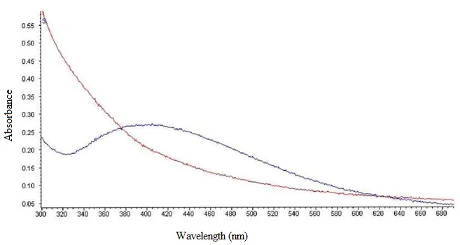
Figure 2: UV- Visible spectra of AgNp (blue) and leaf extract (red).
Studies suggest that a SPR (Surface Plasmon Resonance) peak located between 410 and 450 nm has been observed for AgNP and might be attributed to spherical nanoparticles (Jyoti et al., 2016). The absorption peak of the AgNp may vary from 350-500 nm according to the size of the nanoparticle formed.
Fourier transforms infrared (FTIR) spectroscopic analysis
The IR spectrum of the AgNp reaction mixture and plant extract alone (Fig.3) was collected over a wavelength range of 4000-500cm-1usin KBr. In leaf extract, the peaks are observed at 669.12, 1115.01, 1216.58, 1438.24, 1617.91, 2362.55, 3019.60 and 3409.19cm−1, respectively. After reaction with AgNO3, the peaks are shifted to 668.57, 1078.17, 1400.47, 169.92, 2078.5 and 2360.55 cm−1. The spectrum showed hydroxyl absorption at 3500-3200 cm-1 . The mild peaks present at 3700-3900 cm-1 indicates the O-H stretching in alcohols and phenolic compounds. Sharp absorption peaks observed in the range of 1000-1700 cm-1. Generally, flavonoids show strong absorption at 1600-1700 cm-1 region. From the results obtained after FTIR analysis, it can be inferred that flavonoids play a key role in the reduction and capping of silver ions during the formation of AgNp. The flavonoids in the leaf extract, known for their strong reducing properties, likely facilitate the reduction of silver nitrate into silver nanoparticles while also contributing to their capping and stabilization (Jyoti et al., 2016).

Figure 3: FTIR spectrum of leaf extract (shown in red) and that of silver nanoparticle (blue).
Transmission electron microscopy (TEM)
The morphology and size of the synthesized nanoparticles were analyzed using TEM (Fig. 4a), which revealed a variety of shapes and size distributions. The TEM images indicated that the nanoparticles were predominantly spherical and measured approximately 50 nm in diameter. The selected area electron diffraction (SAED) pattern of the AgNp (Fig. 4b) displayed concentric rings, confirming their highly crystalline nature (Indana et al., 2016). In general, SAED patterns for crystalline nanoparticles can produce either a single spot (dot) image for a single crystal or a ring pattern for polycrystalline materials. This makes SAED a valuable tool for distinguishing crystalline nanoparticles from non-crystalline ones (Bolade et al., 2022).

Figure 4: a) TEM image of AgNp at different nanometer range, b) SAED patterns of the AgNP exhibit concentric rings, indicating the crystallinity of nanoparticle.
Determination of stability of AgNp
The stability of AgNp formed was determined by measuring the OD value of the solution. The spectrophotometric determination of OD value showed no significant difference upon the AgNp even after 24 hours (Fig.5). It indicates that the AgNp formed is stable under standardized conditions at room temperature. This clearly defines the role of plant compounds as stabilizing agents. But the chemical methods of nanoparticles requires an extra stabilizing agent.

Figure 5: OD versus time graph of AgNp. No significant change in OD value at the 0th hour and 24th hour.
Anti-mycobacterial activity of the biosynthesized AgNP
The antibacterial activity of biosynthesized AgNP was tested against M smegmatis by standard agar well diffusion method. Inhibition of the organism tested with leaf and fruit pulp extract of P. campechiana and AgNp is shown in Fig. 6. The leaf extract alone showed inhibition with a zone of 12 mm in diameter. The AgNp inhibited the organism with a zone of 19 mm. The zone formed by the fruit pulp extract was negligible around 3mm.

Figure 6: Anti-mycobacterial activity of leaf extract, AgNp and fruit of Pouteria campechiana (Kunth) Baehni against M smegmatis
Determination of MIC and MBC
After incubation of the dilution tubes containing AgNp and M smegmatis culture, slight turbidity was seen from 10-3 to 10-6 dilutions. The lowest dilution of the extract that inhibited the growth of the microorganism was designated the minimum inhibitory concentration. Thus the MIC observed was 10-2. To determine the MBC, the dilutions were spread-plated on MHA and incubated overnight at 37˚C. Growth was observed from the plates with 10-3 to 10-6 dilution. The result suggest that the biosynthesized AgNP might be effective as anti-mycobacterial agent and can be used as lead in development of anti-tubercular agent.

Figure 7: a) MIC determination by macrodilution method b) MBC. The lowest dilution of AgNp inhibiting M smegmatis is 10-2
Enhanced effect of AgNp in combination with rifampicin
The effect of rifampicin on the action of AgNp was determined by well diffusion method on MHA plates. The rifampicin and AgNp was combined to evaluate the synergistic action. Significant difference in the inhibition zone was observed. The zone of inhibition formed by rifampicin against rifampicin was 30 mm while an enhanced effect on the activity of AgNp was observed when combined with rifampicin. The zone formed by AgNp alone was 19 mm (Fig.6) and rifampicin was 30 mm. A zone of inhibition of 45 mm was formed as a combinatorial effect. There is increased hepatotoxicity for the TB drugs including rifampicin. P. campechiana (Kunth) Baehni is known for its hepatoprotective activity (Aseervatham et al., 2014). Thus, the lethal effect of the drugs can be reduced when used in synergy with the drug. Also, combining AgNp with the anti TB might reduce the development of drug resistance in M tb. Nanoparticles usually serve as drug carriers. So, they can be engineered to selectively target TB infected cells alone, reducing the side effects associated. AgNp are reported to have immune-modulatory activity (Chakraborty et al., 2016). So, they can also increase the immune response to TB infection, thereby increasing the treatment efficacy.

Figure: Inhibition zone of a) Rifampicin b) Zone of inhibition of AgNP + rifampicin.
DISCUSSION
Green synthesis offers an eco-friendly method for producing metal nanoparticles with improved biocompatibility. In this study, silver nanoparticles were successfully synthesized using P. campechiana leaf extract, with FTIR analysis indicating the involvement of flavonoids in the reduction and stabilization of AgNp. The biosynthesized AgNp exhibited significant anti-mycobacterial activity against M. smegmatis, producing a clear inhibition zone of 19 mm, whereas the crude plant extract showed minimal activity, demonstrating the enhanced bioactivity achieved through nanoparticle formation. The MIC value (10⁻²) confirmed effective growth inhibition, while MBC results suggested a primarily bacteriostatic effect at lower concentrations. To the best of our knowledge, this is the first study reporting the anti-mycobacterial activity of silver nanoparticles synthesized using P. campechiana, as no previous studies have specifically explored the antitubercular potential of nanoparticles derived from this plant.
A key novelty of this study is the evaluation of the synergistic interaction between biosynthesized AgNp and rifampicin. The combination produced a markedly enhanced inhibition zone (45 mm) compared to AgNp or rifampicin alone, highlighting the potential of nanoparticle-assisted combination therapy to improve anti-tubercular efficacy and possibly reduce drug dosage.
Despite these promising findings, the known dose-dependent toxicity of silver nanoparticles and the use of an in vitro model organism represent limitations of the study. Further in vivo validation and cytotoxicity assessments are required. Importantly, the reported hepatoprotective property of P. campechiana, along with the drug-carrier and immunomodulatory potential of AgNp, suggests that such combinations may reduce drug-induced toxicity and enhance host immune response. Overall, this study positions P. campechiana-derived AgNp as promising adjuncts in sustainable and effective anti-tubercular therapy.
CONCLUSION
This study demonstrates that Pouteria campechiana leaf extract can be effectively utilized for the green synthesis of silver nanoparticles (AgNPs), with plant-derived flavonoids contributing to nanoparticle reduction and stabilization. The synthesized AgNPs were successfully characterized by UV–visible spectrophotometry, FTIR, and TEM, confirming the formation of stable nanosized particles. The biosynthesized AgNPs exhibited significantly greater antimycobacterial activity than the crude leaf extract and demonstrated enhanced inhibitory activity when combined with rifampicin against Mycobacterium smegmatis. These findings highlight the value of plant-mediated nanoparticle synthesis as a sustainable approach for generating bioactive nanomaterials with improved antimicrobial properties. The study also provides preliminary evidence supporting the exploration of P. campechiana-derived AgNPs as adjunct antimicrobial agents for mycobacterial infections. Nevertheless, comprehensive investigations, including determination of minimum inhibitory concentration (MIC), cytotoxicity profiling, mechanistic studies, activity against Mycobacterium tuberculosis and drug-resistant isolates, and in vivo evaluation, are required to validate their biological efficacy and safety.
Acknowledgement
The authors acknowledge the Department of Biotechnology and Microbiology, Dr Janaki Ammal Campus, Kannur University, Kerala, India, Instrumentation facility at MG University, Kerala, India.
Conflict of Interest
The authors have no conflict of interest.
REFERENCES

Sneha M.V., Ayana N. , Prajina P., Ramesan C.K.V.,Divya Easwaran, Green Synthesis, Characterization, And Antimycobacterial Activity Of Silver Nanoparticles Derived From Pouteria Campechiana (Kunth) Baehni Leaves, Int. J. of Pharm. Sci., 2026, Vol 4, Issue 7, 4199-4211. https://doi.org/10.5281/zenodo.21471446
 10.5281/zenodo.21471446