

We use cookies to ensure our website works properly and to personalise your experience. Cookies policy
Sai Meer college of Pharmacy, Chhibramau, Kannauj-209721
This study investigated the hepatoprotective potential of Nardostachys jatamansi extract (NJE) against kava (Piper methysticum)-induced liver toxicity in Wistar rats. Drug-induced liver injury is a major health concern and is associated with increased morbidity and mortality. To evaluate the protective role of NJE, an experimental model of kava-induced hepatotoxicity was established. Administration of kava extract significantly elevated liver function biomarkers such as AST, ALT, ALP, bilirubin, albumin, cholesterol, and LDH, indicating severe hepatic damage and impaired membrane integrity. Histopathological studies further revealed lipid alterations in hepatocytes, localized necrosis, hepatocyte degeneration, Kupffer cell activation, and inflammatory cell infiltration in liver tissues. These effects are believed to result from toxic metabolites, oxidative stress, glutathione depletion, and inflammatory mediator release. Treatment with NJE markedly reduced the elevated biochemical parameters toward normal levels and improved liver tissue architecture. Rats receiving NJE along with kava extract showed reduced lipid degeneration and improved hepatocyte structure compared to rats treated with kava alone. The hepatoprotective effect was dose-dependent, with 800 mg/kg of NJE showing the most significant protection, comparable to the standard hepatoprotective drug, Silymarin (100 mg/kg). The protective mechanism may involve antioxidant activity and inhibition of hepatotoxic free radical formation through modulation of cytochrome P450 enzymes. Acute toxicity studies conducted according to OECD guideline 423 demonstrated that NJE was safe, with an oral LD50 greater than 5000 mg/kg body weight and no observable toxic effects. Overall, the findings confirm that Nardostachys jatamansi possesses significant hepatoprotective and antioxidant properties against kava-induced liver damage.
The liver is a crucial organ that performs a variety of metabolic, detoxifying, and synthetic tasks necessary to preserve physiological equilibrium. It is essential for the metabolism of proteins, fats, and carbohydrates as well as for the detoxification of exogenous compounds including medications and environmental pollutants (1). The liver regulates the body's chemical levels, stores vitamins, fats, carbohydrates, and minerals, and directly or indirectly affects every metabolism. Because of its involvement in these biochemical pathways and the abundance of metabolic enzymes, the liver is extremely vulnerable to damage from a variety of toxicants, chemicals, and pharmaceutical agents (1). Therefore, maintaining a healthy liver is crucial for its normal functioning as well as for other organs (2).
One in twenty-five persons die from liver cancer, cirrhosis, and viral hepatitis. A third of the nearly two million people who die from liver-related illnesses each year are women. Four percent of individuals globally die from liver disease. Every year, between 600,000 and 900,000 people pass away from liver cancer. Liver disease is currently the tenth most common cause of death, despite the fact that liver-related deaths may be underreported. Cirrhosis-related mortality in Africa are currently ranked tenth, down from thirteenth in 2015. It is ranked seventh in the Eastern Mediterranean and ninth in South-east Asia and Europe. (3).
Nature's gift to humanity, medicinal herbs play a vital role in maintaining, enhancing, and preserving our health (4). A promising source of treatments for a range of illnesses is medicinal plants and their derivatives. Hepatoprotection is greatly aided by these plants' secondary metabolites, which include alkaloids, flavonoids, phenolic compounds, terpenoids, steroids, saponins, tannins, and anthraquinones (5). Antioxidants from both traditional food sources and medicinal herbs can shield the liver from oxidative stress and other substances. There is still a lot of interest in using herbal tonics to treat liver problems, and plants and phytochemicals derived from plants are attractive hepatoprotective medicines since they have less negative effects (6). Numerous plants and preparations have been reported to have hepatoprotective properties. Approximately 160 phytoconstituents from 101 plants have been reported to have liver-protective qualities. Over 87 plants are used in 33 patented and proprietary multi-ingredient plant formulations in India. Consequently, there has been a lot of interest worldwide in the development of plant-based hepatoprotective drugs that are effective against a range of liver illnesses (7).
Traditional medicine makes extensive use of the critically endangered medicinal herb Nardostachys jatamansi. It is a member of the Valerianaceae family and has long been utilized in traditional Indian medicine. There is strong evidence that it can cure illness. The plant's rhizomes are used as a bitter tonic, stimulant, antispasmodic, epileptic therapy, and for hysteria in the Ayurvedic medical system. Nardostachys jatamansi has been shown to have antifungal, hepatoprotective, central nervous system, anticonvulsant, neuroprotective, antiparkinson's, antioxidant, antidiabetic, tranquilizing, and antiestrogenic properties in pharmacological reports. Additionally, jatamansone has been associated with anti-hypertensive, anti-arrhythmic, anti-asthmatic, nematicidal, and antibacterial properties (8). The plant's primary secondary metabolites, nardostachone, jatamansone, and actinidine, are the only ones that have been shown to be effective thus far. They work well as bronchodilators, vasodilators, and platelet aggregation inhibitors (9).
MATERIALS AND METHODS
Plant procurement and authentication: The Nardostachys jatamansi rhizomes (Caprifoliaceae) were gathered from the Kannauj market in Uttar Pradesh. Dr. K.C. Bhatt, a researcher and chief scientist of New Delhi's ICAR-NBPGR, confirmed the authenticity of the plant sample. The NBPGR has a voucher specimen of Nardostachys jatamansi (AC-25/2026) that may be used for future reference.
Preparation of Plant Extract: After being carefully cleansed with distilled water to remove any dirt or soil, the newly collected rhizome of Nardostachys jatamansi was left to shade-dry in a well-ventilated place until it reached room temperature. After the dried plant material was divided into little pieces, Using the cold percolation approach, which avoided heat damage, 50% ethanol was extracted after defatting with petroleum ether at temperatures ranging from 60 to 80°C. Once the extract was dried using a rotavapor (Model noDB-5546, Decibel Instruments, Chandigarh), Purified and concentrated at 40±1°C under decreased pressure to achieve a consistent weight. Next, yield % was computed. For pharmaceutical study, we kept Nardostachys jatamansi extract (NJE) at -20 °C. (10).
Preparation of Toxicant (Piper methysticum) Extract: Twenty minutes of sonication at room temperature (-21 ± 1° F) will be used to extract eight grammes of powdered kava (Piper methysticum) from fifty millilitres of water. Following this, the mixture will be centrifuged at 10,000xg for 20 minutes. The liquid above the solid will be discarded, and the solid will be re-extracted using 25 millilitres of water. It will be necessary to mix the two supernatants before freezing and lyophilizing the filtrate. Doses for administration to rats will be determined using the dry extract containing the natural proportions of the kavalactones, in accordance with the HPLC test of their kavalactone composition. All of the kava samples will come from the same batch so that the results can be reliably reproduced. (11)
Physico-chemical Evaluation: Morphological and microscopic analysis of powdered Nardostachys jatamansi rhizomes was performed which included tests of Powder microscopy, Moisture content, Ash Value, Total Ash, Acid-insoluble ash values, Water-soluble ash values, Extractive Value, Extractive value soluble in alcohol, Estimation of extractive value soluble in water.
Preliminary phytochemical screening: Systematic phytochemical screening was performed on Nardostachys jatamansi rhizomes extract for chemical components. Following the methodology indicated in our earlier research, the newly obtained raw extracts were examined qualitatively for detection in secondary metabolites including glycosides, alkaloid compounds, sugary carbohydrates as components, proteins, saponins, tannins, coumarins, phenols, and flavonoid
Studies for Acute toxicity: Using the OECD-423 criteria as a guide, we determined the extract's LD-50 value (80). Rats ranging in weight from 150 to 200 grammes were given dosages of 5, 50, 500, 1000, 2000, and 5000 milligrammes per kilogram of body weight. We measured the weight of the rats after they were administered the medicine orally. The OECD-423 (12) criteria for acute oral toxicity testing were followed. Three male Wistar rats weighing 150-200 g were used for each treatment. The concentrations that were shown to be effective were 5, 50, 500, 1000, 2000, and 5000 mg/kg/body weight. We calculated the LD-50 of the extract. After one night of fasting and unlimited water, the rats received the medicine orally. Rats' pre- and post-treatment weights were recorded. After one day of therapy, animals were evaluated for toxicity, including behavioural abnormalities, gait abnormalities, convulsions, and death. (13).
Test drug administration: A rat-specific needle was used to gavagely give the amaranth extract in a single dose. The cattle were kept in a situation where they were famished throughout the night. Individuals were weighed after fasting and given oral doses of the test medication at 5, 50, 300, and 2000 mg/kg. Following that, for the next three or four hours, the rat could only eat certain things. Control rats receive 10 mL/kg body weight physiological serum (NaCl) intra-gastrically via gavage. On treatment day, the animal's weight dictated the experimental chemical dose.
Indicators Documented Throughout Toxicity Studies: Animals were monitored daily for the first day, then at 30-minute intervals for the next day, in order to detect any changes in behaviour or disease, the rats were watched at least twice a day. Some of the immediate indicators that might be seen are drowsiness, unconsciousness, lethargy, diarrhoea, and salivation. Somatomotor function, patterns of behaviour, skin, hair, eyes, and mucous membranes are among the things we search for, along with features of the cardiovascular, autonomic, central nervous, and respiratory systems. When available, we have given the date of death. It was informed that participants should not eat for at least another hour or two after the test material was given. Continual daily counting of survivors began twenty-four hours after the tragedy and continued for the subsequent fourteen days.
Experimental Study: Wistar albino rats weighing 150–220 grammes will be utilized. A 23-degree Celsius environment with a 12-hour light-dark cycle might suit the species. Free food and drink abound. Standard practice is to acclimatize animals for seven days before the trial. Qualified CDRIs will provide all animals. The investigation will follow the direction of the institution's animal ethics committee and the CPCSEA, which oversees animal studies.
Assessing the Hepatoprotective Effects of Medications: Five groups of five rats (N=5) will be randomly assigned to the animals. Throughout the trial, Group I will act as the control and receive the vehicle at a concentration of 0.5% CMC. Group II will be given 500 mg of kavalactones daily and will serve as a control group that has been intoxicated with kava (14). The conventional medicine silymarin will be administered to Groups III and IV will get NJE extract at 400 and 800 mg/kg body weight, respectively, while Group V will receive 100 mg/kg. In all cases, with the exception of the rats used as controls, 500 mg of kavalactones will be given orally once daily, two hours after the dosages of plant extract and silymarin, respectively. A total of fourteen days in a row (15).
Table 1: Experimental design for induction of liver toxicity in rats
|
Groups |
No. Of Rats/ Mice |
Treatment |
Dose |
|
I |
6 |
Control |
0.5 % CMC, 1 ml/kg, p.o. |
|
II |
6 |
Kava extract |
500 mg/kg kavalactones per day |
|
III |
6 |
NJE + Kava extract |
400mg/Kg + 500 mg/kg |
|
IV |
6 |
NJE + Kava extract |
800mg/Kg + 500 mg/kg |
|
V |
6 |
Silymarin + Kava extract |
100mg/Kg + 500 mg/kg |
|
VI |
15 Mice |
LD50 |
As per OECD 423 |
We will sacrifice the animals under anaesthesia six hours following their final kava extract administration. Each animal's blood will be drawn using capillary tubes via the retro-orbital plexus and tested for a number of biochemical parameters. For histopathological examinations, the livers of the rats in each group will be collected (10).
Evaluation Based on Histopathology: A puncture was used to draw blood from the retro-orbital plexus and heart after anaesthesia. Both treated and untreated rats were killed after each programme. The liver was excised and fixed in 10% formalin for histology.
Examinations Using Statistical Methods: Prism Pad software (Version 6.05) was used to evaluate the level of significance after analysing the five rats' mean ± S.E.M. using a student t-test and Newman-Keuls test. Probability values below 5% (P < 0.05) were deemed significant.
RESULTS
Extract from Nardostachys jatamansi rhizome: a primary phytochemical screening
Plant samples were phytochemically analysed using ethanol and methanol. Secondary metabolites such alkaloids, tannins, saponins, phenols, sugars, flavonoids, proteins and amino acids, glycosides, and more were found or absent. Nardostachys jatamansi ethanol extracts include proteins, glycosides, carbohydrates, and flavonoids; however, they are devoid of terpenoids, steroids, phenolic compounds, quinones, and saponins.
Table 2: Primary phytochemical screening of Nardostachys jatamansi extract with different constituents
|
SR. NO |
CONSTITUENTS |
Nardostachys jatamansi extract |
|
1 |
Steroids |
-- |
|
2 |
Terpenoids |
- |
|
3 |
Phenolic compounds |
- |
|
4 |
Flavonoids |
++ |
|
5 |
Quinones |
- |
|
6 |
Tannins |
-- |
|
7 |
Carbohydrates |
++ |
|
8 |
Saponins |
-- |
|
9 |
Glycosides |
++ |
|
10 |
Proteins |
++ |
|
11 |
Alkaloids |
++ |
|
Key: +++ =High, ++ =Moderate, + =Normal concentration, - = Absent |
||
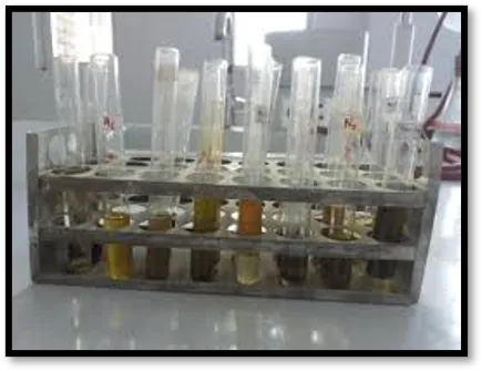
Figure 1: Extraction of Nardostachys jatamansi and its phytochemical analysis
Physico-chemical Evaluation
Table 5.2 shows that powdered Nardostachys jatamansi was used to determine loss on drying, ash values (total, water-soluble, and acid-insoluble), and extractive values (water, alcohol, and non-volatile ether):
Table 3: mechanical, chemical, Assessment of Nardostachys jatamansi powdered medication
|
SR. NO |
Parameters |
Values % W/W Nardostachys jatamansi Ethanol extract |
|
1 |
Moisture Content |
16.62 % |
|
2 |
Total ash |
17.08 % |
|
3 |
Insoluble ash in acid |
5.02 % |
|
7 |
Hydrophobic extractive potential |
8.1 % |
|
8 |
The value of extractives soluble in ethanol |
6.51 % |
|
9 |
Non-volatile ether soluble extractive value |
5.91 % |
Acute toxicity tests: A single dosage of the aqueous extract was safe since its oral LD50 was above 5000 mg/kg body weight. Skin, eye, and mucous membrane colour remained constant. The oral acute toxicity test followed OECD standards 423 (the acute toxicity class approach). Table shows no pre- or post-experimental weight changes or toxicity markers. The oral aqueous extract was safe in a single dosage since its LD50 was above 5000 mg/kg body weight. The skin, eyes, and mucous membranes stayed the same colour.
Liver, kidney, and cardiac gross morphology are shown in the picture. The kidneys are encased in a fibrous pea-shaped sac, which gives them a smooth, reddish-brown surface. The diaphragmatic surface is a smooth dome that lies next to the diaphragm; the lower boundary of the liver is formed by the circumference around the visceral surface, the flat visceral surface, and the projecting diaphragmatic surface. With its three red sides and prominent atrioventricular groove that divides the atrium and ventricle, the heart resembles a strong, three-pronged pyramid. The three sides of the liver form a triangle, with the base serving as a boundary between the flat and projecting diaphragmatic surfaces as well as the visceral surface. Adjacent to it, The liver has a smooth dome on its diaphragmatic surface and a horizontal H-shaped groove on its visceral surface that separates its lower surface into lobes. Smooth, reddish-brown kidneys are in a fibrous, pea-shaped sac.
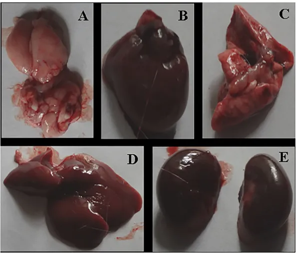
Figure 2: Anatomical view of animal organs of acute toxicity studies: (A) Brain; (B) Heart; (C) lungs; (D) Liver; and (E) Kidneys.
Table 4: Effect of ethanolic extracts of Nardostachys jatamansi in a rat study of acute oral toxicity
|
Response |
Unmarked |
Head |
Body |
Tail |
||||
|
|
Before |
After |
Before |
After |
Before |
After |
Before |
After |
|
Alertness |
N |
N |
N |
N |
N |
N |
N |
N |
|
Pain response |
A |
A |
A |
A |
A |
A |
A |
A |
|
light reflex |
A |
A |
A |
A |
A |
A |
A |
A |
|
Gripping strength |
N |
N |
N |
N |
N |
N |
N |
N |
|
Corneal reflux |
P |
P |
P |
P |
P |
P |
P |
P |
|
Pupils |
N |
N |
N |
N |
N |
N |
N |
N |
|
Urination |
N |
N |
N |
N |
N |
N |
N |
N |
|
Salivation |
N |
N |
N |
N |
N |
N |
N |
N |
|
Skin colour |
N |
N |
N |
N |
N |
N |
N |
N |
|
Lacrimation |
N |
N |
N |
N |
N |
N |
N |
N |
Table 5: Various animals' body weights after being treated with an ethanolic extract of Nardostachys jatamansi
|
Weekly weather: Weight in (g) |
1st day |
3rd day |
6th day |
8th day |
12th day |
14th day |
|
Untreated control animals |
109±2.04 112±3.03 113±2.06 |
109±3.03 112±2.06 114±3.07 |
114±2.06 116±3.01 118±3.04 |
117±2.03 119±4.03 121±3.02 |
118±4.01 119±3.03 121±2.05 |
120±3.03 121±2.01 124±4.03 |
|
animals treated at the dose of 5 mg/kg of body weight |
112±3.01 113±2.02 114±3.05 |
114±2.02 115±4.03 116±2.04 |
115±4.02 116±3.03 118±2.04 |
120±2.02 119±2.04 121±3.05 |
123±2.03 123±3.01 124±2.06 |
126±2.06 126±3.03 128±2.01 |
|
animals treated at the dose of 50 mg/kg of body weight |
111±2.02 112±3.03 113±2.01 |
113±2.05 114±3.03 116±2.01 |
115±2.01 116±3.03 118±2.02 |
119±2.06 120±3.02 121±2.03 |
123±2.03 121±2.04 123±3.05 |
125±2.03 124±3.02 126±2.06 |
|
animals treated at the dose of 300 mg/kg of body weight |
113±2.03 114±3.04 115±3.01 |
115±3.02 116±2.04 117±3.06 |
116±2.03 118±3.06 119±3.01 |
119±3.02 122±2.06 124±3.02 |
122±2.02 124±3.02 126±2.01 |
126±2.02 127±4.05 129±3.07 |
|
animals treated at the dose of 2000 mg/kg of body weight |
113±2.03 116±2.02 118±3.04 |
115±3.03 117±2.02 119±2.02 |
116±2.03 121±3.02 123±4.01 |
120±4.03 125±3.04 126±2.05 |
123±3.04 128±4.05 129±2.06 |
129±4.02 132±3.03 135±2.01 |
Table 6: Rats' organ weights as measured in grams during an acute toxicity test
|
Groups |
Treatment and dose |
Organs weights (gms) |
|||
|
|
|
Liver |
Heart |
Kidney 1 |
Kidney 2 |
|
Control
|
Weight in (g) of untreated control animals
|
3.75±0.32 3.83±0.31 3.81±0.24 |
0.46±0.01 0.47±0.04 0.48±0.02 |
0.34±0.025 0.33±0.022 0.37±0.026 |
0.35±0.022 0.38±0.026 0.39±0.027 |
|
A. cruentus
|
Weight in (g) of animals treated at the dose of 2000 mg/kg of body weight
|
3.85±0.31 3.87±0.25 3.86±0.27 |
0.49±0.06 0.47±0.02 0.48±0.03 |
0.42±0.023 0.45±0.025 0.47±0.024 |
0.44±0.025 0.47±0.027 0.45±0.023 |
Comparative liver gross architecture of experimental and control animals.
Figure 5.2 shows that control and experimental animals had comparable liver architecture. Figure 5.2A depicts a typical Group I control's anatomy. Group II and III treated with ethanol and 400 mg/kg NJE had liver damage (Figure 5.2B, C). Group IV (800 mg/kg) treated with NJE exhibited normal liver architecture (Figure 5.2D, E). Similar to group V (Silymarin) with normal liver morphology.
Ethanolic effects Nardostachys jatamansi extract and kava-induced hepatotoxicity on body, liver, and relative liver weight
The table shows the beginning, ending, and liver weights of control and experimental animals. After kava injection, the end weight of normal group I rats was 202.1 ± 10.3 g, substantially lower than group II rats' 166.8 ± 10.7 g (P < 0.01). NJE-treated rats in groups III (400 mg/kg) and IV (800 mg/kg) had considerably greater body weights than the risky group II, with 190.5 ± 11.4 g and 202.4 ± 12.5 g increases, respectively (P < 0.05).
In group II (kava treatment), liver weight increased significantly to 4.94 ± 0.55/100 g body weight (P < 0.001), compared to the control group (3.19 ± 0.17/100 g body weight). When given 400 and 800 mg/kg NJE, liver weight considerably decreased (P < 0.01) to 3.75 ± 0.42 and 2.89 ± 0.28 g, respectively, compared to 4.94 ± 0.55 g with kava therapy. Group IV rats administered NJE exhibited less activity than group V rats given normal silymarin at the dosage employed.
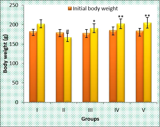
Figure 3: Effect of Nardostachys jatamansi extract on body weight of control and group II.
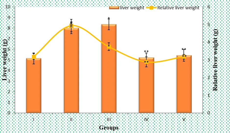
Figure 4: Effect of Nardostachys jatamansi extract on liver weight and relative liver weight of control and toxic group.
Mean ± SEM of six rats in each group is used to represent the values.
Numbers #<0.01 and ##<0.001 were the p-values when contrasted with the same control group I.
Comparing group II (Ethanol) against group I (Control) yielded p-values ranging from *<0.05 to ***<0.001.
Impact of Nardostachys jatamansi ethanolic extract on hepatotoxicity and control rats' liver damage.
The effect of NJE is depicted in the tables. In group II rats treated with kava, there was a notable increase in serum AST (318.88 ± 31.56 U/l, P < 0.001), ALT (186.65 ± 12.72 U/l, P < 0.001), ALP (202.52 ± 14.52 U/l, P < 0.001), LDH (702.37 ± 27.33 U/l, P < 0.001), ALB (3.87 ± 0.09 gm/dl, P < 0.001), CHL (77.58 ± 5.76 mg/dl, P < 0.001), and Bilirubin (3.78 ± 0.28, P < 0.001) when compared to control group I rats (94.43 ± 11.82 U/l, 41.32 ± 7.24 U/l, 75.59 ± 8.55 U/l, 372.15 ± 32.02 U/l, 5.07 ± 0.13 gm/dl, 33.15 ± 6.12 mg/dl, and 0.63 ± 0.14 mg/dl), respectively.
The NJE treatment for groups III and IV of rats at dosages of 400 and 800 mg/kg resulted in a significant reduction of AST levels (242.86 ± 18.86 and 122.22 ± 12.84 U/l, P < 0.01 and P < 0.001), ALT levels (99.05 ± 11.24 and 51.83 ± 4.42 U/l, P < 0.01 and P < 0.001), ALP levels (103.15 ± 12.23 and 91.18 ± 16.24 U/l, P < 0.01 and P < 0.001), LDH levels (523.46 ± 15.25 and 398.52 ± 18.16 U/l, P < 0.01 and P < 0.001), CHL levels (53.19 ± 5.88 and 38.29 ± 3.02 mg/dl, P < 0.01 and P < 0.001), and Bilirubin levels (2.37 ± 0.18 and 0.88 ± 0.13 mg/dl, P < 0.01 and P < 0.001). Additionally, the levels of ALB were noticeably greater in group II animals compared to group I animals (4.73 ± 0.12 and 45.92 ± 0.09 gm/dl, P < 0.01 and P < 0.001, respectively).
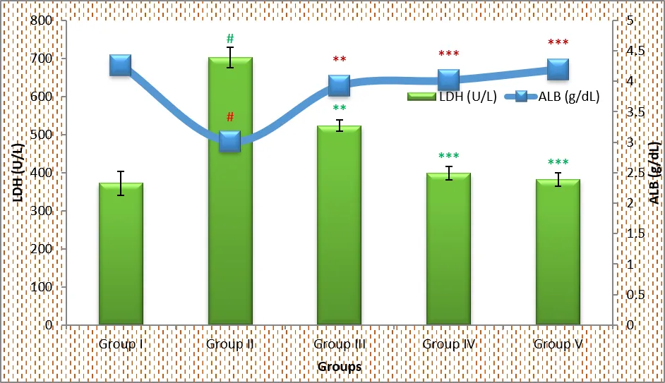
Figure 5: Effect of Nardostachys jatamansi on serum LDH (U/L) and ALB (g/dl) against Kava induced rats toxicity.
Results are based on the mean ± S.E.M. of six rats per group.
Considered in relation to the matching control groups, the p-values are below 0.001.
The p-values are as follows when comparing group II (Ethanol): *<0.05, **<0.01, ***0.001.
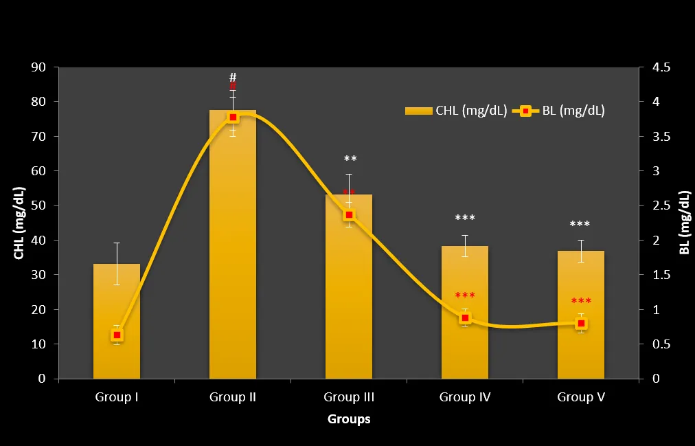
Figure 6: Effect of Nardostachys jatamansi on serum CHL (mg/dl) and Bilirubin level (mg/dl) against kava induced rats’ toxicity.
Mean ± S.E.M. of 5 rats in each group is used to represent the values.
With regard to the corresponding control groups, the p-values are less than 0.001.
In comparison to group II (Ethanol), the p-values are as follows: *<0.05, **<0.01, ***<0.001.
Histopathological Observation
Figure 5.7. shows that hematoxylin and eosin staining is used in liver tissue histopathology. (A) Hepatocytes in normal control rats are uniform, the nuclei are tiny and vesicular, and the architecture is well-preserved. (B) Liver slices from rats that were given Kava have eosinophilic cytoplasm with fuzzy cell borders, which suggests cell growth. (C) After being administered kava and 400 mg/kg of NJE, rats' liver sections exhibit minor localised necrosis and sinusoidal dilatation. (D) When rats are given kava plus 800 mg/kg of NJE, the liver sections of the rats exhibit comparable abnormalities. (A) The architecture of homogeneous hepatocytes with tiny vesicular nuclei is well-preserved in a healthy rat's liver. (B) A liver slice from a rat treated with kava shows eosinophilic cytoplasm, which indicates cell growth, as well as smaller hepatocytes and fuzzy cell borders. The building may be in a state of deterioration, but at least some vascularity has grown. (C) Rats given 400 mg/kg of NJE plus kava had fewer inflammatory cells, somewhat more localised necrosis, and sinusoidal dilatation in their liver tissues. (D) Rats liver slices that were exposed to 800 mg/kg of NJE and kava had well defined cell borders, an abundance of eosinophilic cytoplasm, and normal vesicular nuclei (Fig. 4). architecture that has regular interstitial cells and robust vascularity. The liver sections of rats that were given kava plus 100 mg/kg silymarin showed normal hepatocyte morphology (E).
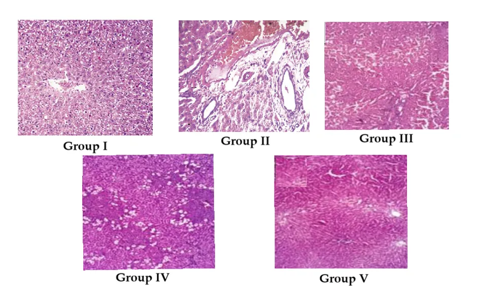
Figure 7: Histopathological changes in liver tissues (Hematoxylin and eosin) of control, standard and treated rats.
DISCUSSION
Experimentally, the kava-induced liver damage model was set up to represent Nardostachys jatamansi's protective effect. This research used kava-induced hepatotoxicity in Wistar rats to find that Nardostachys jatamansi extract (NJE) had a hepatoprotective effect. The use of medicinal medications has led to liver disease, which is a leading cause of mortality in adults, and there is medical backing for drug-induced liver dysfunction. To demonstrate the protective effect of Nardostachys jatamansi, we designed an experiment using a kava-induced liver damage model. This study shown that Nardostachys jatamansi extract (NJE) protected the livers of Wistar rats when exposed to kava-induced hepatotoxicity.
Due to Piper methysticum-induced liver damage, Group II rats had substantially higher levels of AST, ALT, ALP, BIL, ALB, CHL, and LDH. Bilirubin, AST, ALT, ALP, ALB, LDH, CHL, and CHL levels reduced considerably to normal levels, indicating liver tissue healing and plasma membrane integrity. Rats given a Piper methysticum extract with kavalactones showed a change in hepatocyte lipids in histological analyses of liver sections, but rats given a combination of NJE and Piper methysticum extract showed less hepatocyte lipid change.
The detrimental effects of (Piper methysticum) kava extract on the livers of rats after prolonged administration were demonstrated in a study conducted by Kholy et al., (2011) (16). But other research confirmed what this one had suspected: localised necrosis and hepatocyte degradation (17). Activated Kupffer cells in the sinusoidal region and cellular infiltration of the liver parenchyma were also noted in the analysis. Other studies found that liver macrophages release inflammatory mediators and reactive oxygen species that harm the liver (18).
Furthermore, several studies have shown that kava preparation may indirectly or directly induce inflammation due to toxic metabolites, cycloxygenase enzyme inhibition, and liver glutathione depletion (19) (20).
The acute toxicity studies on Nardostachys jatamansi has revealed that a single dosage of the aqueous extract was safe since its oral LD50 was above 5000 mg/kg body weight. Skin, eye, and mucous membrane colour remained constant. The oral acute toxicity test followed OECD standards 423 (the acute toxicity class approach). Table shows no pre- or post-experimental weight changes or toxicity markers. The oral aqueous extract was safe in a single dosage since its LD50 was above 5000 mg/kg body weight. The skin, eyes, and mucous membranes stayed the same colour.
Liver, kidney, and cardiac gross morphology are shown in the picture. The kidneys are encased in a fibrous pea-shaped sac, which gives them a smooth, reddish-brown surface. The diaphragmatic surface is a smooth dome that lies next to the diaphragm; the lower boundary of the liver is formed by the circumference around the visceral surface, the flat visceral surface, and the projecting diaphragmatic surface. With its three red sides and prominent atrioventricular groove that divides the atrium and ventricle, the heart resembles a strong, three-pronged pyramid. The three sides of the liver form a triangle, with the base serving as a boundary between the flat and projecting diaphragmatic surfaces as well as the visceral surface. Adjacent to it, The liver has a smooth dome on its diaphragmatic surface and a horizontal H-shaped groove on its visceral surface that separates its lower surface into lobes. Smooth, reddish-brown kidneys are in a fibrous, pea-shaped sac.
Kava's toxic effects were minimised by boosting liver function in rats given 400 and 800 mg/kg Nardostachys jatamansi ethanolic extract. It seems to prevent liver damage (Table and). Compared to the gold standard medicine Silymarin (100 mg/kg), a rise in blood bilirubin levels suggests jaundice severity, which is a clear sign of cellular leakage and cell membrane dysfunction. The fact that Nardostachys jatamansi ethanolic extract reduces tissue damage may explain this protective effect. Traditional medication Silymarin (100 mg/kg) was used to compare the results. Rising blood bilirubin levels indicate cellular leakage and membrane damage, worsening jaundice. Hepatotoxicant poisoning lowered protein synthesis in both silymarin-treated and Nardostachys jatamansi-ethanolic extract groups. In kava users, blood albumin was substantially lower and serum bilirubin elevated. Protein synthesis decreases with hepatotoxicant dosage. All aberrant values recovered to normal in the silymarin or Nardostachys jatamansi ethanolic extract group. Nardostachys jatamansi extract's dose-dependent activity peaks at 800 mg/kg, p.o., equivalent to the control and Silymarin-treated groups (Standard). This study found that 800 mg/kg Nardostachys jatamansi extract was most effective when taken orally. Increases activity similarly to the control and Silymarin groups (Standard).
CONCLUSION
All things considered, our findings suggest that Wistar rats may be protected from the oxidative liver damage caused by Piper methysticum if they are given an ethanolic extract of Nardostachys jatamansi as a supplement. Taking 400 or 800 mg/kg of NJE significantly mitigated the kava-induced liver damage. The fact that NJE prevented the increase of liver enzymes including AST, ALB, CHL, and LDH caused by Piper methysticum was an extra perk of utilising it. Finally, supplementing with NJE increased the albumin production by Piper methysticum. The liver damage caused by kava was greatly reduced when 400 or 800 mg/kg of NJE was injected. In addition, the increase in liver enzyme production caused by Piper methysticum was avoided by the addition of NJE. Among the enzymes that showed an uptick were lactate dehydrogenase (LDH), albumin (ALB), Bilirubin, AST, and ALT. NJE increased Piper methysticum's albumin-lowering effects. Hepatocellular necrosis and inflammatory cell infiltration were reduced by NJE, which may explain its hepatoprotective benefits as shown in liver histology pictures.
Nardostachys jatamansi ethanolic extract may mitigate Piper methysticum-induced liver damage, according to the available research. Not only that, it boosts liver function and acts as an antioxidant. An ethanolic extract of Nardostachys jatamansi may reduce the liver-harming effects of Piper methysticum, according to new research.
REFERENCES

Hemlata, Dr. Arpit Katiyar, Anuj Kumar, Hepatoprotective Properties of Ethanolic Extract of Nardostachys jatamansi Rhizome Against Kava-Intoxicated Albino Wistar Rats, Int. J. of Pharm. Sci., 2026, Vol 4, Issue 7, 6242-6255. https://doi.org/10.5281/zenodo.21727722
 10.5281/zenodo.21727722