

We use cookies to ensure our website works properly and to personalise your experience. Cookies policy
Department of Pharmaceutical Quality Assurance , Late Shrimati Hanjabai Gahlot Institute of Pharmacy, Maharashtra State, India
Hirschsprung’s disease (HSCR), also known as congenital aganglionic megacolon, is a rare congenital disorder of the gastrointestinal tract characterized by the absence of enteric ganglion cells in a variable segment of the colon. This neuronal deficiency results from abnormal development, migration, or differentiation of enteric neural crest cells during embryogenesis, leading to impaired relaxation and coordinated movement of the affected intestinal segment. Consequently, patients may develop intestinal obstruction and difficulty in passing stool. Clinical manifestations vary with the length of the aganglionic segment and may include delayed passage of meconium, abdominal distension, constipation, vomiting, and enterocolitis. The disease has a complex genetic basis, with several susceptibility genes implicated, particularly RET, EDNRB, SOX10, and GDNF. Among these, mutations in the RET gene are strongly associated with familial and syndromic forms of HSCR. Diagnosis is based on clinical evaluation, contrast studies, anorectal manometry, and confirmation by rectal biopsy demonstrating the absence of ganglion cells. Treatment primarily involves surgical removal of the aganglionic segment and restoration of bowel continuity. Early diagnosis and appropriate management are essential to prevent severe complications and improve patient outcomes. This review summarizes the epidemiology, pathophysiology, classification, clinical manifestations, causes, risk factors, diagnostic evaluation, and current treatment approaches for Hirschsprung’s disease.
Hirschsprung’s disease (HD) is a condition where nerve cells (called ganglion cells) are missing in a part of the intestine. About 75% of all cases of Hirschsprung's disease happen in the rectosigmoid area (the part of the intestine near the rectum). [1]. In 1904, Hirschsprung explained about ten patients who were born with an abnormally wide (dilated) colon. [2]. The prevalence of Hirschsprung’s disease is approximately 1.09 per 10,000 live births, with males being affected more frequently [3, 4]. The syndrome is sporadic in 80% of cases, with only 18% related with other congenital defects. [5].
There is a lack of inhibitory enteric ganglia results in continuous contraction Loss of peristalsis in the afflicted bowel segment, including the internal anal sphincter. This causes a functional blockage due to impaired stool propulsion [6, 7]. The majority of cases (80%) present within the first year of life. [5]. Rectal biopsy is still the gold standard for diagnosis. [8]. Operation intervention is the mainstay of treatment and seeks to ascertain the extent of a ganglionosis, resect the bowel in question, and maintain the sphincter complex [1].
Historically, surgical management was phased; however, modern practice favors single-stage procedures where clinical conditions allow [3, 9]. Surgical methods have improved over time, starting with open surgery, then using laparoscopic help, and now using fully trans nasal (through the nose) techniques.Although 4 to 6 months is the ideal age for surgery, there is still disagreement over the best approach for rectal dissection [3].
Treatment for HD has greatly evolved since de la Torre-Mondragon and Ortega initially described the whole transanal endorectal pull-through (TERPT) in 1998. TERPT offers several advantages, including avoidance of intraperitoneal contamination, reduced risk of adhesions and pelvic injury, better pain control, Compared to traditional trans abdominal methods, these offer faster recovery and better cosmetic results [10].This study aims in a tertiary hospital in Oman, HD patients who had total transanal endorectalpull-through surgery were evaluated for complications and long-term bowel function.
Introduction:
Hirschsprung's disease (HSCR), which is also referred to as congenital ganglionic megacolon, is the Danish pediatrician Herald Hirschsprung is honoured by the name. It alludes to neurocristopathy, a group of illnesses characterized by aberrant migration, proliferation, and differentiation of neural crest cells. [13] These anomalies cause the absence of autonomic nerve plexuses inside a section of the colon. [11] HSCR is the most frequent neurodevelopmental condition of the enteric nervous system (ENS), accounting for roughly one in every 1 in every 5,000 live births worldwide, around [14] Males are primarily affected.[15] Derived from Latin term megacolon congenitum refers to the typical dilatation of the colon next to a ganglionic segment [12].In the classic form, which accounts for over 80% of cases, a ganglionosis is limited to the area of the rectosigmoid. It might, however, occasionally spread to the distal small intestine.
Complete colonic ganglionosis is uncommon and has a significant death rate. [16] Usually, symptoms appear during the new born stage. Multiple endocrine neoplasia type 2B (MEN 2B), sympathoadrenal neuroblastoma, adrenal medullary tumor (pheochromocytoma), auditory-pigmentary syndrome, von Recklinghausen disease, autosomal dominant non-syndromic sensorineural hearing loss, and trisomy 21are among the disorders that are frequently linked to Hirschsprung's illness. Mutations in a number of genes, most notably RET, GDNF, EDNRB, and SOX10, can cause HSCR genetically. Particularly linked to familial variants of the disease is RET, which codes for a nerve growth factor receptor.
Epidemiology
A deficiency of ganglion cells in the intestinal wall is the main feature of Hirschsprung's disease (HD).Rectosigmoid ganglionitis accounts for approximately 75 percent of all instances [15]. In 1904, Hirschsprung reported ten cases of babies born with an enlarged colon. [16].Hirschsprung’s disease affects about 1.09 out of every 10,000 live births and is more common in males [17, 18] Around 80% of cases occur randomly, while about 18% are linked with other birth defects.[19]
The lack of intestinal ganglia that inhibit results in continuous contraction and the affected part of the bowel, including the internal anal sphincter, loses its ability to move food through normally. This causes a blockage because the stool can't move properly [20,21]. Most cases (about 80%) are detected in the first year of life.[19]. A rectal biopsy is still the most reliable way to diagnose it. [22]
Surgical intervention it is the primary treatment approach, focused on identifying how much of the bowel lacks nerve cells, removing that part, and keeping the sphincter muscles intact. [15]. While surgical management was historically staged, current practice favors single-stage procedures when the clinical condition allows [17, 23]. Surgical procedures have progressed from open to laparoscopic-assisted, and most recently, totally transanal approaches.
Although 4 to 6 months is the ideal age for surgery, there is still disagreement over the best approach for rectal dissection [17]. Treatment for HD has greatly evolved since de la Torre-Mondragón and Ortega initially described the whole transanal endorectal pull-through (TERPT) in 1998. TERPT offers several advantages, including avoidance of intraperitoneal contamination, reduced risk of adhesions and pelvic injury, better pain control, shorter hospital stays, and improved cosmetic outcomes compared to traditional trans abdominal techniques [24].This study aims to assess the complications and long-term bowel function in HD patients who had total transanal endorectal pull-through surgery at a major hospital in Oman.
Pathophysiology
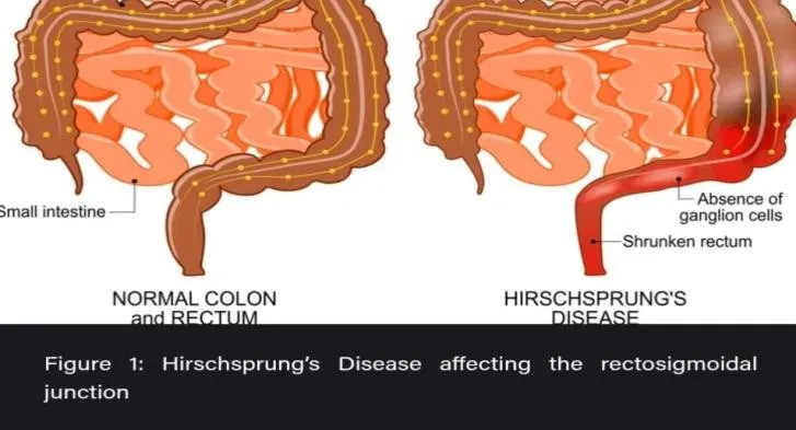
Hirschsprung's disease is distinguished the absence of ganglion cells in the myenteric (Auerbach) and submucosal (Meissner) plexuses of the intestine, which extend proximally from the anus to varying lengths of the colon. The enteric nervous system is formed by neural crest cells that migrate from the vagus nerve to the foregut mesenchyme in a cranio-caudal orientation. The most widely accepted cause of Hirschsprung's disease is a stop in the migration of neuroblasts formed from neural crest cells during development of the fetus, usually between the 8th and 12th weeks of gestation. In some cases, although migration occurs normally, the neuroblasts may fail to survive, differentiate properly, or proliferate due to intrinsic cellular defects such as apoptosis or impaired signaling [26].A persistent constriction of the affected intestine segment due to a deficiency of enteric ganglion cells results in impaired bowel motility and disturbed peristalsis. Specifically, the ganglionic section fails to relax in response to rectal distension, resulting in functional blockage. Stool accumulation in the rectosigmoid area is a defining feature of this obstruction, and it causes several clinical symptoms such as chronic constipation, abdominal distention, and vomiting.Prolonged stasis can cause proximal bowel dilatation, increased intraluminal pressure, and impaired mucosal perfusion. These changes compromise the mucosal barrier and promote bacterial overgrowth, which can lead to Hirschsprung's enterocolitis (HAEC) a potentially life-threatening condition with a death rate of 25–30% if not promptly recognized and treated. In severe cases, HAEC may progress to sepsis and death [25].
Categories:
Hirschsprung’s disease is categorized based on how much of the intestine lacks nerve cells:
This is the most common type, making up around 75–80% of cases. The nerve-free area is limited to the rectum and The sigmoid colon's distal portion
Found in about 10% of cases, this type involves a longer section without nerve cells, stretching from the rectum up to the splenic flexure.
This is the rarest and most severe type, seen in roughly 5% of cases. In this form, the entire colon is missing nerve cells.
Abnormalities
The impacted section of the large intestine remains constricted due to the lack of enteric ganglion cells. The most typical clinical signs of Hirschsprung's disease include increasing abdominal distension, eating intolerance, and impaired peristalsis. During the first several months of life, these characteristics are seen in about 80% of affected infants. Up to 90% of newborns with HSCR have delayed meconium transit beyond the first 24 hours following delivery. The illness usually manifests in older children as growth retardation, fecal impaction, persistent progressive constipation, and malnutrition. [27].
In new-borns, Hirschsprung’s disease may show up as:
In children and older individuals, signs may include:
Causes
The exact cause of this condition is not always known, but it is associated with genetic mutations that affect nerve cell development. In some cases, Hirschsprung's disease runs in families and is more commonly seen in individuals with certain genetic syndromes, such as Down syndrome.
Table 1. Major Genetic, Developmental, Environmental, and Associated Factors Involved in Hirschsprung’s Disease
|
Factor |
Description |
|
Genetic Factors |
Hirschsprung’s disease can run in families and is associated with mutations in several genes, particularly the RET gene. |
|
Developmental Problem in the Womb |
During fetal development, neural crest cells fail to migrate properly into the lower portion of the colon, resulting in the absence of enteric nerve cells (ganglion cells). |
|
Environmental Factors |
Certain factors during pregnancy, such as maternal smoking, may slightly increase the risk, although their exact contribution is not fully established. |
|
Associated Conditions |
Hirschsprung’s disease may occur along with genetic or congenital conditions such as Down syndrome and Waardenburg syndrome. |
CONCLUSION
Hirschsprung disease affects about one in every 5,000 live newborns and is a genetic condition, occurring three times more frequently in males. If one parent has the disease, there is a 1% chance it will be passed on to their child. The disorder is characterized by the absence of nerve cells in the colon, which causes difficulty passing feces. It typically starts from the internal anal sphincter and spreads upward through the intestine. Diagnosis involves several methods, including barium enema, tissue sampling from the rectum, and abdominal imaging (X-ray) as well as anal manometry. Among these, abdominal X-ray alone is not sufficient for an accurate diagnosis. Surgical interventions such as ostomy, pull-through procedures, and other corrective surgeries are the primary treatments. Laparoscopically assisted pull-through surgery results in fewer complications. Rectal suction biopsy is a cost-effective, rapid, and safe diagnostic method, offering both simplicity and high accuracy.
REFERENCES

Sakshi Humane, Preeti Kulkarni, Shrinath Satpute Hirschsprung's Disease Contingency for Novel Therapies and Diagnosis, Int. J. of Pharm. Sci., 2026, Vol 4, Issue 8, 3827-3833, https://doi.org/10.5281/zenodo.22076563
 10.5281/zenodo.22076563