

We use cookies to ensure our website works properly and to personalise your experience. Cookies policy
1,2Department of Medical Laboratory Science, College of Medicine and Health Science, Afe Babalola University Ado-Ekiti, Ekiti State, Nigeria
3Department of Physics, Faculty of Physical Science,Federal University of Technology Akure,Ondo state.
This study investigates the histological and biochemical changes in Wistar rats following exposure to amoxicillin oral suspension. Amoxicillin, a commonly prescribed antibiotic, is known for its efficacy against a wide range of bacterial infections. However, its impact on non-target organs remains inadequately understood. In this experiment, adult Wistar rats were administered amoxicillin at a dose of 200 mg/kg body weight for 14 days. Histological examinations of liver and kidney tissues were conducted using standard staining techniques, revealing significant alterations, including cellular degeneration and inflammatory infiltrates. Biochemical analyses of serum samples demonstrated elevated levels of liver enzymes (AST, ALT) and renal markers (creatinine, urea), indicating potential hepatotoxicity and nephrotoxicity. These findings suggest that while amoxicillin is effective in treating infections, its administration can lead to detrimental effects on liver and kidney functions in Wistar rats. The results underscore the need for careful consideration of antibiotic use and further investigation into the long-term consequences of amoxicillin exposure. This study contributes valuable insights into the safety profile of amoxicillin, highlighting the importance of monitoring biochemical markers and histological changes in the evaluation of drug safety
Amoxicillin is a semi-synthetic, broad-spectrum beta-lactam antibiotic belonging to the penicillin class. It exhibits bactericidal activity against a wide range of Gram-positive and Gram-negative bacteria and is widely prescribed as a first-line therapy for various infections, including those affecting the respiratory, gastrointestinal, genitourinary, cutaneous, and central nervous systems (Suarez, 2009). In dentistry, amoxicillin is commonly recommended for the treatment of alveolar abscesses, soft tissue infections, and maxillofacial sinus tract infections. It is also frequently administered as a prophylactic agent against infective endocarditis (Bascones, 2014).
Despite its widespread therapeutic applications, amoxicillin use is associated with adverse effects, primarily hypersensitivity reactions and gastrointestinal disturbances. More recently, growing evidence from epidemiological and clinical studies has suggested a possible association between amoxicillin exposure and dental pathologies such as dental fluorosis. This condition, primarily attributed to excessive fluoride intake during tooth development, is histologically marked by enamel hypomineralization and clinically identified by white mottling and brown discoloration of teeth. Although the pathogenesis remains unclear, some studies report that children exposed to amoxicillin during early development may have a higher risk of developing enamel defects, highlighting the need for further investigation (Hong, 2005).
Animal models have been employed to explore the potential adverse effects of amoxicillin on dental development. For example, Laisi et al. (2008) reported alterations in enamel thickness in mice exposed to amoxicillin. Given the anatomical and developmental similarities between rodent and human dentition, studies involving laboratory animals offer a controlled and reproducible means to assess drug-induced developmental effects. Kumazawa (2012) emphasized the value of in vivo animal models for evaluating the safety of pharmaceutical agents during critical developmental periods, including the prenatal stage.
Amoxicillin’s pharmacokinetic profile contributes to its extensive use. It is stable in the gastrointestinal tract and exhibits greater oral bioavailability compared to naturally occurring penicillins (Sousa, 2005). However, it is susceptible to inactivation by bacterial β-lactamases. In human medicine, it is often combined with clavulanic acid—a β-lactamase inhibitor—to broaden its spectrum of activity. In veterinary practice, however, amoxicillin is typically used alone.
The antibiotic is extensively utilized in veterinary medicine for treating and preventing bacterial infections in domestic and food-producing animals, including dogs, cats, poultry, pigs, goats, sheep, and cattle. In small animals, it is used primarily for respiratory, urinary, and soft tissue infections (Pfizer, 2004), while in livestock, it addresses a range of respiratory, gastrointestinal, and urinary tract pathogens, such as Escherichia coli, Streptococcus suis, Pasteurella multocida, and Mannheimia haemolytica (Hernandez et al., 2005 and Reyns et al., 2008). Furthermore, its use extends to pre-ruminating calves and lactating dairy cows for the treatment of enteritis, pneumonia, and mastitis (FDA, 2011; Schering-Plough, 2007).
Considering the growing concerns regarding the potential developmental toxicity of amoxicillin, especially during critical periods such as prenatal and early postnatal stages, there is a need for further research into its systemic effects. This study therefore aims to evaluate the histological and biochemical changes in Wistar rats exposed to amoxicillin oral suspension, providing insights into its safety and potential risks during developmental stages.
.
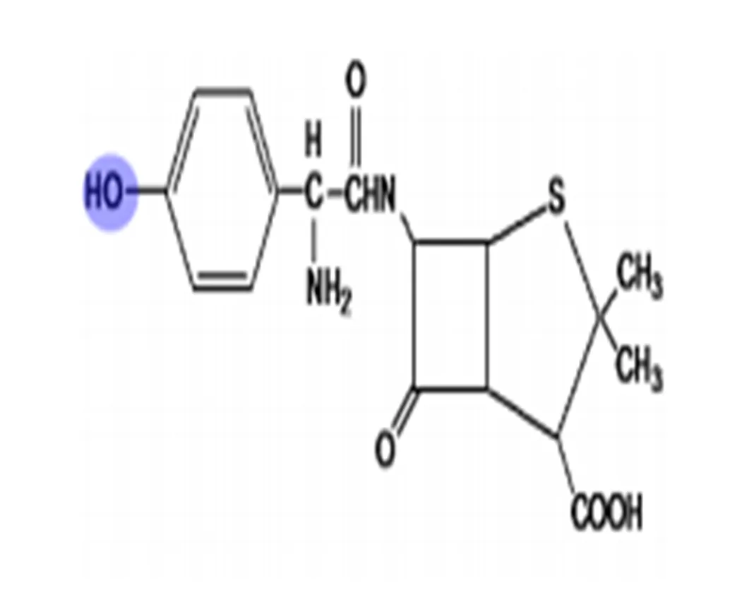
Fig. 1: It shows the chemical structure of Amoxicillin (Durand, 2003)
Antibiotics have revolutionized the treatment of infectious diseases, but their overuse and misuse have introduced new clinical challenges, including toxicity to vital organs. Amoxicillin, a widely used β-lactam antibiotic, is frequently administered due to its broad-spectrum activity and favorable pharmacokinetic profile. However, concerns have emerged over its safety profile, particularly regarding hepatic and renal function during prolonged or high-dose exposure (Al-Attar, 2017; Diniz et al., 2020).
The liver plays a central role in drug metabolism, while the kidneys are essential for drug excretion. Both organs are highly susceptible to toxic insults, and antibiotic-induced injury to these organs can result in significant morbidity. Although amoxicillin is generally considered safe, there are documented cases of hepatotoxicity and nephrotoxicity following its administration (Chalasani et al., 2008). This study investigates the histological and biochemical effects of amoxicillin in an experimental rat model to provide empirical evidence of its potential toxicity.
Although amoxicillin is primarily administered to treat bacterial infections, it is crucial to assess its potential impact on non-target organs such as the liver, kidneys, and heart. These organs are integral to drug metabolism, detoxification, and systemic regulation, making them susceptible to off-target toxicity, particularly during prolonged or high-dose exposure (Olayemi et al., 2020). Hepatotoxicity and nephrotoxicity have been reported with various beta-lactam antibiotics, including amoxicillin, which can induce oxidative stress, inflammatory cytokine production, and histopathological alterations in non-target tissues (Elkomy et al., 2021; Ezeja et al., 2023). Recent animal studies have demonstrated significant changes in biochemical markers and tissue architecture following antibiotic exposure, indicating possible systemic toxicity beyond the intended site of action (Adeleye et al., 2022). Therefore, investigating these effects in non-target organs is essential for understanding the broader safety profile of amoxicillin and for supporting evidence-based recommendations regarding its therapeutic use in both humans and animals.
2.0 MATERIALS AND METHODS
2.1 Experimental Animals
Adult male Wistar rats (weighing 150–200 g) were obtained from the Animal House Facility of Babcock University, Ilishan-Remo, Nigeria. The animals were housed under standard laboratory conditions with a 12-hour light/dark cycle, temperature of 22 ± 2°C, and relative humidity of 50–60%. Rats had free access to standard rodent chow and clean drinking water. The animals were allowed to acclimatize for one week before the commencement of the experiment. All experimental procedures were conducted in accordance with institutional ethical guidelines for animal care and use.
2.2 Materials
Aqueous solution of clindamycin hydrochloride, dissecting sets, Haematoxylin and Eosin stains, 10% Neutral Buffered formalin, Absolute alcohol, 95% alcohol, 1% acid solution, xylene, DPX, rat cage and feeds, cotton wool, hand gloves and universal bottles, needles and syringes, weighing balance, lithium heparin bottles, EDTA bottle, oral gavage feeding tubes, micropippettes, non-heparinized capillary tubes, spectrophotometer. Randox AST assay kit, Randox ALT assay kit, Agape albumin kit, Agape total protein kit, Randox urea kit and Randox creatinine kit.
2.3 Source of Animals
Twenty (36) Wistar rats weighing averagely between 100-120grams were purchased from the laboratory animal facility at Babcock University. The animals were acclimatized under laboratory conditions for a week at the animal house before the commencement of the experiment. Wooden cages with sufficient ventilation was used to house the rats. Saw dust was provided for bedding of the animals which will be replaced by new ones on a daily basis so as to provide a hygienic environment
2.4 Study Area
This research was conducted at the Animal House of the Department of Anatomy, Babcock University, Ilishan-Remo, Ogun State, Nigeria. Babcock University is a first class Seventh - day Adventist Institution of Higher learning, with a student population of about six thousand, located in the South-Western Region of Nigeria, coordinates; 6.8862ºN, 3.7055ºE.
2.5 Duration of Study
This study was carried out within a period of four (4) weeks.
3.5 Ethical Consideration
Ethical approval for the use and sacrifice of experimental animals for research was obtained from the Babcock University Health Research Ethics Committee (BUHREC) before the commencement of the study.
2.6 Sample Collection
5mL of blood was collected through cardiac puncture with a syringe into EDTA bottle for hematological analysis and lithium heparin bottle for biochemical analysis. The lithium heparin bottles were centrifuged and the plasma was transferred into plain bottles and then subjected to tests.
The liver, kidney, heart and testis of rats in each batch were harvested into and fixed in 10% Neutral Buffered Formalin to prevent tissue autolysis and bacterial decomposition after which it was then excised properly for each of the rats.
2.7 Source of the Amoxicillin
Amoxicillin was purchased from the Pharmacy and certified by a well-trained pharmacologist in Babcock University.
2.8 Experimental Design
2.8.1 Analysis of dosage
Amoxicillin was administered according to the body weight of Wistar rats using a standard value of 500mg twice a day for a period of 14 days according to Akhavan et al., (2023).
Group I: Dissolve 500mg of Amoxicillin in 50ml of distilled water.
Group II: Under-dose, was administered 0.5ml of Amoxicillin suspension twice daily
Group III: Normal dose was administered 1ml of Amoxicillin suspension twice daily
Group IV: Over dose was administered 2ml of Amoxicillin suspension twice daily
Group V: Max dosage was administered 2.5ml of Amoxicillin suspension twice daily
2.9 Biochemical Assay
2.9.1 Estimation of Serum Activity of ALT (Reitman and Frankel 1957)
Principle for ALT Estimation
α-ketoglutarate reacts with L-alanine in the presence of alanine aminotransferase (ALT) to give L-glutamate and pyruvate, glutamate pyruvate transferase is measured by monitoring the concentration of pyruvate hydrazone formed with 2, 4-dinitrophenyl hydrazine. The absorbance is measured at a wavelength of 546 nm which is directly proportional to the concentration of ALT present in the serum.
Reagent composition for ALT
R1; Phosphate buffer, 100 mmol/L pH 7.4; L-alanine, 100 mmol/l; α-oxoglutarate 2 mmol/l
R2; 2, 4-dinitrophenylhydrazone, 2 mmol/L.
All reagents was brought to room temperature before use. The sample was thawed at room temperature and mixed well before analysis.
2. 9..2 Estimation of Serum Activity of AST (Reitman and Frankel 1957)
Assay principle of AST
α-ketoglutarate reacts with L-aspartate in the presence of aspartate aminotransferase (AST) to give L-glutamate and oxaloacetate, AST activity is measured by monitoring the concentration of oxaloacetate hydrazone formed with 2, 4-dinitrophenyl hydra-zine. The absorbance is measured at a wavelength of 546 nm which is directly proportional to the concentration of AST present in the serum.
Reagent composition for AST
R1: Phosphate buffer, 100 mmol/L pH 7.4; L-aspartate, 100 mmol/L; α-oxoglutarate 2 mmol/L.
R2: 2, 4-dinitrophenylhydrazone, 2 mmol/L.
Assay procedure for AST
Wavelength: 546 nm, Cuvette: 1cm light path, Temperature: 37ºC
All reagents was brought to room temperature before use. The sample was thawed at room temperature and mixed well before analysis.
2.9.3 Estimation of Alkaliine Phosphatase
METHOD: Colourimetric method
Principle: Paranitrophenol which is colourless, is hydrolyzed by alkaline phosphatase at pH10.5 & 37C to form paranitrophenol which is coloured yellow. The addition of NaOH stops the enzymes activity and final colour shows maximum absorbance at 410nm.
Reagent1:
Buffer: diethanolamine buffer 1mol/l, MgCl2 0.5mmol/l
Reagent2: p-nitrophenylphospate 10mmol/l.
3.9.4: Estimation of plasma albumin using Bromocresol green method as described by Doumaset al., (1971)
Principle of plasma albumin estimation
Bromocresol green is an indicator which is yellow between pH 3.5 – 4.2. When it binds to albumin the colour changes from yellow to blue-green that is proportional to albumin concentration. The absorbance of the colour produced is measured in a spectrophotometer at 630 nm wavelength.
Reagent composition for albumin estimation
Succinate buffer (pH 4.20) 75 mmol/L
Bromocresol green (BCG) 0.14 g/L
Albumin standard 3 g/L
2.9.5: Biuret method for the estimation of plasma TP as described by Gomallet al., (1949)
Principle of Biuret method for TP
Biuret method is based on the reaction which occurs between cupric ions in the reagent and peptide bonds of the protein molecules in alkaline solution to form blue-violet or purple coloured complexes. The absorbance of the colour is measured in a spectrophotometer at 546 nm.
Reagents composition for TP
Content composition
Potassium sodium tartarate 21 mmol/L
Potassium iodide 6 mmol/L
Copper sulphate 6 mmol/L
Sodium hydroxide 58 mmol/L
Standard protein concentration 6 g/dl
2.9.6 Estimation of plasma urea as described by (Henry, 1963; Wheatherburn, 1967; Searcy et al., 1967)
Principle of urea estimation
Enzymatic determination of urea according to the following reaction:
Urea + H2O 2NH3 + CO2
NH3 + Salicylate > 2,2-Dicarboxy Indophenol
Reagent composition for urea estimation
Urea B colour reagent R1 2 × 53 ml
Sodium salicylate 80mmol/l
Sodium nitroprusside 4mmol/l
Sodium hypochlorite 45mg/dl
Urea B colour reagent R2 10 ×10 ml
Phosphate buffer 60mmol/l
Urease 20KU/L
Distilled Water
Urea B standard 1 × 4 ml
Urea Standard Concentration 40mg/dl
2.9.7 Estimation of Triglycerides
Method: enzymatic colourimetric method
Principle: triglyceride is hydrolyzed to glycerol and fatty acids in the presence of lipase, the glycerol is phosphorylated in the presence of glycerokinase to glycerol phosphate which is then oxidized by an oxidase to dihydroacetone phosphate and hydrogen peroxide. The products so formed reacts with chlorophenol and aminoantipyrine to produce a coloured compound called quinoneimine.
Reagents
1) Standard
2) Buffer chromogen: 4-chlorophenol with pH 7.5
3) Enzymes: lipase, glycerokinase, glycerol-3-phospate oxidase, peroxidase, 4-aminoantipyrine.
2.10 Histopathological Investigation
The histological staining technique used for this research was the routine Hematoxylin and Eosin staining method to ascertain the change in general structure and cytoarchitechture of the liver, kidney, heart and testis. Representative samples were placed in cassettes and processed using the Automatic Tissue Processor. After processing, the tissues were embedded in molten paraffin wax to form a solid support for microtomy after cooling. Upon an initial fixation process, the sample was then processed for a Microscopic view in an automated tissue processing machine which lasted for 18 hours. Automatic Tissue Processing (Avwioro, 2014).
Table 2.10.1 Tissue Processing Schedule
|
Beaker 1 |
10% Formol Saline |
1Hr 30 Minutes |
|
Beaker 2 |
70% Alcohol |
1 Hour |
|
Beaker 3 |
80% Alcohol |
1 Hour |
|
Beaker 4 |
90% Alcohol |
1 Hour |
|
Beaker 5 |
95% Alcohol |
1 Hour |
|
Beaker 6 |
95% Alcohol |
1Hr 30 Minutes |
|
Beaker 7 |
Absolute Alcohol |
2 Hours |
|
Beaker 8 |
Absolute Alcohol |
2 Hours |
|
Beaker 9 |
Xylene |
1 Hour 30 Minutes |
|
Beaker 10 |
Xylene |
1 Hour 30 Minutes |
|
Beaker 11 |
Wax (Molten) |
2 Hours |
|
Beaker 12 |
Wax (Molten) |
2 Hours |
3.0 RESULTS AND DISCUSSION
3.1 Biochemical Results.
Table 2.0 Mean comparisons of serum electrolytes level between Na, K, Cl, HCO3, Urea, Creatinine and Control (Analysis Variance).
|
GROUP |
DOSAGE (mg/ml) |
N |
Na+ MEAN±SD |
K+ MEAN±SD |
Cl- MEAN±SD |
HCO3 MEAN±SD |
Urea MEAN±SD |
Creatinine MEAN±SD |
|
I |
CONTROL |
5 |
135.000±2.000 |
3.533±0.19 |
100.833±2.23 |
23.500±1.05 |
25.000±5.480 |
1.233±0.210 |
|
II |
0.5 |
5 |
135.833±1.170 |
3.533±0.19 |
100.833±2.23 |
23.500±1.05 |
22.000±5.621 |
1.133±0.250 |
|
III |
1.0 |
5 |
135.670±1.211 |
3.533±0.19 |
100.833±2.23 |
23.500±1.05 |
18.333±6.531 |
0.933±0.301 |
|
IV |
2.0 |
5 |
136.140±1.140 |
3.540±0.21 |
101.000±2.45 |
23.400±1.14 |
13.800±1.640 |
0.700±0.090 |
|
V |
2.5 |
5 |
136.170±1.147 |
3.533±0.19 |
100.833±2.23 |
23.500±1.05 |
13.833±4.750 |
0.750±0.242 |
|
|
P-value |
|
0.550 |
1.00 |
1.00 |
1.00 |
0.003 |
0.001 |
P<0.05= statistically significant
Table 2.1 Mean comparison of the serum hormonal level between AST, ALT, ALP and control (Analysis of Variance)
|
GROUP |
DOSAGE (mg/Kgbwt) |
N |
AST MEAN±SD |
ALT MEAN±SD |
ALP MEAN±SD |
|
I |
CONTROL |
5 |
93.833±19.994 |
70.500±21.040 |
36.500±3.45 |
|
II |
0.5 |
5 |
97.500±22.331 |
69.670±27.134 |
36.500±3.45 |
|
III |
1.0 |
5 |
84.500±25.982 |
63.000±46.891 |
36.500±3.45 |
|
IV |
2.0 |
5 |
75.000±22.34 |
57.200±28.960 |
36.200±3.77 |
|
V |
2.5 |
5 |
87.833±21.664 |
47.833±17.960 |
36.500±3.45 |
|
P |
|
|
0.530 |
0.680 |
1.00 |
P<0.05 = statistically significant
In table 1.0, it was observed that the serum electrolytes (Na+, K+ Cl-, HCO3) levels were statistically insignificant compared to the control P= 0.550, P= 1.000. However, the serum urea and creatinine levels in the test groups were significantly decreased (P=0.003, P= 0.001) when compared to the control. The serum liver enzymes (AST, ALT& ALP) levels were observed to decrease insignificantly (P= 0.530, P= 0.680, P= 1.000) when compared to the control (Table 2.0)
3.2 Histopathogical Results
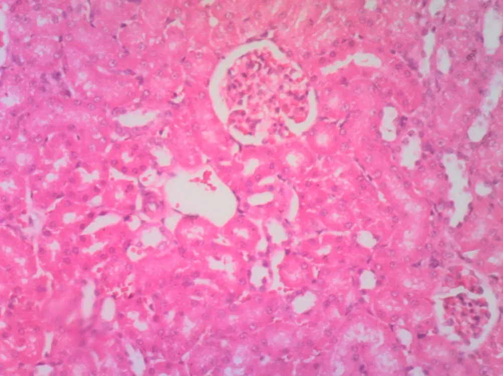

Control (a) Group II (a) Group III (a)
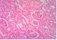

Group IV(a) Group V(a) Control (b)
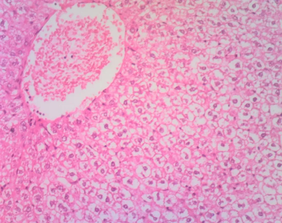
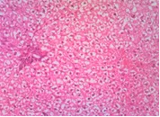
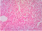
Group II (b) Group III (b) Group IV (b)
Group V (b)
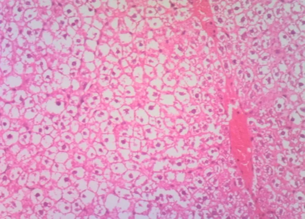
Figure 2.0: Photomicrograph showing organs architecture following oral adminisration of amoxicillin in wistar rats
Control Group (a and b) Kidney and Liver sections of Wistar rat showing normal architecture: glomeruli, distal and proximal convoluted tubules and hepatocytes with basophilic nuclei, sinusoids, and the hepatic vein are arranged with no pathological alteration. In group II(a), Kidney section of Wistar rat showing normal architecture: glomeruli, distal and proximal convoluted tubules following oral administration of 0.5mg/Kgbwt twice daily for 14days.While Group II(b) of Liver section of Wistar rats showing massive necrosis in the hepatocytes consistent of ballooning degeneration and hemorrhagic central vein following oral administration of 0.5mg/Kgbwt Amoxicillin suspension twice daily for 14 days.Kidney section of Wistar rat in Group III (a) showing normal architecture: glomeruli, distal and proximal convoluted tubules following oral administration of 1.0mg/Kgbwt twice daily for 14days. While Liver section of Wistar rats of Group III (b) showing massive necrosis in the hepatocytes consistent of ballooning degeneration and portal triads following oral administration of 1.0mg/Kgbwt Amoxicillin suspension twice daily for 14 days. Kidney section of Wistar rat Group IV (a) showing normal architecture: glomeruli (Black arrow), distal and proximal convoluted tubules following oral administration of 2.0 mg/Kgbwt twice daily for 14days. Liver section of Wistar rats of Group IV (b) showing severe necrosis in the hepatocytes consistent of ballooning degeneration, loss of hepatocytes and the hepatic vein following oral administration of 1.5 mg/Kgbwt Amoxicillin suspension twice daily for 14 days. Kidney section of Wistar rat in Group V (a) showing normal architecture: glomeruli,, distal and proximal convoluted tubules following oral administration of 2.5mg/Kgbwt twice daily While Liver section of Wistar rats in Group V (b) showing severe necrosis in the hepatocytes consistent of ballooning degeneration, loss of hepatocytes hemorrhagic hepatic vein following oral administration of 2.0mg/Kgbwt Amoxicillin suspension twice daily for 14 days. Stained by Haematoxylin and Eosin. Magnification. X1000.
3.2 DISCUSSION
Amoxicillin is a semi-synthetic, acid-stable β-lactam antibiotic within the penicillin class, widely utilized to manage a broad spectrum of Gram-positive and Gram-negative bacterial infections in both humans and animals (Bush, 2003). Although generally regarded as safe, Amoxicillin has been reported, in rare cases, to exert adverse effects on visceral organs and hematological parameters.
In this study, varying doses of Amoxicillin oral suspension were administered to Wistar rats to assess potential biochemical and histological alterations. The analysis of serum electrolytes—including sodium (Na⁺), potassium (K⁺), chloride (Cl⁻), and bicarbonate (HCO₃⁻)—revealed no statistically significant differences between treated and control groups, indicating that Amoxicillin did not markedly disrupt electrolyte balance under the experimental conditions.
However, significant changes in serum urea and creatinine levels were observed, suggesting potential renal impairment at certain dosages. These findings raise concern regarding Amoxicillin-induced nephrotoxicity, warranting further investigation. Despite the biochemical alterations, histopathological examination of renal tissues (Figures 4.0–4.4) showed preserved glomerular and tubular architecture, indicating minimal structural damage.
Liver function markers—AST, ALT, and ALP—did not differ significantly across experimental groups (p=0.530, p=0.680, p=1.000). This supports previous findings by Garcia et al. (2011), who reported that liver injury is rare in patients treated with Amoxicillin alone. In a study involving 422,646 individuals, only 14 cases (0.003%) of acute liver injury were associated with Amoxicillin monotherapy, compared to 21 cases (0.017%) among those administered Amoxicillin/clavulanate.
Interestingly, this study recorded a significant decrease (p<0.005) in serum urea and creatinine levels in Amoxicillin-treated groups. This contrasts with the findings of Zeller et al. (2020), who observed acute kidney injury following high-dose Amoxicillin therapy in hospitalized patients in France. The discrepancy may be attributed to dosage variations and differences in treatment duration.
Histological evaluation of liver sections revealed varying degrees of hepatocellular changes, particularly in groups receiving higher doses of Amoxicillin, suggesting potential dose-dependent hepatic effects.
CONCLUSION
In conclusion, the findings suggest that while Amoxicillin is generally safe at therapeutic doses, higher concentrations may pose biochemical risks to renal function, despite limited histopathological evidence of structural damage. The hepatic effects appear minimal, reinforcing the low incidence of Amoxicillin-induced liver toxicity.
While amoxicillin remains a vital therapeutic agent, the findings of this study highlight the importance of monitoring liver and kidney function during its use, especially in patients with preexisting conditions or those receiving long-term treatment.
This study demonstrates that high-dose or prolonged exposure to amoxicillin can result in significant hepatic and renal toxicity, as evidenced by altered biochemical markers and tissue damage. Rational prescription practices and regular monitoring are crucial to mitigate the risks associated with amoxicillin use.
LIST OF ABBREVIATION
ALT- Alanine transaminase
AST- Aspartate transaminase
ALP- Alkaline phosphatase
LH- Luteinizing hormone
FSH- Follicle stimulating hormone
IDS- Infectious disease
BRL-Beecham research laboratory
6-APA- 6- Aminopenicillanic
PBP-1-A- Penicillin-binding protein 1A
AHA- American heat association
ADA-American dental association
AAOS-American academy of orthopedic surgeons
MIC-Minimum inhibiting concentration.
VLDL- Very low-density lipoprotein
IDL- Intermediate density lipoprotein
LDL- Low density lipoprotein
HDL-High density lipoprotein
IVC- Inferior vena cava
RBC-Red blood cell
IgG- ImmunologlubulinG
DPX- Dibutylphthalate Polytyrene xylene
EDTA- Ethylenediamine tetraacetic acid
BCG- Bromocresol green
Declarations
Ethical Approval and Participation Consent
All procedures carried out in this research were reviewed and authorized by the Babcock University Health Research Ethics Committee (BUHREC), under protocol number BUHREC/2023/056. Animal handling and experimentation adhered strictly to institutional ethical standards and national guidelines for laboratory animal care.
Consent for Publication:
This article does not include any human data.
Competing Interests:
The authors affirm that there are no financial or personal conflicts of interest related to the content of this manuscript.
Funding:
This research did not benefit from any dedicated grant from funding agencies in the public, commercial, or not-for-profit sectors.
Authors’ Roles and Contributions
AKEREDOLU Florence Funke,designed the study framework, performed the experimental work, carried out data analysis, and drafted the initial version of the manuscript.
EKUNDINA V O, AKINLEYE O.P, OLUWALOYE T.G and AGBOMHERE H.M contributed to supervision, methodological guidance, and critical manuscript revisions.
Acknowledgements:
The research team extends sincere appreciation to the technical staff at the Laboratory Animal Facility, Babcock University, for their support during the animal care and experimental phases.
REFERENCES


Akeredolu F F, Ekundina V O, Akeredolu E I, Histological and Biochemical Changes in Wistar Rats Exposed to Amoxicillin Oral Suspension, Int. J. of Pharm. Sci., 2026, Vol 4, Issue 7, 4238-4248, https://doi.org/10.5281/zenodo.21471844
 10.5281/zenodo.21471844