We use cookies to ensure our website works properly and to personalise your experience. Cookies policy
1, 3 AKRG college of pharmacy, Nallajela, Andhra Pradesh, India
2HOD, Department of Pharmacology, Sri Vasavi Institute of Pharmaceutical Sciences, Pedatadepalli, Andhra Pradesh, India.
Urolithiasis is a multifactorial disorder characterized by urinary stone formation associated with oxidative stress, inflammation, and calcium oxalate (CaOx) supersaturation. This study evaluated the antiurolithiatic potential of the aqueous extract of Triticum aestivum leaves (AETA) using in vitro CaOx crystallization assays and an ethylene glycol (0.75%)-ammonium chloride (1%)-induced urolithiasis model in Wistar rats. Phytochemical screening confirmed the presence of flavonoids, phenolics, alkaloids, saponins, tannins, steroids, and glycosides. AETA significantly inhibited CaOx crystal nucleation, growth, and aggregation, with maximum inhibitions of 73.88%, 74.11%, and 79.76%, respectively, at 600 ?g/mL, comparable to Neeri. Oral administration of AETA (100, 300, and 700 mg/kg) for 28 days produced dose-dependent protection by reducing urinary calcium, oxalate, and electrolyte excretion while restoring urine volume. The 700 mg/kg dose significantly improved body weight (209.36 vs. 155.46 g), reduced kidney weight (0.69 vs. 0.98 g), and normalized serum creatinine (3.53 vs. 4.78 mg/dL), blood urea nitrogen (0.65 vs. 0.84 mg/dL), and uric acid (3.24 vs. 3.74 mg/dL) compared with the disease control. Histopathology confirmed marked reductions in renal crystal deposition and preservation of renal architecture. These findings demonstrate that Triticum aestivum possesses significant antiurolithiatic and nephroprotective activities, supporting its potential as a natural therapeutic agent for urolithiasis
Urolithiasis (kidney stone disease) is one of the most prevalent urological disorders worldwide and represents a major public health concern because of its increasing incidence, high recurrence rate, and substantial health care burden. The disease is characterized by the formation of crystalline deposits within the urinary tract resulting from urinary supersaturation with stone-forming constituents, predominantly calcium oxalate and calcium phosphate, followed by uric acid, struvite, and cystine stones[1]. Stone formation is initiated when physicochemical alterations in urine, including reduced urinary volume, increased concentrations of lithogenic solutes, abnormal urinary pH, and depletion of endogenous crystallization inhibitors, create a favorable environment for crystal precipitation.
The pathogenesis of urolithiasis is a complex, multifactorial process involving urinary supersaturation, crystal nucleation, growth, aggregation, and retention within the renal tubular epithelium, ultimately resulting in clinically significant stone formation (Tamborino et al., 2024). These events are further amplified by oxidative stress, inflammation, renal tubular epithelial injury, metabolic abnormalities, dietary factors, inadequate fluid intake, recurrent urinary tract infections, and genetic susceptibility, all of which facilitate crystal adherence and promote recurrent stone formation[2,3]. Among these mechanisms, oxidative stress-induced epithelial damage has emerged as a critical event that enhances crystal attachment and retention, thereby providing an attractive therapeutic target for preventing the progression of stones.
Urinary stone formation is governed by a delicate balance between crystallization promoters and inhibitors. Promoters such as calcium, oxalate, urate, sodium, cystine, and acidic urinary pH accelerate crystal nucleation and growth, whereas endogenous inhibitors, including citrate, magnesium, pyrophosphate, nephrocalcin, Tamm-Horsfall protein, glycosaminoglycans, and urinary prothrombin fragments, inhibit crystal aggregation, adhesion, and retention on the renal epithelium[4]. Disruption of this protective balance shifts the urinary milieu toward crystallization and recurrent stone formation.
Clinically, patients with urolithiasis commonly present with renal colic, hematuria, dysuria, nausea, vomiting, or urinary tract infection, although many stones remain asymptomatic until obstruction occurs. Diagnosis relies on urinalysis, biochemical investigations, 24-hour urinary metabolic evaluation, stone composition analysis, and imaging modalities, including ultrasonography and non-contrast computed tomography, the latter being the current diagnostic gold standard[5].
Current therapeutic strategies include pharmacological interventions such as potassium citrate, thiazide diuretics, allopurinol, and pyridoxine, along with dietary modifications and surgical procedures, including extracorporeal shock wave lithotripsy (ESWL), ureteroscopy, and percutaneous nephrolithotomy (PCNL)[6]. Although these approaches effectively remove existing stones or reduce stone burden, they do not adequately prevent recurrence in many patients and are associated with several limitations, including adverse drug reactions, poor long-term adherence, high treatment costs, procedure-related complications and persistent metabolic abnormalities. Consequently, recurrence remains a major clinical challenge, emphasizing the need for novel therapeutic agents capable of targeting multiple stages of stone pathogenesis while preserving the renal function.
Medicinal plants have emerged as valuable sources of novel therapeutic agents owing to their multitarget pharmacological actions, favorable safety profiles, and long-standing use in traditional medicine. A growing body of evidence indicates that phytoconstituents with antioxidant, anti-inflammatory, nephroprotective, and diuretic activities can modulate several critical events involved in urolithiasis, including oxidative renal injury, crystal nucleation, aggregation, growth, and retention within the renal tubules[7]. Consequently, phytopharmaceuticals are being increasingly explored as safer and more effective alternatives for the prevention and management of urinary stone disease.
Triticum aestivum L. (Family: Poaceae), commonly known as wheatgrass, is a nutritionally and pharmacologically important medicinal plant containing a wide spectrum of bioactive constituents, including chlorophyll, phenolic acids (ferulic, caffeic, chlorogenic, and gallic acids), flavonoids (apigenin, luteolin, quercetin, rutin, orientin, and tricin), antioxidant enzymes, vitamins, essential minerals, amino acids, saponins, tannins, alkaloids, glycosides, and terpenoids. These phytochemicals collectively confer potent antioxidant, anti-inflammatory, nephroprotective, and diuretic properties that may attenuate renal oxidative stress, preserve tubular epithelial integrity, enhance urinary flow, reduce urinary supersaturation of lithogenic constituents, and facilitate the clearance of calcium oxalate crystals [8,9]. Despite its broad pharmacological profile and established therapeutic potential in oxidative stress-related disorders, the antiurolithiatic efficacy of T. aestivum has not been thoroughly investigated. Therefore, the present study aimed to evaluate the protective effects of an aqueous extract of T. aestivum against ethylene glycol-induced urolithiasis by assessing urinary biochemical parameters, renal function markers, crystal deposition, and histopathological alterations, thereby elucidating its possible mechanisms of action[10].
MATERIALS & METHODS
Chemicals
Neeri tablets (Aimil Pharmaceuticals (India) Ltd., India), Ethylene Glycol, Ammonium Chloride and All Other Reagents Used Were of Analytical Grade. The diagnostic Kits Used in This Study Were Procured from Span Diagnostics Ltd., India and Excel Diagnostics Ltd., India.
Instruments:
UV-Visible Spectrophotometer (Analytical Systems, Model No: AUV 2060), Electronic Balance (Shimadzu, Model No: DS-852 J), Metabolic Cage (Inco, Model No: CL-157), Homogenizer (Ever Shine, Model No: 607), Centrifuge (Remi, Model No: KKLO-9013). Microscope (Inco.).
2.2. Animals
Adult Wistar albino rats of either sex weighing 200–220 g were procured from Vyas Labs (Hyderabad, India). The animals were housed under standard laboratory conditions with a 12 h light/dark cycle, controlled temperature (22 ± 2°C), and relative humidity (50 ± 10%), with free access to standard pellet diet and water ad libitum. The animals were acclimatized to the laboratory environment for at least seven days prior to the initiation of the experimental procedures[11]. All experimental protocols were conducted in accordance with the guidelines of the Institutional Animal Ethics Committee (IAEC) and were approved by the IAEC of A.K.R.G. College of Pharmacy, Nallajerla, Andhra Pradesh, India (Approval No.: IAEC-2026/AKRG/MPC/010).
2.1. Plant Material and Preparation of the Aqueous Extract
Fresh leaves of Triticum aestivum were collected from the West Godavari District, Andhra Pradesh, India. The collected leaves were thoroughly washed to remove adhering dirt, shade-dried at room temperature, and powdered coarsely. Approximately 250 g of the powdered material was macerated with 500 mL of distilled water for 14 d with intermittent stirring. A few drops of chloroform were added as a preservative to prevent microbial and fungal growth during extraction. The mixture was then filtered through a muslin cloth, followed by a Whatman No. 1 filter paper. The filtrate was concentrated under reduced pressure using a rotary evaporator at 50–60°C, then further dried in a water bath, and finally stored in a vacuum desiccator to obtain the dried aqueous extract[12]. The dried extract was weighed, transferred to an airtight container, and stored at 4°C until further pharmacological evaluation[13].
Preliminary phytochemical analysis
The aqueous extract of Triticum aestivum was subjected to preliminary phytochemical screening using standard qualitative methods to identify major classes of phytoconstituents. The analysis was performed to detect the presence of alkaloids, flavonoids, phenolic compounds, tannins, saponins, glycosides, carbohydrates, proteins, amino acids, and terpenoids, which may contribute to the observed pharmacological activity.
Dose selection rationale
Previous toxicological investigations have established the safety of Triticum aestivum (Wheatgrass). Acute oral toxicity studies demonstrated no mortality or treatment-related toxic effects at doses up to 2000 mg/kg, with no significant changes in body weight or biochemical and haematological indices, indicating an oral LD₅₀ greater than 2000 mg/kg[14]. Furthermore, repeated-dose studies have reported no evidence of hepatic, renal, or histopathological toxicity, supporting its favorable safety profile[15]. Accordingly, oral doses of 100, 300, and 700 mg/kg were selected for the present study to evaluate the antiurolithiatic potential of the aqueous extract of Triticum aestivum, as these doses were well within the established safety range.
In vitro Antiurolithiatic Studies
Calcium Oxalate Crystal Nucleation Assay
The inhibitory effect of the aqueous extract of Triticum aestivum (AETA; 50, 100, 200, 400, and 600 µg/mL) on calcium oxalate (CaOx) crystal nucleation was evaluated using a turbidimetric spectrophotometric method. Calcium chloride (5 mM) and sodium oxalate (7.5 mM) solutions were prepared in 0.05 M Tris buffer containing 0.15 M NaCl (pH 6.5). Briefly, 1 mL of AETA was mixed with 1 mL of calcium chloride solution, followed by the addition of 1 mL sodium oxalate solution to initiate crystal formation. After incubation at 37°C for 30 min, the absorbance was measured at 620 nm using a UV–visible spectrophotometer. The percentage inhibition of crystal nucleation was calculated using the following equation:[15,16].
% Inhibition=1ODTestODControl×100

Calcium Oxalate Crystal Growth Assay
The effect of AETA on CaOx crystal growth was determined by measuring the depletion of free oxalate ions during crystal formation. The reaction mixture consisted of 1.5 mL of Tris-HCl buffer (10 mM) containing 90 mM NaCl (pH 7.4), to which 1 mL each of 4 mM calcium chloride and sodium oxalate solutions was added. Crystal growth was initiated by adding 30 µL of CaOx crystal slurry (1.5 mg/mL), followed by AETA (50, 100, 200, 400, and 600 µg/mL) addition. The decrease in oxalate concentration was monitored at 214 nm for 600 s using UV–visible spectrophotometry. Crystal growth inhibition was expressed as relative inhibitory activity using the following equation:[17].
Relative Inhibitory Activity (%)=C-SC×100

Calcium Oxalate Crystal Aggregation Assay
The inhibitory effect of AETA on CaOx crystal aggregation was assessed using preformed CaOx seed crystals. Equal volumes of 50 mM calcium chloride and sodium oxalate were mixed, heated at 60°C for 1 h, incubated overnight at 37°C, and the resulting crystals were collected, dried, and suspended (0.8 mg/mL) in Tris-HCl buffer (0.05 M Tris and 0.15 M NaCl, pH 6.5). The crystal suspension (3 mL) was treated with 1 mL of AETA (50, 100, 200, 400, and 600 µg/mL), vortexed, and incubated at 37°C for 30 min. Absorbance was then recorded at 620 nm, and the percentage inhibition of crystal aggregation was calculated as follows[15].
% Inhibition=1ODTestODControl×100

Microscopic Evaluation of Crystal Aggregation
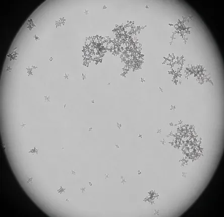
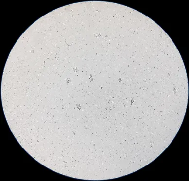
Following the aggregation assay, aliquots of the crystal suspension were mounted on glass slides and examined under a light microscope at 100× magnification. Representative images were captured to evaluate the morphology and aggregation of calcium oxalate crystals[18].
Ethylene Glycol & Ammonium Chloride-Induced Urolithiasis
Experimental urolithiasis was induced by administering 0.75% (w/v) ethylene glycol (EG) and 1% (w/v) ammonium chloride (AC) orally for 28 consecutive days to all animals, except those in the vehicle control group. Animals in the vehicle control group received distilled water, whereas those in the standard group were treated with Neeri tablets (70 mg/kg, p.o.). The aqueous extract of Triticum aestivum (AETA) was administered orally at doses of 100, 300, and 700 mg/kg once daily from day 1 to 28. At the end of the treatment period, 24-h urine samples were collected using metabolic cages to determine the urinary volume and biochemical parameters, including calcium, magnesium, phosphate, and oxalate, using commercially available diagnostic kits according to the manufacturers' instructions. Blood samples were subsequently collected, and serum was separated for the estimation of blood urea nitrogen (BUN), creatinine, and calcium levels using standard diagnostic kits following the respective kit protocols. The kidneys were excised for histopathological examination to assess the extent of crystal deposition and renal tissue damage[19] The experimental groups and treatment regimens are presented in Table 3.
|
Group |
Treatment |
|
I (Vehicle Control) |
Distilled water (p.o.) |
|
II (Lithiatic Control) |
EG (0.75%, p.o.) + AC (1%, p.o.) |
|
III (Standard) |
Lithiatic regimen + Neeri (7 mL/kg, p.o.)* |
|
IV (AETA-100) |
Lithiatic regimen + AETA (100 mg/kg, p.o.)* |
|
V (AETA-300) |
Lithiatic regimen + AETA (300 mg/kg, p.o.)* |
|
VI (AETA-700) |
Lithiatic regimen + AETA (700 mg/kg, p.o.)* |
*The lithiasis regimen consisted of EG (0.75%, p.o.) and AC (1%, p.o.) to induce urolithiasis.
Estimation of total urinary volume
All animals were kept in individual metabolic cages, and urine samples were collected over 24 h on the 28th day. The animals had free access to drinking water during the urine collection period. All animals were kept in individual metabolic cages, and urine samples were collected for 24 h on the 28th day. The animals had free access to drinking water during the urine collection period. A drop of concentrated HCl was added to the urine before being storage at 4 °C. Urine was analyzed for magnesium, calcium, phosphate, oxalate, and creatinine content using kits[20].
Histopathological analysis of the kidneys
After serum and urinary parameters estimation, the rats were euthanized by cervical dislocation after blood samples were collected. Both kidneys were removed by incising the abdominal wall of each rat, and each kidney was weighed. The isolated kidneys were preserved in 10% formalin solution. Slides were prepared using hematoxylin and eosin staining solutions. Slides were observed under a light microscope, and images were captured at 400X. Kidney samples were weighed and rapidly fixed in 10% neutralized formalin (pH 7.4). A section of the kidney was prepared in paraffin, stained with hematoxylin and eosin, and observed for pathological changes[21].
Statistical analysis
Results are expressed as mean ± S.E.M. Data were analysed using one-way ANOVA followed by Dunn's test using GraphPad Prism version 8.0. p < 0.05 [22].
RESULTS AND DISCUSSION
From the successive maceration extraction of AETA, the percentage yield, consistency, and color were as follows:
Table 5.1: Percentage yield of AETA
|
S.NO |
Extracts |
Color |
Consistency |
Yield |
|
1 |
Aqueous Extract of Triticum aestivum (AETA) |
Dark green |
Solid powder |
39.50% |
Table 5.2: Results of phytochemical screening of AETA
|
Test conducted |
AETA |
|
|
Alkaloids |
Mayer’s test |
+ |
|
Wagner’s test |
+ |
|
|
Tannin by 1% lead acetate |
+ |
|
|
Tannin by the FeCl3 and KOH method |
+ |
|
|
Protein |
+ |
|
|
Flavonoid |
+ |
|
|
Phenol |
+ |
|
|
Steroid |
+ |
|
|
Saponin |
+ |
|
|
Glycoside |
+ |
|
|
Carbohydrate |
Benedict’s test |
+ |
|
Fehling’s test |
+ |
|
|
Amino acid |
+ |
|
Inhibitory Effect of Neeri and AETA on Calcium Oxalate Crystal Nucleation Assay
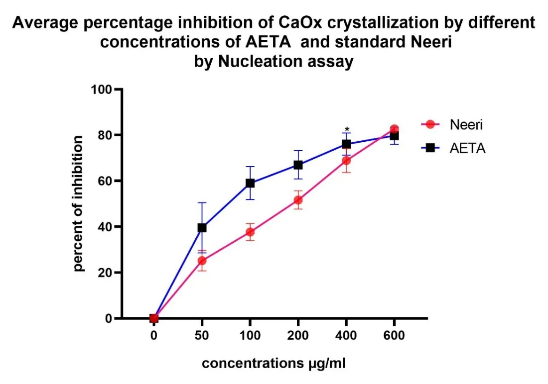
Graph 1: Average percentage inhibition of CaOx crystallization by different concentrations of AETA and the standard Neeri by the nucleation assay.
The nucleation assay demonstrated that both Neeri and AETA inhibited calcium oxalate crystal nucleation in a concentration-dependent manner. Neeri exhibited a higher percentage of inhibition than AETA at all tested concentrations. The maximum inhibition was observed at 600 µg/mL, where Neeri and AETA showed 80.50 ± 0.58% and 73.88 ± 3.09% inhibition, respectively. These findings indicate that AETA possesses significant antiurolithiatic activity by preventing the initial formation of calcium oxalate crystals, although its effect was slightly lower than that of the standard drug, Neeri. These results suggest that Triticum aestivum extract can effectively interfere with crystal nucleation, a crucial step in urolithiasis pathogenesis.
Inhibitory Effect of Neeri and AETA on Calcium Oxalate Crystal Growth Assay
Similar to the nucleation assay, both Neeri and AETA exhibited concentration-dependent inhibition of CaOx crystal growth. Neeri showed slightly greater inhibition than AETA at all the tested concentrations. At 600 µg/mL, Neeri produced a maximum inhibition of 77.98 ± 0.74%, whereas AETA exhibited 74.11 ± 0.86% inhibition, indicating considerable antiurolithiatic potential by preventing further growth of preformed CaOx crystals.
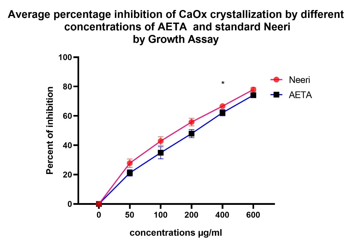
Graph 2: Average percentage inhibition of CaOx crystallization by different concentrations of AETA and the standard Neeri by Growth Assay.
Inhibitory Effect of Neeri and AETA on Calcium Oxalate Crystal Aggregation Assay
The aggregation assay demonstrated that both the standard drug Neeri and the aqueous extract of Triticum aestivum (AETA) inhibited calcium oxalate crystal aggregation in a concentration-dependent manner. Crystal aggregation is a critical step in urolithiasis, as the coalescence of smaller crystals leads to the formation of larger stone aggregates[23]. Therefore, the inhibition of crystal aggregation is considered an important indicator of antiurolithiatic activity.
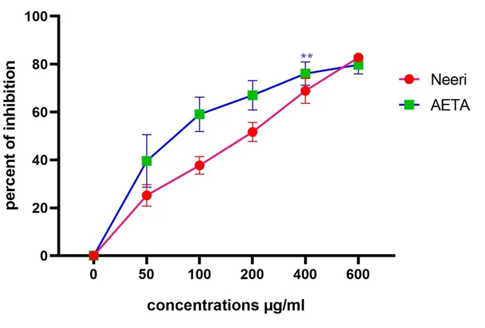
Neeri exhibited a progressive increase in the percentage inhibition of crystal aggregation from 25.19 ± 2.59% at 50 µg/mL to 82.75 ± 0.52% at 600 µg/mL concentration. Similarly, AETA showed substantial inhibition of crystal aggregation, increasing from 39.58 ± 6.32% at 50 µg/mL to 79.76 ± 2.20% at 600 µg/mL concentration.
Interestingly, AETA demonstrated greater inhibitory activity than Neeri at lower and intermediate concentrations (50–400 µg/mL). At 400 µg/mL, AETA produced 76.09 ± 2.79% inhibition compared to 68.88 ± 3.02% observed with Neeri, indicating a stronger ability to prevent crystal aggregation. However, at the highest concentration tested (600 µg/mL), Neeri exhibited slightly greater inhibition (82.75 ± 0.52%) than AETA (79.76 ± 2.20%).
The observed anti-aggregation activity of AETA may be attributed to the presence of flavonoids, phenolic compounds, saponins, and other phytoconstituents that interfere with crystal–crystal interactions and reduce the adhesion of CaOx crystals. By preventing the formation of larger crystal aggregates, AETA may reduce stone retention within the urinary tract and facilitate the elimination of smaller crystals through the urine.
Overall, these results indicate that AETA possesses significant anti-aggregation activity and effectively inhibits calcium oxalate crystal aggregation in a dose-dependent manner. The observed activity was comparable to that of the standard drug Neeri, suggesting that Triticum aestivum has promising antiurolithiatic potential through the inhibition of crystal aggregation.
Microscopic Observation of Calcium Oxalate Crystals
|
|
|

|
|
In the absence of plant extract (Control) |
200μg/mL concentration of AETA |
600μg/mL concentration of AETA |
|
|
|
|
|
|
200μg/mL concentration of NEERI |
|
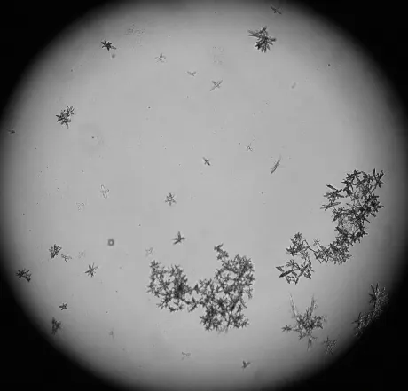
Figure 1: Micrograph of Calcium oxalate crystal inhibition, 10X
Microscopic examination of the control group revealed abundant calcium oxalate crystals with marked crystal formation and aggregation, confirming uninhibited nucleation. Treatment with the aqueous extract of Triticum aestivum (AETA) resulted in a concentration-dependent reduction in crystal number, size, and aggregation. At 200 μg/mL, AETA moderately inhibited crystal formation, as evidenced by the presence of fewer and smaller crystal aggregates. A more pronounced inhibitory effect was observed at 600 μg/mL, with only a few small dispersed crystals remaining. The standard drug, Neeri (200 μg/mL), exhibited a comparable reduction in crystal density and aggregation. These findings indicate that AETA effectively suppresses calcium oxalate crystal nucleation in a dose-dependent manner, with higher concentrations showing activity comparable to that of the standard treatment.
Effect of Neeri and AETA on Urinary Biochemical Parameters
Table 6: Effect of Neeri and AETA on urinary biochemical parameters in ethylene glycol–ammonium chloride-induced urolithiasis.
|
Gr |
Calcium (mg/L) |
Magnesium (mg/ L) |
Phosphate (mg/L) |
Sodium m (mmol /L) |
Potassium (mmol/L) |
Urine volume (mL) |
Oxalates mg/L |
|
I |
11.05±2.01 |
27.06±0.02 |
46.06±0.02 |
0.15±1.35 |
32.05±0.02 |
17.06±1.13 |
4.05±2.17 |
|
II |
19.04±1.01# |
38.05±0.02# |
35.08±0.02# |
0.25±0.62# |
36.07±0.03# |
10.05±0.02# |
8.07±2.22# |
|
III |
12.07±1.21* |
31.03±1.41* |
44.05±0.02# |
0.16±1.32* |
31.25±1.02* |
16.11±2.03* |
5.34±1.21* |
|
IV |
18.35±2.33ns |
35.05±2.51ns |
37.05±0.02ns |
0.19±1.84* |
34.12±1.04ns |
10.05±1.01ns |
7.57±1.06ns |
|
V |
17.06±1.31ns |
29.04±0.02* |
41.06±0.02* |
0.16±1.22* |
34.05±1.02ns |
12.06±1.02* |
7.61±0.14ns |
|
|
13.06±1.12* |
27.05±0.02* |
44.05±0.02* |
0.13±0.01* |
33.15±1.02* |
14.04±1.42* |
6.41±022* |
The values are expressed as mean ± SEM (n=6). ns, non-significant; # p < 0.05 compared to the normal control; * p < 0.05 compared to the disease control; ns, not significant.
Administration of ethylene glycol and ammonium chloride markedly altered the urinary biochemical profile compared to that of the normal control group (Table 6). The lithiatic control group showed significantly increased urinary calcium (19.04 vs. 11.05 mg/L), oxalate (8.07 vs. 4.05 mg/L), sodium (0.25 vs. 0.15 mmol/L), potassium (36.07 vs. 32.05 mmol/L), and magnesium (38.05 vs. 27.06 mg/L) levels, together with a reduction in urinary phosphate (35.08 vs. 46.06 mg/L) and urine volume (10.05 vs. 17.06 mL/24 h), confirming successful induction of urolithiasis.
Treatment with Neeri (70 mg/kg) significantly restored all altered urinary parameters towards normal values. Similarly, AETA produced a dose-dependent improvement in the urinary biochemical profile. The 100 mg/kg dose produced only modest changes, whereas 300 mg/kg significantly improved urinary magnesium, phosphate, sodium, and urine output, with partial reductions in calcium and oxalate excretions. The 700 mg/kg dose produced the greatest protective effect, significantly normalizing the urinary calcium, magnesium, phosphate, sodium, potassium, urine volume, and oxalate levels, with responses comparable to those observed in the standard-treated group.
Effect of Neeri and AETA on Body Weight and Kidney Weight
As shown in Figure 2, ethylene glycol administration resulted in a marked reduction in body weight (155.46 ± 0.53 g) and a significant increase in kidney weight (0.98 ± 0.01 g) compared with the normal control group (210.05 ± 0.02 g and 0.68 ± 0.01 g, respectively), indicating renal injury associated with the crystal deposition.
Treatment with Neeri (70 mg/kg) effectively restored the body weight (212.58 ± 1.13 g) and significantly reduced the kidney weight (0.70 ± 0.01 g). AETA treatment had a dose-dependent protective effect. Although the 100 mg/kg dose produced only partial improvement, the 300 mg/kg dose significantly increased the body weight and reduced the kidney weight. The 700 mg/kg dose almost completely restored both parameters (body weight: 209.36 ± 1.87 g and kidney weight: 0.69 ± 0.01 g), demonstrating marked protection against ethylene glycol-induced renal damage.
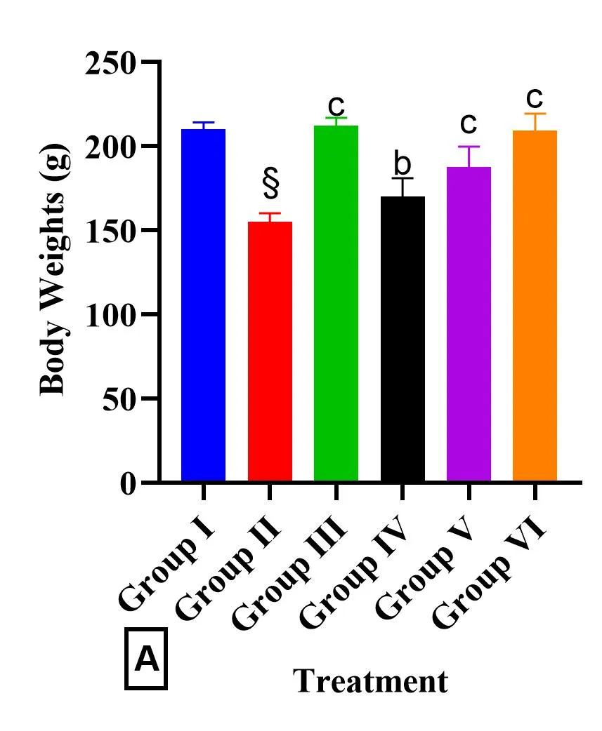
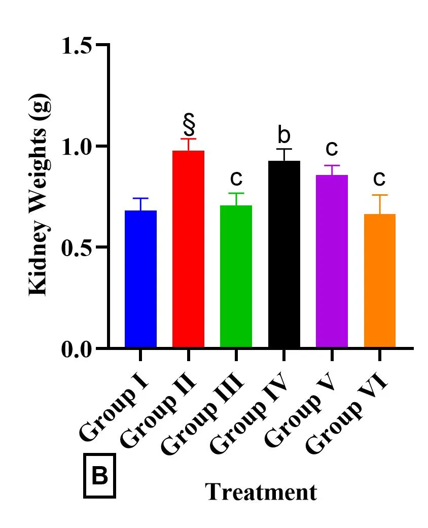
Figure 2 : Effect of Neeri and AETA on body weight and kidney weight in ethylene glycol–ammonium chloride-induced urolithiasis.
Effect of the Neerin and AETA extract on Serum Parameters
Table 7: Effect of AETA extract on the serum parameters.
|
Groups |
Serum parameters |
||||
|
Creatinine (mg/dL) |
BUN (mg/dL) |
Serum protein (g/ dL) |
Serum albumin (g/dL) |
Uric acid (mg/dL) |
|
|
Normal control |
2.75 ± 0.25 |
0.56 ± 0.32 |
9.14 ± 1.25 |
5.44 ± 2.82 |
3.06 ± 1.32 |
|
Disease control |
4.78 ± 1.22# |
0.84 ± 0.02 # |
14.17 ± 0.02# |
6.14 ± 0.02# |
3.74 ± 0.03# |
|
Standard |
3.24 ± 2.02* |
0.64 ± 0.01* |
10.08 ± 0.04* |
5.45 ± 0.02* |
3.13 ± 0.02* |
|
AETA100 mg/kg |
4.26 ± 3.05* |
0.75 ± 0.2* |
13.05 ± 0.02* |
5.94 ± 0.01* |
3.45 ± 0.01* |
|
AETA300 mg/kg |
3.75 ± 1.21* |
0.74 ± 0.02* |
12.15 ± 0.02* |
5.74 ± 0.02* |
3.44 ± 0.02* |
|
AETA700 mg/kg |
3.53 ± 2.13* |
0.65 ± 0.02* |
11.05 ± 0.02* |
5.55 ± 0.02* |
3.24 ± 0.02* |
The serum biochemical parameters of the disease control group showed significant alterations compared to those of the normal control group, indicating severe renal dysfunction following ethylene glycol-induced urolithiasis. A significant increase in serum creatinine, blood urea nitrogen (BUN), serum protein, serum albumin, and uric acid levels was observed in the disease control group. These changes reflect impaired glomerular filtration, renal tubular damage, and reduced excretory capacity associated with crystal deposition and with nephrolithiasis.
Treatment with the standard drug significantly reversed these biochemical abnormalities, bringing the values closer to those observed in the normal control group. Reductions in serum creatinine, BUN, serum protein, serum albumin, and uric acid levels indicate effective restoration of renal function and protection against stone-induced renal injury.
Administration of AETA at 100 mg/kg produced a significant, albeit modest, improvement in all serum parameters compared to the disease control group. Although reductions in serum creatinine, BUN, serum protein, serum albumin, and uric acid levels were observed, the values remained markedly higher than those of the normal control group, suggesting that the rats received partial protection against renal damage.
AETA at 300 mg/kg demonstrated a greater nephroprotective effect, as evidenced by a further reduction in serum creatinine, BUN, serum protein, serum albumin, and uric acid levels. These findings indicate improved renal filtration and reduced metabolic disturbances compared to lower-dose treatment.
The highest dose of AETA (700 mg/kg) exhibited the most pronounced protective effect, substantially normalizing all serum biochemical parameters. The values obtained were comparable to those observed in the standard-treated group, indicating significant preservation of renal function and attenuation of ethylene glycol-induced nephrotoxicity in the test group.
Overall, the results demonstrate that AETA treatment effectively ameliorated the alterations in serum biochemical markers induced by urolithiasis in a dose-dependent manner. The observed reductions in serum creatinine, BUN, and uric acid levels suggest improved renal clearance and decreased crystal-induced renal damage, whereas the normalization of serum protein and albumin levels indicates the restoration of normal renal physiology. These findings support the nephroprotective and antiurolithiatic potential of Triticum aestivum extract, particularly at a dose of 700 mg/kg.
Histopathological findings
|
|
|
|
Normal control |
Disease control |
|
|
|
|
Standard NEERI 70 mg/kg |
|
|
|
|
|
AETA100 mg/kg |
AETA700 mg/kg |
Figure 3: Histopathological Changes in Kidney Tissue Following AETA Treatment
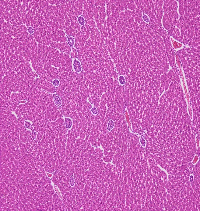
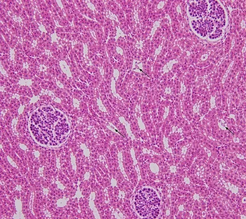
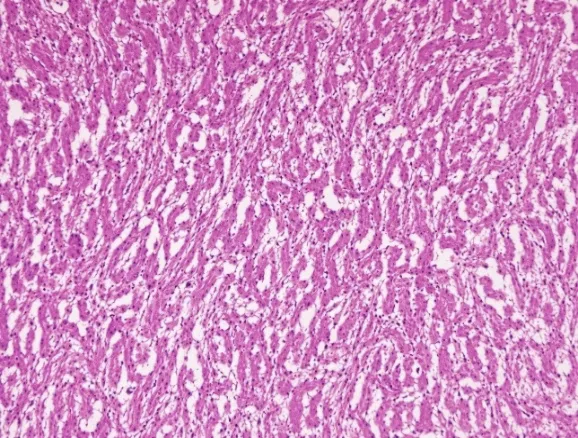
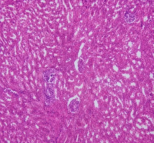
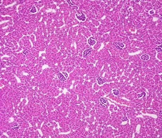
In the normal control group (Group I), the kidney sections exhibited normal histological architecture with intact glomeruli (Bowman's capsules) and well-organized renal tubules. No signs of tissue damage, inflammation, or crystal deposition were observed in the kidneys. In the disease control group (Group II), treated with EG and AC, marked alterations in the renal architecture were observed. The kidney sections showed tubular degeneration, widened interstitial spaces, cellular swelling, and numerous crystal deposits within the renal tubules, indicating crystal-induced renal injury. In the standard treatment group receiving Neeri (70 mg/kg), considerable protection against renal damage was observed, with preservation of the normal renal architecture and a marked reduction in crystal deposition. Treatment with AET at 100 mg/kg (Group IV) and AETA at 500 mg/kg (Group VI) produced dose-dependent nephroprotective effects. Kidney sections showed improved histological architecture, with reduced tubular damage, minimal interstitial swelling, intact Bowman's capsules, and markedly fewer crystal deposits than those in the disease control group. Histopathological findings of the AETA 300 mg/kg group (Group V) are not included in the present figure.
SUMMARY AND CONCLUSION
Urolithiasis is a multifactorial disorder characterized by the formation of stones within the urinary tract and does not have a single specific cause. The disease results from complex interactions between genetic, metabolic, dietary, environmental, and anatomical factors. The supersaturation of urine with stone-forming constituents, such as calcium, oxalate, phosphate, and uric acid, initiates crystal nucleation, growth, aggregation, and retention within the kidneys[24]. Oxidative stress, renal tubular epithelial injury, inflammation, urinary tract infections, low urine volume, dehydration, and metabolic abnormalities also contribute significantly to stone formation and recurrence. Because multiple pathogenic mechanisms are involved, effective management of urolithiasis requires therapeutic agents that simultaneously target multiple stages of stone development[25].
This study aimed to evaluate the antiurolithiatic potential of the aqueous extract of Triticum aestivum leaves (AETA) using in vitro calcium oxalate crystallization assays and an ethylene glycol-ammonium chloride-induced urolithiasis model in rats. The extract was obtained by aqueous maceration, yielding a dark green, 39.50% solid powder. Preliminary phytochemical screening revealed the presence of bioactive constituents, including flavonoids, phenolics, saponins, and alkaloids, as well as other phytoconstituents known for their antioxidant, anti-inflammatory, nephroprotective, and diuretic properties.
In vitro, AETA exhibited significant concentration-dependent inhibition of calcium oxalate crystal nucleation, growth, and aggregation. The extract effectively reduced crystal formation and prevented the enlargement and aggregation of the preformed crystals. At 600 μg/mL, AETA demonstrated inhibition comparable to that of the standard drug Neeri. Microscopic observations further confirmed a marked reduction in crystal density, size, and aggregation in the extract-treated samples compared to the control group, indicating interference with the critical stages of stone formation.
In the in vivo study, the administration of ethylene glycol and ammonium chloride successfully induced urolithiasis, as evidenced by increased urinary excretion of calcium, oxalate, sodium, potassium, and magnesium; reduced urine volume; increased kidney weight; weight loss; altered serum biochemical parameters; and extensive renal crystal deposition. These changes were associated with impaired renal function and histopathological damage characteristic of nephrolithiasis.
Oral AETA treatment for 28 days provided dose-dependent protection against urolithiasis. The low dose (100 mg/kg) showed mild improvement, whereas the medium dose (300 mg/kg) produced moderate restoration of the urinary and serum biochemical parameters. The highest dose (700 mg/kg) demonstrated the most significant antiurolithiatic activity, effectively normalizing urinary calcium, oxalate, phosphate, and electrolyte levels, urine output, body weight, and kidney weight. Furthermore, AETA significantly reduced elevated serum creatinine, blood urea nitrogen, uric acid, serum protein, and albumin levels, indicating improved renal function and nephroprotection. The effects observed at 700 mg/kg were comparable to those of the standard drug, Neeri.
Histopathological examination supported these biochemical findings. Kidneys from disease-control animals showed extensive tubular degeneration, crystal deposition, and interstitial damage, whereas AETA-treated groups exhibited marked preservation of renal architecture, reduced crystal deposition, and minimal tissue injury. The protective effect was most pronounced at the highest dose.
The observed antiurolithiatic activity of AETA may be attributed to its diverse pharmacological effects. Antioxidant constituents may protect renal epithelial cells from oxidative stress-induced injury, thereby reducing crystal adherence and retention. Its anti-inflammatory properties may attenuate crystal-induced renal inflammation, while its diuretic effect increases urine output, dilutes stone-forming constituents, and facilitates the elimination of crystals from the urinary tract[26–28]. Additionally, the mineral-rich composition of wheatgrass may help maintain the urinary electrolyte balance and reduce urinary supersaturation.
Overall, the findings of the present study demonstrate that the aqueous extract of Triticum aestivum possesses significant antiurolithiatic and nephroprotective activities. The extract effectively inhibited calcium oxalate crystallization in vitro and prevented ethylene glycol-induced renal-stone formation in vivo. These results support the traditional and nutraceutical use of wheatgrass and suggest that Triticum aestivum may serve as a promising natural therapeutic agent for preventing and managing urolithiasis.
CONCLUSION
The present study demonstrated that the aqueous extract of Triticum aestivum leaves (AETA) possesses significant anti-urolithiatic activity in both in vitro and in vivo experimental models. The extract effectively inhibited calcium oxalate crystal nucleation, growth, and aggregation in a concentration-dependent manner, indicating its ability to interfere with the key stages of stone formation. Microscopic observations further confirmed a marked reduction in crystal size, number, and aggregation following AETA treatment.
In the ethylene glycol and ammonium chloride-induced urolithiasis model, AETA significantly ameliorated alterations in urinary and serum biochemical parameters, improved urine output, restored body weight, reduced kidney hypertrophy, and protected against renal tissue damage. Histopathological studies revealed a substantial reduction in crystal deposition and preservation of normal renal architecture in the extract-treated animals. Among the tested doses, 700 mg/kg AETA showed the greatest protective effect, comparable to that of the standard drug Neeri.
The antiurolithiatic activity of Triticum aestivum may be attributed to its rich content of flavonoids, phenolic compounds, minerals, and other bioactive constituents possessing antioxidant, anti-inflammatory, nephroprotective, and diuretic properties. These mechanisms collectively prevent crystal formation, reduce oxidative renal injury, enhance urinary flow, and maintain normal kidney function.
Overall, the findings of this study suggest that Triticum aestivum is a safe and promising natural antiurolithogenic agent. Its ability to target multiple factors involved in urolithiasis pathogenesis highlights its potential as a nutraceutical and therapeutic adjunct for the prevention and management of renal stones.
Conflict of Interest declaration: The authors declare that they have no affiliations with or involvement in any organization or entity with any financial interest in the subject matter or materials discussed in this manuscript.
Author Contributions: MS and NS contributed to the design and implementation of the research, and RBK contributed to the analysis of the results and to the writing of the manuscript. NS conceived the original idea and supervised the project.
REFERENCES



Bharath Kumar Rajaboina, Srinivasu Matta, Saikrishana Nallajerla, Investigation of the In Vitro and In Vivo Antiurolithiatic Activity of the Aqueous Extract of Triticum aestivum Against Ethylene Glycol-Induced Urolithiasis in Rats, Int. J. of Pharm. Sci., 2026, Vol 4, Issue 8, 9-26, https://doi.org/10.5281/zenodo.21734981
 10.5281/zenodo.21734981