
We use cookies to ensure our website works properly and to personalise your experience. Cookies policy
Department of Pharmaceutics, College of Pharmaceutical Sciences, Govt Medical College Kannur, 670503
Diabetes mellitus is a major burden worldwide to the socioeconomic structures and the healthcare systems. Although they are effective, the clinical utility of traditional oral antidiabetic drugs is often compromised by narrow therapeutic indices, rapid elimination from the body and related systemic toxicities. Plant-derived secondary metabolites represent an interesting multi-target polypharmacological alternative, but their actual utilization is seriously hampered by problems of poor solubility in aqueous media, instability in the gastrointestinal tract, and low bioavailability. Lipid-based drug delivery systems (LBDDS) including liposomes, solid lipid nanoparticles (SLNs), nanostructured lipid carriers (NLCs) and self-nanoemulsifying drug delivery systems (SNEDDS) are a game changer intervention based on the material science. This review provides a comprehensive overview of the classification of plant-derived active compounds, their molecular mechanisms of action in terms of antidiabetic effects, and the physicochemical principles governing the design of lipid carriers. It reviews systematically, progressive processing methods, parameters for scale-up at macro level and multi-dimensional biopharmaceutical evaluation approaches from in vitro to in vivo methodologies. Finally, the regulatory, clinical and technological contexts, highlighting advanced prospective configurations such as hybrid systems, stimuli-responsive platforms and lipid formulations optimized by artificial intelligence to drive green endocrinology into validated therapeutic applications for human use will be discussed.
Diabetes mellitus is a signature metabolic crisis of the twenty-first century. The global prevalence has increased from an estimated 30 million cases in 1985 to 537 million cases in 2021, and is projected to reach 783 million cases by 2045. This increase is driven by the aging population, rapid urbanization, and increased obesity rates. Macroeconomic modeling indicates that this epidemic will result in a 0.22% annual tax on global GDP from 2020 to 2050, with a total global cost of $10.2 trillion without informal care, and an astonishing $78.8 trillion when informal care costs are fully internalized(1).
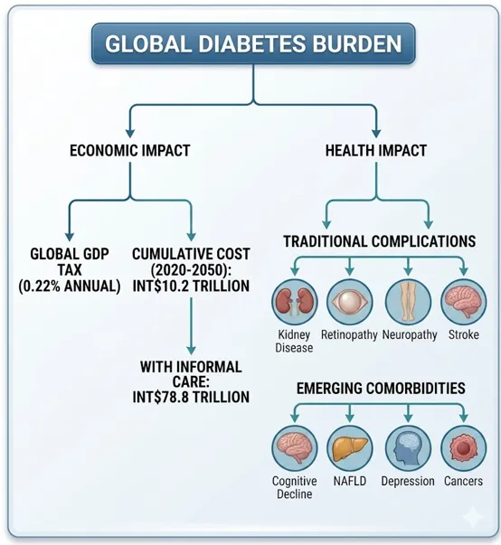
Fig 1. Diabetes burden
The orthodox front-line treatments (exogenous insulin, insulin secretagogues, and synthetic sensitizers (biguanides, thiazolidinediones)) are effective in stabilizing acute glycemic fluctuations but they are not a permanent cure. They are also often limited by secondary failure, weight gain, severe hypoglycemia, and cardiorenal risks. This therapeutic gap has led to an increasing interest in plant-derived alternatives. Historically, modern metabolic medicine has been based on natural product chemistry, e.g. metformin derived from biguanides extracted from Galega officinalis (2). More than 1,200 flowering species are known with specific hypoglycemic indices. These botanicals employ complex networks of co-existing secondary metabolites to simultaneously target multiple metabolic pathways—lowering peripheral insulin resistance, increasing pancreatic β-cell regeneration, reducing localized oxidative stress, and flattening small-intestinal glucose absorption curves. However, their poor aqueous solubility, low gastrointestinal stability and rapid systemic clearance often severely hinder therapeutic translation of these botanicals. To address these biopharmaceutical challenges, advanced lipid- and biopolymer-based drug delivery systems, especially gel-based networks, have emerged as a game-changing formulation approach. This review highlights the synergy of phytomedicine and pharmaceutical material science in the design, characterization and therapeutic evaluation of lipid-based plant alternatives as controlled-release, highly bioavailable vehicles for the modern management of diabetes mellitus (3).
2. CLASSIFICATION AND ANTIDIABETIC MECHANISMS OF PLANT ACTIVES
Therapeutic integration requires classifying plant-derived inputs into distinct chemical and operational domains, moving away from traditional single-target pharmacology toward multi-systemic metabolic networks:
Crude Extracts: Unpurified whole-plant matrices processed into basic aqueous infusions, tinctures, or powders. They frequently display superior efficacy compared to isolated components due to natural component synergy.
Standardized Fractions: Purified fractions that concentrate specific groups of active compounds to guarantee batch-to-batch therapeutic consistency. For instance, under regulatory frameworks (e.g., AYUSH/CDSCO), a defined phytopharmaceutical drug must comprise a standardized fraction isolating a minimum of four distinct bio-active phytoconstituents.
Isolated Phytochemicals: Pure, single molecules extracted directly from the plant matrix. These pure entities (e.g., specific flavonoids or alkaloids) eliminate tracking variations and provide clean, modern delivery routes(4,5).
Phytochemical Families and Molecular Mechanisms
Natural antidiabetic agents possess diverse structural properties that match specific physiological targets:
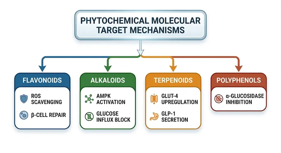
Fig 2. Phytochemical Target Mechanisms
1. Flavonoids (e.g., Quercetin, Morin, Rutin)
Flavonoids act as powerful antioxidants that neutralize reactive oxygen species (ROS), preserving pancreatic β-cell viability and architectural integrity. At the receptor level, specific molecules like morin activate insulin receptor autophosphorylation, improving downstream metabolic pathways and reducing endoplasmic reticulum (ER) stress. Others show direct competitive inhibition against mammalian carbohydrate-digesting enzymes.
2. Alkaloids (e.g., Trigonelline, Berberine)
Alkaloids regulate glucose profiles through multiple structural pathways. Trigonelline, found in defatted fenugreek fractions, mediates strong glucose-lowering effects. This class systematically upregulates insulin receptor sensitivity, promotes endogenous pancreatic insulin secretion, and blocks dietary intestinal glucose absorption.
3. Terpenoids and Saponins (e.g., Gymnemic Acids, Ginsenosides)
Ginsenosides (triterpene saponins) operate across multiple target organs: Ginsenovide Rb2 reduces excessive hepatic gluconeogenesis, while Rb1, Rg3, and Compound K enhance peripheral glucose clearance in skeletal muscle and adipose tissue by upregulating the expression and membrane translocation of GLUT-1 and GLUT-4 transporters. Thermally degraded saponins like ginsenoside Rg3 also stimulate glucagon-like peptide-1 (GLP-1) release from enteroendocrine cells. Gymnemic acids induce reversible cell permeabilization in pancreatic islets to stimulate endogenous insulin release, while simultaneously blocking small-intestinal glucose transport and hepatic glycogenolysis.
4. Polyphenols and Tannins (e.g., Charantin, Epicatechin, Gallic Acid)
These molecules prevent secondary microvascular complications like cataracts through systemic free radical scavenging. Molecules like epicatechin promote pancreatic islet regeneration, while broader polyphenolic fractions block α-amylase and α-glucosidase pathways, flattening postprandial glucose absorption spikes(6–8).
Target Profiles of Core Botanicals (9–12)
Table 1. Properties of Phytoconstituents
|
Plant Species/ Common Name |
Classification |
Bioactive Components / Active Markers |
Key Mechanisms & Specific Effects |
|
Eugenia caryophyllata (Clove) |
Phenols |
Eugenol |
Activates AMPK via CaMKK; suppresses the nuclear CRTC2-CREB complex; decreases hepatic glucose production, lowers blood glucose/insulin levels, and improves glucose tolerance. |
|
Zingiber officinale (Ginger root) |
Phenols |
[6]-Gingerol |
Promotes AMPK activation, GLUT4 translocation, and intracellular Ca2+ increase via CaMKK2; upregulates PGC-1α reduces pancreatic β-cell ROS, suppresses G6Pase/PEPCK gluconeogenesis, and lowers serum TNF-α |
|
Coffee, various fruits |
Polyphenols |
Chlorogenic acid |
Activates AMPK signaling and increases GLUT4 translocation; downregulates and inhibits glucose-6-phosphatase (G6Pase); lowers blood glucose, reduces gluconeogenesis, and decreases fatty acid synthesis. |
|
Echinacea purpurea, Chicory root |
Polyphenols |
Cichoric acid |
Activates AMPK, PI3K/Akt, and Nrf2-Keap1 pathways; inhibits hepatic injury, reduces gluconeogenesis, increases glycogen synthesis, and suppresses oxidative stress. |
|
Rosmarinus officinalis (Rosemary) |
Polyphenols |
Rosmarinic acid |
Activates AMPK and promotes reverse cholesterol transport (RCT); upregulates ABCG5/8, CYP7A1, and CPT1a; promotes fatty acid oxidation, decreases plasma/hepatic cholesterol/triglycerides, and improves glucose homeostasis. |
|
Terminalia arjuna (Leaves) |
Polyphenols |
Ellagic acid |
Activates the AMPK-ERK-atypical PKC (aPKC) pathway without affecting Akt; stimulates GLUT4 translocation to increase glucose uptake and improve glucose homeostasis. |
|
Tea, berries, fruits, plants |
Polyphenols |
Gallic acid |
Activates AMPK and the Sirt1/PGC-1α pathways; promotes autophagy and thermogenesis (via UCP1); enhances mitochondrial function, reduces fat accumulation, and improves glucose/insulin homeostasis. |
|
Curcuma longa (Turmeric) |
Polyphenols |
Curcumin |
Activates AMPK and SIRT1; suppresses NF-κB, acetyl-CoA carboxylase (ACC) phosphorylation, and gluconeogenic genes (PEPCK, G6Pase); deacetyleates FOXO1; improves glucose/lipid metabolism, reduces myocardial infarction size, and decreases apoptosis. |
|
Rhodiola rosea |
Polyphenols |
Salidroside |
Inhibits respiratory chain complex I to alter the AMP/ATP ratio; activates the AMPK/PI3K/Akt/GSK3β pathway and phosphorylates ACC; suppresses PEPCK and G6Pase; improves glucose uptake, alleviates insulin resistance, and reduces liver steatosis. |
|
Polygonum cuspidatum, Grapes |
Polyphenols |
Resveratrol |
Inhibits cAMP-dependent phosphodiesterases (PDEs) to trigger CaMKK-β/AMPK phosphorylation; inhibits mTOR and p70 S6K; reverses palmitate-induced serine phosphorylation of IRS-1; enhances oral glucose sensitivity, muscle mitochondrial respiration, and protects health via AMPK. |
|
Rosmarinus officinalis |
Terpenoids |
Carnosic acid |
Activates AMPK; suppresses gluconeogenic genes G6PC and PCK1; inhibits lipogenic genes (FAS, ACC1, SREBP-1c); enhances fatty acid oxidation via PGC-1α and CPT1A. |
|
Platycodon grandiflorum (Root) |
Terpenoids |
Platycodin D |
Activates AMPK, downregulates PCK1 and G6Pase; upregulates ACC phosphorylation and CPT-1 expression; directly interacts with glycolipid metabolic proteins; reduces hyperglycemia, mitigates hepatic fat via the AMPK/ACC/CPT-1 pathway, and upregulates GLUT4. |
|
Bupleurum chinense (Root) |
Terpenoids |
SaikosaponinA (SSA) & Saikosaponin D (SSD) |
Enhances phosphorylation of AMPK and ACC; inhibits MAPK pathway branches (ERK, p38 for SSA; ERK, JNK for SSD); downregulates PPAR-γ C/EBP α SREBP-1c, FABP4, FAS, and LPL; inhibits lipid accumulation and suppresses early-stage adipogenesis. |
|
Nelumbo nucifera (Lotus seed embryo) |
Alkaloids |
Isoliensinine |
Activates AMPK phosphorylation, increases GLUT4 translocation, downregulates PPARγ, SREBP-1c, and ACC phosphorylation; reduces blood glucose by ~50% and body weight by ~19%, increases insulin, and improves hyperlipidemia. |
|
Coptis chinensis (Huanglian) |
Alkaloids |
Berberine |
Binds directly to the AMPK γ subunit inducing allosteric activation; inhibits mitochondrial respiratory complex I in gut/liver; blocks IKKe/adrenergic/cAMP pathway and downregulates TBK1; lowers glucose, HbA1c, total cholesterol, and triglycerides; suppresses tissue apoptosis. |
|
Uncaria rhynchophylla |
Alkaloids |
Hirsutine |
Activates PI3K/Akt and AMPK/ACC signaling cascades; downregulates gluconeogenic markers (PEPCK, G6Pase) and transcription factors (PGC-1α FOXO1); upregulates GLUT4; decreases glucose production, increases fatty acid oxidation, and reduces hepatic steatosis. |
|
Black Cumin (Nigella sativa) |
Monoterpene quinone
|
Thymoquinone (TQ)
|
Stimulates and normalizes glucose-induced endogenous insulin secretion from pancreatic β-cells. Inhibits expression of rate-limiting gluconeogenic enzymes (Glucose-6-phosphatase / G6Pase), reducing excessive hepatic glucose production. Re-oxidizes metabolic NADH back into NAD+ via intracellular redox cycling. Suppresses pro-inflammatory signals (TNF-α, IL-6, IL-1β CRP) and apoptotic cascades (caspase-3, p53)(13). |
|
Cynodon grandis (Bark) |
Alkaloids |
Laurolitsine (LL) |
Inhibits ATP production and alters the ADP/ATP ratio to activate the LKB1/AMPK pathway; regulates mitochondrial oxidative phosphorylation; reduces blood glucose, weight gain, and inflammatory markers; modulates gut microbiota. |
|
Salvia miltiorrhiza |
Quinones |
Tanshinone IIA |
Enhances AMPK activity and inhibits the transcriptional activity of PPARγ; reduces PERK and JNK phosphorylation while increasing insulin-mediated Akt activation; mitigates insulin resistance caused by ER stress, and lowers blood glucose. |
|
Arctium lappa (Great burdock) |
Phenylpropanoids |
Total Lignans from Burdock Fruit (TLFA), Arctigenin |
Indirectly activates AMPK via mitochondrial complex I inhibition; stimulates the CaMKK- and LKB1-dependent AMPK cascades; activates PI3K/Akt; suppresses mitochondrial respiration, improves HbA1c and glucose tolerance, stimulates insulin, and reduces lipid accumulation (10). |
|
Spatholobus suberectus |
Flavonoids/ Phenylpropanoids |
Formononetin-rich extract |
Activates the Akt-AMPK signaling axis; inhibits advanced glycation end-products (AGEs) cross-linking and downregulates RAGE; upregulates Nrf2 and Glo1; decreases fasting blood glucose, HbA1c, lipid profiles, and urinary albumin-to-creatinine ratios to protect kidney function. |
|
Momordica charantia (Bitter melon) |
Saponins, peptides, triterpenoids |
Charantin, polypeptide-p, cucurbitane-type triterpenoids |
Charantin stimulates insulin secretion from pancreatic β-cells; polypeptide-p mimics insulin actions to promote peripheral glucose uptake; cucurbitane triterpenoids suppress hepatic gluconeogenesis; increases hexokinase activity and inhibits glucose-6-phosphatase. |
|
Trigonella foenum-graecum (Fenugreek) |
Fiber, amino acids, alkaloids |
Galactomannan, 4-hydroxyisoleucine, trigonelline, diosgenin |
Galactomannan forms viscous gels slowing carbohydrate absorption; 4-hydroxyisoleucine facilitates glucose-stimulated insulin release and GLUT4 translocation; trigonelline upregulates IRS proteins, activates PI3K/Akt, and blocks gluconeogenic enzymes; inhibits α-amylase/α-glucosidase. |
|
Zanthoxylum alatum |
Polyphenols/ Terpenes |
Polyphenolic and flavonoid derivatives |
Inhibits PTP1B (Protein Tyrosine Phosphatase 1B) activity and stimulates glucose uptake in skeletal muscle, lowering blood sugar. |
Physicochemical, Pharmacokinetic, and Stability Obstacles
Translating crude botanicals or isolated phytochemicals into reliable clinical therapies is hindered by several major biopharmaceutical limitations:
1. Large Molecular Mass and High Polarity: Many potent plant metabolites (e.g., polar glycosides, high-molecular-weight tannins) possess structures that prevent them from passively crossing the lipid-rich biological membranes of the intestinal mucosa, restricting absorption.
2. Gastrointestinal Instability: Orally ingested phytochemicals face rapid destruction inside the strongly acidic environment of the stomach and undergo extensive metabolic modification by native gut microflora or digestive enzymes.
3. Poor Aqueous Solubility: Lipophilic terpenoids and volatile fractions dissolve poorly within aqueous gastrointestinal fluids, causing highly erratic dissolution profiles and low baseline plasma availability.
4. Rapid Systemic Clearance: The combination of low intestinal absorption, high pre-systemic clearance, and short metabolic half-lives prevents molecules from maintaining therapeutic plasma levels unless high, frequent doses are administered—which increases the risk of local gastrointestinal side effects(14).
To resolve these limitations, modern formulation design leverages specialized delivery networks to encapsulate and shield these molecules, improving their metabolic stability and biological absorption.
3. LIPID-BASED DRUG DELIVERY PLATFORMS: CORE STRUCTURAL PRINCIPLES
Lipid-based drug delivery systems (LBDDS) function as biomimetic nanoscale carriers. They present a superior safety profile compared to synthetic polymer matrices due to their high biocompatibility and biodegradability, as they utilize lipid components identical to endogenous biological membranes. By completely encapsulating active payloads, these systems insulate them from external degradation, modulate their release kinetics, and unlock versatile administration routes (oral, transdermal, mucosal, and parenteral)(15,16).
Structural Classification of Lipid Carriers
Liposomes
Spherical vesicles featuring an aqueous core enclosed by one or more concentric phospholipid-cholesterol bilayers. This unique structural layout allows them to concurrently host hydrophilic compounds in the internal water compartment and lipophilic drugs within the middle hydrophobic bilayers. Conventional configurations can suffer from self-leakage or structural aggregation, which is typically managed using stabilizers like DSPE-MPEG 2000(17).
Solid Lipid Nanoparticles (SLNs)
Particulate carriers composed of a solid hydrophobic lipid core (e.g., tristearin or purified stearic acid) stabilized by an outer surfactant layer. Because the core lipid remains strictly solid at both room and body temperatures, SLNs form a highly ordered crystalline matrix that excels at loading lipophilic molecules (log P > 3). When applied to the skin, they form a dense, occlusive film that restricts transepidermal water loss. However, their highly ordered solid lattices can undergo crystal restructuring during storage, potentially squeezing the internal matrix and causing drug expulsion.
Nanoemulsions
Advanced, submicron colloidal dispersions consisting of a liquid lipid core wrapped within a surfactant monolayer shell. Composed of oils, surfactants, and water-soluble co-solvents, they are highly effective at enhancing the solubility, intestinal dissolution rate, and oral bioavailability of intensely hydrophobic compounds while shielding them from first-pass hepatic elimination.
Nanostructured Lipid Carriers (NLCs)
Developed as second-generation lipid nanoparticles to overcome the drug expulsion flaws of SLNs. Their core matrix is formed by intentionally blending solid lipids with liquid oils (e.g., oleic acid, medium-chain triglycerides). This fluid-fat blend yields a highly disorganized, less-ordered crystalline framework with numerous imperfect nano-compartments that accommodate higher drug payloads and prevent leakage. They are classified into:
Type 1 (Imperfect): High drug loading capacity paired with quick release profiles.
Type 2 (Amorphous): Prevents crystallization completely.
Type 3 (Multiple): Microscopic liquid oil droplets dispersed inside a solid fat matrix(18,19).
Ethosomes
Modified liposomal architectures containing high concentrations of ethanol (20%-50%) alongside phospholipids and water. This hydro-ethanolic solution imparts exceptional elasticity and a net negative charge to the vesicle membrane, facilitating continuous transdermal flux via a dual mechanism:
1. The Ethanol Effect: Disrupts and fluidizes the highly organized intercellular lipid bilayers of the stratum corneum, opening narrow pathways.
2. The Ethosome Effect: Malleable vesicles deform to squeeze through these newly opened channels into deep epidermal and dermal layers, fusing with cell membranes to release their payload(20).
Transferosomes
Ultra-deformable vesicular systems containing phospholipids combined with a single-chain surfactant acting as an edge activator. The edge activator destabilizes the lipid bilayer at specific stress points, making the vesicle flexible and highly responsive to osmotic forces. Applied non-occlusively to the skin, transferosomes respond to the natural hydration gradient, changing shape to pass intact through narrow skin pores that would stop conventional liposomes.
Physicochemical Framework of Lipid Carriers
The loading capacity, encapsulation parameters, and transport dynamics of LBDDS are governed by precise chemical principles:
Lipophilicity: Hydrophobic compounds (log P > 3) partition seamlessly into the internal fatty acid domains of SLNs, NLCs, and nanoemulsions, ensuring high payload capacity and diffusion-controlled release.
Amphiphilicity: Phospholipids feature a zwitterionic hydrophilic polar head group and two lipophilic fatty acid acyl chains. In aqueous media, they spontaneously self-assemble into closed bilayers to hide their hydrophobic tails from water, creating distinct compartments that can carry polar and non-polar drugs simultaneously.
Encapsulation & Charge Dynamics: Perfect, uniform solid fats can push out drugs over time, whereas adding liquid oils disrupts molecular packing to maximize encapsulation space. Furthermore, introducing a positive surface charge (zeta potential) creates strong electrostatic attraction with negatively charged biological membranes, increasing skin adhesion and cellular internalization(15,21).
4. BIOAVAILABILITY AND STABILITY IMPROVEMENTS FOR PLANT ACTIVES
1. Solubility Enhancement: Embedding molecules like curcumin or quercetin into lipid bilayers or disorganized NLC cores significantly improves their saturation solubility and water dispersion, preventing the particle aggregation seen with raw botanical extracts.
2. Protection from Degradation: Lipid nanocarriers form a protective physical barrier that completely insulates plant actives from acid and enzymatic hydrolysis in the stomach. For photo-sensitive molecules like curcuminoids, encapsulation in an SLN matrix keeps 88%-96% of the active components completely intact after six months of storage.
3. Bioavailability Multipliers: In animal models, curcumin-loaded SLNs demonstrate a 39- to 155-fold increase in plasma bioavailability compared to free suspensions. Quercetin-loaded SLNs provide a 5-fold increase in bioavailability by shifting the primary absorption zone to the small intestine. Puerarin-loaded SLNs achieve a 3-fold increase in absolute bioavailability with a shortened Tmax. For skin applications, vesicular ethosomes increase the tissue deposition of psoralen by 6.56-fold, while curcumin delivered via nanoemulsions improves local epidermal drug accumulation by 2.4- to 3.3-fold(22).
5. FORMULATION DESIGN, OPTIMIZATION, AND INDUSTRIAL SCALE-UP
Developing a stable, clinically viable LBDDS requires a structured formulation workflow, moving from preformulation profiling to continuous industrial manufacturing.
5.1 Preformulation and Excipient Selection
Rational formulation begins by establishing the physicochemical profile of the plant active, tracking its intrinsic aqueous solubility, ionization constant (pKa), partition coefficient (log P/log D), and solid-state compatibility with lipid matrices.
Mapping the pH-solubility curve predicts precipitation risks across the gastrointestinal tract, while tracking the pKa reveals the ratio of ionized to unionized species at gastric (~pH 1.2) versus intestinal (~pH 6.8) conditions, directly influencing encapsulation strategies. High log P compounds (>3) are assigned to self-emulsifying or solid lipid cores, while lower log P fractions require vesicular assembly or phospholipid complexation (phytosomes). Solid-state interactions are screened using FTIR, DSC, and XRD to prevent phase separation or drug degradation in the final vehicle.
Excipient composition is guided by the Lipid Formulation Classification System (LFCS), which groups formulations from Type I (pure oils) to Type IV (pure hydrophilic surfactants and co-solvents).
Table 2. Excipient selection
|
Excipient Class |
Core Functions |
Representative Examples |
|
Lipid Core / Oils |
solubilization, lymphatic transport, structural matrix |
Long-chain triglycerides (LCT) like soybean oil; Medium-chain triglycerides (MCT) like Captex 355; Mono-/diglycerides (Capmul MCM, Geleol) |
|
Surfactants |
Interfacial tension reduction, self-emulsification, steric stabilization |
High HLB: Tween-80, Cremophor EL (Kolliphor EL), TPGS Low HLB: Span-80, Pluronic blocks |
|
Co-Solvents |
Secondary solubilization, phase transition optimization |
Ethanol, propylene glycol, polyethylene glycol (PEG 400) |
Surfactant selection balances Hydrophilic-Lipophilic Balance (HLB) values against behavior during gastrointestinal lipolysis. High-HLB surfactants promote spontaneous emulsification into nanoscale droplets (<200 nm) upon contact with gastric fluids. Surfactants like Vitamin E TPGS are widely utilized because they function as both structural stabilizers and active p-gp efflux inhibitors, directly boosting oral absorption(23,24).
5.2 Processing Methodologies and Loading Strategies(25,26)
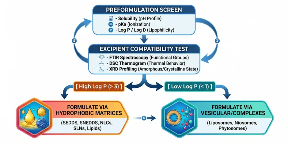
Fig 3. Preformulation and processing methodologies
Processing methods dictate the internal structure, droplet size distribution, and encapsulation efficiency of the lipid carrier. These methods are divided into high-energy and low-energy techniques:
High-Energy Methods: High-Pressure Homogenization (HPH) and Microfluidization apply intense shear, cavitation, and impact forces to break down macro-emulsions into uniform nano-emulsions or solid lipid matrices. While highly efficient for scalable manufacturing, they generate local temperature spikes that can degrade heat-sensitive phytochemicals. Ultrasonication is suitable for rapid bench-scale screening but carries risks of titanium probe shedding and uneven energy distribution.
Low-Energy Methods: Thin-Film Hydration (TFH) dissolves lipids and actives in an organic phase, evaporates it into a thin film, and hydrates it with an aqueous buffer. This method achieves high encapsulation for amphiphilic compounds but requires secondary extrusion for uniform particle sizing. Phase Inversion Temperature (PIT) and Solvent Diffusion utilize the chemical energy of the system; by adjusting temperature or shifting water-miscible solvents, the system spontaneously reorganizes into nanoscale structures under gentle stirring(27).
Loading Architecture
Encapsulating plant antidiabetics requires unique loading strategies based on whether the active payload is a complex whole-plant extract or an isolated pure molecule:
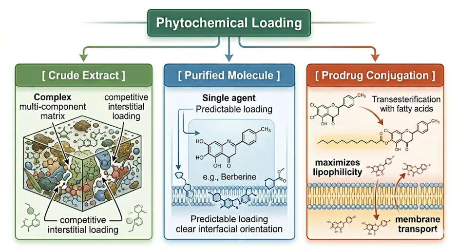
Fig 4. Phytochemical loading
Complex crude extracts contain mixtures of competing polarities, resulting in lower overall encapsulation efficiency due to competition for space within the lipid matrix. Pure isolated compounds behave predictably, enabling formulators to tailor the lipid matrix to the molecule's specific log P. Highly lipophilic compounds embed deeply within the fatty acid core of SLNs/NLCs, whereas weakly hydrophobic compounds position themselves at the surfactant-water interface (surface adsorption), which can lead to a rapid initial burst release rather than a sustained release profile. For highly hydrophilic compounds, reversibly coupling the active to a fatty acid chain via an ester or amide linkage creates a lipophilic lipid-prodrug conjugate that dissolves efficiently into the lipid carrier and releases the parent drug via endogenous esterases after crossing the intestinal barrier(28,29).
5.3 Scale-Up and Manufacturing Validation
Fig 5. Scale up of LNP
Transitioning from small lab-scale batches to industrial production introduces significant chemical engineering challenges. Traditional techniques like thin-film hydration are limited by batch volume limits and high batch-to-batch variability. Industrial production relies on continuous manufacturing setups, such as cross-flow microfluidic chips or multi-stage continuous high-pressure homogenizers, to maintain consistent Critical Quality Attributes (CQAs).
During scale-up, operators must strictly control engineering variables (Critical Process Parameters, or CPPs), including volumetric flow rate ratios in microfluidic channels, precise homogenization pressures, and cooling/solidification rates during lipid crystallization. Standardized marker assays must be established to manage the natural chemical variability of plant extracts and guarantee consistent drug-to-lipid ratios. Finally, complete removal of residual organic solvents (e.g., chloroform, methanol) requires industrial-scale vacuum drying, spray drying, or lyophilization to strictly satisfy international regulatory thresholds (ICH Q3C)(21,30,31).
6. BIOLOGICAL EVALUATION AND MECHANISTIC STUDIES(32)
Validating the performance of plant-derived antidiabetic LBDDS requires a systematic, multi-tiered testing hierarchy, transitioning from in vitro characterization to in vivo pharmacokinetic and biodistribution tracking.
6.1 In Vitro Characterization Models
Physicochemical Characterization: Evaluation of key CQAs begins with Dynamic Light Scattering (DLS) to determine mean particle size (Z-average) and Polydispersity Index (PDI), where a PDI <0.3 indicates a highly stable, monodisperse population. Surface charge is verified via zeta potential, where values outside the range confirm strong electrostatic repulsion that prevents particle aggregation. High-resolution imaging (TEM, SEM) is utilized to confirm spherical morphology and rule out co-existing structures. Encapsulation Efficiency (EE%) and Drug Loading (DL%) must be systematically quantified.
Solid-State Properties: DSC and XRD track lipid crystallinity and polymorphic phase transitions (e.g., transition from highly ordered β-forms to less ordered α or β polymorphs), confirming the presence of an imperfect matrix desirable for preventing drug expulsion. FTIR spectroscopy tracks specific vibrational frequencies to confirm the chemical integrity of the active and verify the absence of incompatible drug-excipient chemical interactions(31,33).
Dissolution Metrics: Drug release is mapped using the dialysis bag method, Franz diffusion cells, or flow-through setups (USP Dissolution Apparatus 4). To generate predictive in vitro-in vivo correlations (IVIVC), standard USP setups are modified with biorelevant media—such as Fasted State (FaSSIF) or Fed State (FeSSIF) Simulated Intestinal Fluids—to replicate physiological lipid digestion, lipolysis, and micellar solubilization.
Permeability & Cellular Safety: Human epithelial Caco-2 cell monolayers serve as the benchmark for forecasting intestinal mucosal permeability, tracking apparent permeability coefficients driven by cell-membrane fluidization or the direct inhibition of ABC efflux transporters (P-gp) by lipid surfactants. Enzyme stability assays expose formulations to simulated gastric/pancreatic enzymes to verify that the lipid core physically insulates sensitive botanicals from premature breakdown.
Cell-Based Functional Assays: Insulin secretion kinetics are tracked in pancreatic β-cell lines (RIN-m5F, MIN6) to verify changes in glucose-stimulated insulin secretion (GSIS). Peripheral glucose uptake is assayed in skeletal muscle cell lines (C2C12 myotubes) using fluorescent glucose analogues (2-NBDG) to quantify insulin-mimetic transport. Cultured 3T3-L1 adipocytes provide insights into lipid transport and the upregulation of PPARγ(34–36).
6.2 Ex Vivo and In Situ Models
Intestinal Perfusion (In Situ): Single-Pass Intestinal Perfusion (SPIP) models in rodents maintain an active, intact mesenteric blood supply and mucus layers. These studies confirm that lipid carriers significantly increase the effective permeability (Peff) of poorly soluble compounds across the natural intestinal barrier compared to unformulated active controls.
Skin Permeation (Ex Vivo): Franz diffusion cells utilizing excised human or porcine skin are deployed to evaluate transdermal systemic systems or localized wound-healing configurations. Highly elastic vesicular assemblies (transfersomes) and lipid nanoparticles blend directly into the stratum corneum, temporarily reorganizing extracellular lipid structures to achieve a high steady-state flux (Jss) into the deep dermal capillaries.
6.3 In Vivo Pharmacokinetics, Lymphatic Transport, and Biodistribution
Preclinical validation requires tracking plasma concentration-time curves (Cmax, Tmax, AUC) in induced diabetic animal models, where lipid nanoformulations systematically improve absolute oral bioavailability (F%) for BCS Class II and IV phytochemicals by keeping them pre-solubilized within the gut lumen(37).
A core advantage of utilizing lipid vehicles containing long-chain triglycerides (LCTs) is their ability to access the intestinal lymphatic transport pathway, bypassing first-pass hepatic elimination:
Fig 6. Physiological fate of lipid carried actives
When highly lipophilic plant compounds (log P > 5), with high solubility in triglycerides) are co-administered with LCTs, they trigger the assembly of chylomicrons within the enterocyte's endoplasmic reticulum and Golgi apparatus. Because of their large size, these chylomicron-phytochemical complexes are selectively absorbed into the highly permeable lacteals of the intestinal lymphatic system rather than entering the blood capillaries of the portal vein. By routing through the thoracic duct directly into the systemic circulation, the active antidiabetic compounds completely bypass first-pass hepatic metabolism. This minimizes pre-systemic clearance, extends the plasma half-life (t1/2), and maintains steady, long-term glycemic control(38,39).
6.4 Pharmacodynamics and Safety Profile
In Vivo Pharmacodynamics: Enhanced systemic absorption leads to sustained plasma concentrations of active phytochemicals, downregulating postprandial glucose spikes via the competitive inhibition of intestinal carbohydrate-digesting enzymes (α-glucosidase and α-amylase). At the cellular level, these protected formulations promote deep tissue penetration into skeletal muscle and adipose tissues, maintaining the activation of the AMPK pathway and triggering the translocation of GLUT-4 vesicles to the cell membrane to reverse insulin resistance. Steady bioavailability prevents the structural collapse of the islets of Langerhans under glucotoxic conditions, preserving β-cell morphology and maintaining baseline GSIS. Furthermore, LBDDS exhibit powerful antioxidant properties by downregulating intracellular lipid peroxidation, and suppress inflammation by inhibiting the NF-κB pathway, which reduces the expression of pro-inflammatory cytokines like TNFα and IL-6.
Safety and Toxicity Metrics: Acute oral toxicity tests establish the Median Lethal Dose (LD50), which shifts higher upon lipid encapsulation due to the lower peak tissue toxicity of shielded drugs. Subchronic toxicity profiles (repeated daily oral dosing for 28 to 90 days) monitor body weight, hematological parameters, and biomarkers for hepatic (ALT, AST) and renal (creatinine, BUN) function to confirm the metabolic safety of the lipid vehicles. Complement activation tests ensure that the nanoformulations do not trigger unexpected hypersensitivity reactions; using highly purified, naturally derived lipids avoids the immune clearance or complement-activation-related pseudoallergy (CARPA) issues linked to synthetic coatings(40).
7. CASE STUDIES OF PHYTOCHEMICAL-LOADED LIPID FORMULATIONS
Curcumin-Loaded Solid Lipid Nanoparticles (SLNs)
The Bioavailability Hurdle: Curcumin exhibits extraordinary multi-targeted therapeutic potential but possesses sub-microgram water solubility, faces rapid crystalline degradation in the intestinal lumen, and undergoes extensive phase-II glucuronidation in the liver.
Lipid Vehicle Strategy: Encapsulation within SLNs using biocompatible solid lipids (e.g., Compritol 888 ATO) stabilized by a hydrophilic surfactant coat (e.g., Poloxamer 188 / Tween 80).
In Vivo Outcomes: In vivo oral dosing demonstrates a 12- to 15-fold increase in absolute bioavailability compared to unformulated native curcumin suspended in water. In STZ-induced diabetic rats, oral administration significantly restores fasting blood glucose and lowers HbA1c. Nanoscale lipid particle accumulation within the vascular endothelium yields a profound reduction in oxidative myocardial remodeling and tissue fibrosis, offering protection against diabetic cardiomyopathy(41,42).
Chitosan-Coated Berberine Solid Lipid Nanoparticles
The Bioavailability Hurdle: The alkaloid berberine is a potent activator of AMPK, but its absolute oral bioavailability is lower than 1% because it dissolves poorly in physiological lipids, possesses a strong positive charge that limits mucosal absorption, and is actively rejected by intestinal P-gp efflux transporters.
Lipid Vehicle Strategy: Core encapsulation of berberine into a lipid nanoparticle matrix, surface-modified with a coating of the mucoadhesive polymer chitosan.
In Vivo Outcomes: Excipients physically mask berberine from P-gp detection while the cationic chitosan shell electrostatically binds to the negatively charged intestinal mucosa, extending intestinal residence time (Tmax) and maximizing transcellular influx. In vivo screening in models of gestational diabetes and db/db mice reveals that low-dose Berberine-SLNs significantly lower HOMA-IR scores, halt hepatic triglyceride deposition (steatosis), and normalize liver antioxidant enzymes far more effectively than unformulated, high-dose native berberine(43) (44).
Quercetin-Loaded Self-Nanoemulsifying Drug Delivery Systems (SNEDDS)
The Bioavailability Hurdle: The flavonoid quercetin acts as a competitive inhibitor of α-glucosidase and α-amylase to control postprandial glucose spikes, but its clinical development is restricted by poor water solubility and aggressive first-pass hepatic metabolism.
Lipid Vehicle Strategy: Formulation into isotropic anhydrous mixtures of long-chain/medium-chain triglycerides, surfactants, and co-surfactants that spontaneously emulsify into ultra-fine droplets (<50 nm) upon contact with gastric fluids.
In Vivo Outcomes: The massive interfacial surface area maintains quercetin in a pre-solubilized state within the gut lumen, while long-chain lipid carrier excipients trigger enterocyte chylomicron assembly, forcing the absorbed droplets into the intestinal lymphatic pathway. In vivo oral glucose tolerance tests in diabetic rats confirm that Quercetin-SNEDDS achieve smooth, immediate postprandial glycemic control, preserve pancreatic β-cell morphology within the islets of Langerhans, and slow down microvascular complications, specifically mitigating diabetic nephropathy(45,46).
8. CLINICAL TRANSLATION, REGULATORY STATUS, AND TECHNICAL GAPS
8.1 Clinical Data and Regulatory Landscapes
A significant translational gap persists between rodent data and human confirmation. Human trials exploring lipid-encapsulated plant nanocarriers remain limited, with active registered clinical trials focusing primarily on Nano-Curcumin preparations (micellar or lipid-lecithin complexes). These early clinical evaluations confirm that nano-lipid delivery significantly stabilizes fasting blood glucose and improves serum lipid profiles (lowering LDL-C and free fatty acids) in patients diagnosed with metabolic syndrome and Type 2 Diabetes, without inducing gastrointestinal distress(42,47,48).
Regulatory approval enforced by the FDA and EMA requires navigating a demanding dual framework:
Fig 7. Regulatory compliance
The combination of complex herbal matrices and nanoscale lipid vehicles requires a comprehensive quality control strategy. Regulatory approval demands strict documentation of Critical Quality Attributes (CQAs), including mean droplet size, PDI bounds, zeta potential, encapsulation efficiency, lipid phase-transition parameters, and residual solvent clear bounds. Furthermore, lipid excipients must ideally hold Generally Recognized as Safe (GRAS) status to streamline Investigative New Drug (IND) application pathways(49,50).
8.2 Technical Challenges and Commercial Realities
Extract Complexity & Phase Separation: Loading crude, multi-component plant extracts into a single lipid matrix presents physical challenges. The wide range of polarities and molecular weights within an extract can disrupt the formation of organized lipid bilayers or solid lattices, leading to low encapsulation efficiency, high particle aggregation, or rapid phase separation (syneresis) during storage.
Analytical Limitations: Standard single-marker HPLC methods are insufficient for checking multi-component stability. Researchers must implement multi-component fingerprinting and advanced dissolution tracking to monitor the release of multiple phytochemical markers from the lipid matrix simultaneously.
Biological Uncertainties: Our understanding of the long-term metabolic fate and tissue distribution of empty lipid carriers after cargo release remains incomplete. Chronic toxicity evaluations are necessary to confirm that daily administration does not cause lipid accumulation in liver tissue or trigger low-grade inflammatory responses in the reticuloendothelial system (RES).
Intellectual Property and Cost Pressures: Securing robust patent protection for plant-based nanoformulations is uniquely challenging due to strict regulatory limits regarding the non-obviousness and prior art of traditional natural medicines. Commercially, the high cost of purified lipid excipients, specialized cold-chain logistics, and advanced GMP-compliant manufacturing equipment significantly inflates production costs. To justify these development expenses, researchers must demonstrate that the nanoformulation delivers a substantial increase in clinical efficacy and safety compared to conventional, low-cost generic options.
9. FUTURE HORIZONS: ADVANCED DELIVERY ARCHITECTURES
To advance past conventional lipid platforms, modern pharmaceutical material science explores smart, functionalized configurations:
Hybrid Lipid-Polymer Nanoparticles: Combining a structurally stable polymeric core (e.g., PLGA) with a biomimetic lipid shell creates hybrid nanostructures that offer the mechanical strength and controlled release profile of polymers alongside the excellent biocompatibility and high epithelial absorption of lipid layers.
Targeted and Ligand-Decorated Carrier Platforms: Grafting specific targeting ligands (such as galactose or GalNAc moieties) onto the surface of lipid carriers enables direct, receptor-mediated delivery to hepatocytes, concentrating active compounds directly in the liver to regulate hepatic gluconeogenesis more effectively.
Stimuli-Responsive Configurations: Engineering environmental sensitivity into the lipid matrix allows for targeted drug release. Incorporating ROS-cleavable linkages or pH-sensitive lipid backbones ensures that the antidiabetic cargo is released preferentially in tissues experiencing high oxidative stress or localized inflammation, such as a chronic diabetic wound bed.
Phytochemical Prodrug Approaches: Chemically conjugating hydrophobic plant actives directly to fatty acid chains (such as oleic or palmitic acid) creates lipophilic prodrugs that self-assemble into stable lipid nanoparticles, maximizing drug loading and significantly reducing premature leakage(12).
The Paradigm Shift: AI, DoE, and Integrated Pharmacology
The traditional, trial-and-error approach to optimizing lipid formulations is inefficient and resource-intensive. Implementing automated Design of Experiments (DoE) frameworks—such as central composite or Box-Behnken designs—allows researchers to systematically map the mathematical relationships between critical preparation variables (e.g., lipid-to-surfactant ratios, homogenization pressures) and key product outcomes (e.g., particle size, encapsulation efficiency).
Furthermore, integrating machine learning algorithms and artificial intelligence (AI) predictive models can streamline development by simulating thousands of virtual formulations, allowing researchers to accurately predict physical stability and screen out unstable lipid-surfactant combinations before performing a single laboratory experiment(48).
Ultimately, successful clinical translation will rely on establishing collaborative, multi-disciplinary research frameworks that combine advanced phytochemistry (to isolate pure, active fractions) directly with rational lipid formulation engineering and rigorous translational pharmacology. This integrated approach ensures that lipid carrier architectures are custom-tailored to the specific chemical properties of the botanical compounds, establishing a reliable path toward validated, human-ready clinical therapies.
10. CONCLUSION
Lipid-based drug delivery systems represent a highly versatile and scalable technological platform that successfully addresses the poor solubility, low bioavailability, and high pre-systemic clearance that have historically limited plant-derived antidiabetic agents. By loading these natural actives into structured lipid matrices—such as liposomes, solid lipid nanoparticles, or self-nanoemulsifying systems—researchers can reliably control release kinetics, protect sensitive botanical structures from enzymatic breakdown, and exploit pathways like the intestinal lymphatic system to bypass first-pass hepatic metabolism.
For transition of these promising laboratory discoveries into viable clinical therapies, future research must move beyond iterative, trial-and-error formulation methods. Instead, focus should shift towards adopting systematic Quality by Design (QbD) principles, leveraging AI-driven optimization tools, and establishing robust analytical standards to manage extract variability. By committing to rigorous quality control and conducting thorough in vivo pharmacokinetic and safety studies, researchers can successfully translate lipid-encapsulated plant actives into reliable, evidence-based options for global diabetes care.
REFERENCES

Deepa Parayil, K. Premaletha, Lipid-Based Delivery of Plant Antidiabetic Agents: Material Design to Clinical Translation, Int. J. of Pharm. Sci., 2026, Vol 4, Issue 6, 7475-7496. https://doi.org/10.5281/zenodo.21054131
 10.5281/zenodo.21054131