View Article

  • Lymphatic Filariasis: Current Status, Pathogenesis, And Global Elimination Strategies

  • SVPM college of pharmacy Malegaon, Baramati

Abstract

Lymphatic filariasis (LF) is a parasitic disease caused by filarial nematodes — primarily Wuchereria bancrofti, Brugia malayi, and Brugia timori — transmitted to humans through the bites of infected mosquitoes. (NCBI) Infection is typically acquired in childhood, where it silently damages the lymphatic system, and later in life manifests as painful and profoundly disfiguring conditions including lymphoedema, elephantiasis, and scrotal swelling. ((CDC) Recurrent secondary bacterial infections of the affected extremity, characterized by severe pain, fever, and chills, hasten the progression of lymphedema to its advanced stage, known as elephantiasis. An additional complication is tropical pulmonary eosinophilia (TPE) syndrome, a potentially serious, progressive lung disease characterized by fever and nocturnal cough. (CDC) Diagnosis is typically made using blood smears and serologic tests, including antigen assays. (CDC) Lymphatic filariasis is endemic in 72 countries worldwide, and the resulting disfigurement and disability can lead to physical impairment, social stigmatization, and loss of employment. (medrxiv) Since 2000, more than 9 billion treatments of antifilarial medicines — combinations of albendazole, diethylcarbamazine, and/or ivermectin — have been distributed through mass drug administration (MDA) programmes in 72 endemic countries, and 17 countries have reached elimination as a public health problem. (PLOS) The WHO's Global Programme to Eliminate Lymphatic Filariasis (GPELF) aims to stop transmission through MDA and to alleviate suffering through morbidity management and disability prevention.

Keywords

Lymphatic Filariasis, Wuchereria bancrofti, Brugia malayi, Brugia timori, Mosquito-borne Disease, Lymphatic System, Lymphedema, Elephantiasis, Tropical Pulmonary Eosinophilia, Mass Drug Administration (MDA), Antifilarial Therapy

Introduction

× Popup Image

INTRODUCTION

History Of Filariasis

Ancient medical records indicate that Lymphatic Filariasis (LF), particularly elephantiasis, has been recognized for thousands of years. Around 600 B.C., the Indian text Sushruta Samhita described Shleepada, a condition characterized by severe swelling of the limbs, closely resembling modern bancroftian filariasis [8,9]. Persian physicians of the same era also documented similar symptoms, suggesting early recognition of the disease [11,15].

Evidence from artifacts and historical depictions indicates that LF may have existed as early as 1500 B.C. In ancient Egypt, reliefs from the funeral temple of Hatshepsut at Thebes—now displayed in the Egyptian Museum—depict individuals with swollen limbs consistent with elephantiasis [11,15]. These findings demonstrate that LF affected early civilizations and was interpreted according to prevailing medical theories of bodily fluid imbalances.

Overall, historical descriptions from India, Persia, and Egypt confirm the long-standing presence and recognition of LF in ancient societies [8–15].

Figure 2.Ancient illustrations depicting cases of elephantiasis adapted. (a) The statue of Pharaoh Mentuhotep depicting a swollen limb, (b) the princess of Punt with elephantiasis.

 
  • Signs & Symptoms of Lymphatic Filariasis

Stage


Signs & Symptoms

Description

Asymptomatic Stage

No visible symptoms

Many infected individuals show no external signs but have microscopic microfilariae in blood and lymphatic damage.


Lymphatic vessel dilation

Silent damage to lymphatic system detectable through imaging.

Acute Stage (Acute Adenolymphangitis – ADL)

Fever

Recurrent episodes of high fever.


Painful lymph nodes

Tender, swollen lymph nodes (lymphadenitis).


Red, inflamed skin


Inflammation along affected lymph vessels.


Limb pain and swelling


Temporary swelling of arms, legs, or scrotum.

Chronic Stage


Lymphedema


Persistent swelling of limbs due to lymph blockage.


Elephantiasis

Thickened skin and massive enlargement of limbs.


Hydrocele

Swelling of the scrotum in males due to fluid accumulation.


Skin changes

Hardening, darkening, thickening, fissures, and nodules.


Reduced mobility

Difficulty walking or using affected limb.

Complications

Secondary bacterial infections

Recurrent infections worsen swelling.


Psychological distress

Social stigma, depression, disability.

Cause and transmission

Lymphatic Filariasis is caused by infection with parasites classified as nematodes (roundworms) of the family Filarioidea.

There are three types of these thread-like filarial worms:

  • Wuchereria bancrofti, which is responsible for about 90% of cases;
  • Brugia malayi, which causes most of the remaining cases;
  • Brugia timori, which also causes the disease [16 - 17].

Adult worms reside in the lymphatic vessels and disrupt the normal function of the lymphatic system. The worms can live for approximately 6–8 years and, during their lifetime, produce millions of microfilariae (immature larvae) that circulate in the blood [17-18].

Mosquitoes are infected with microfilariae by ingesting blood when biting an infected host. Microfilariae mature into infective larvae within the mosquito. When infected mosquitoes bite people, mature parasite larvae are deposited on the skin, from where they can enter the body. The larvae then migrate to the lymphatic vessels where they develop into adult worms, thus continuing a cycle of transmission.

Lymphatic filariasis is transmitted by different types of mosquitoes, for example by the Culex mosquito, widespread across urban and semi-urban areas, Anopheles, mainly found in rural areas, and Aedes, mainly in endemic islands in the Pacific

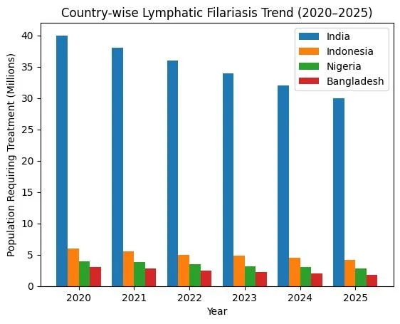
Global epidemiology of LF

Lymphatic filariasis (LF) affects approximately 120 million people across 72 countries globally [19]. In Southeast Asia and India alone, about 45.5 million people are infected [19]. LF is endemic in parts of Africa, Asia, the Pacific and the Americas, including Haiti, the Dominican Republic, Guyana, and Brazil [20].

The disease is mainly caused by Wuchereria bancrofti (widely distributed in tropical and subtropical regions) and Brugia malayi (primarily in South and Southeast Asia) [21–23]. Transmission occurs through mosquito vectors such as Anopheles, Culex, Aedes, and Mansonia species, which thrive in warm, humid climates [24].

LF is classified as a Neglected Tropical Disease (NTD) under the London Declaration (2012) [25]. Control strategies include Mass Drug Administration (MDA), vector control, improved sanitation, and community collaboration to eliminate LF as a public health problem [26–28].

Pathophysiology

Lymphatic Filarisis  

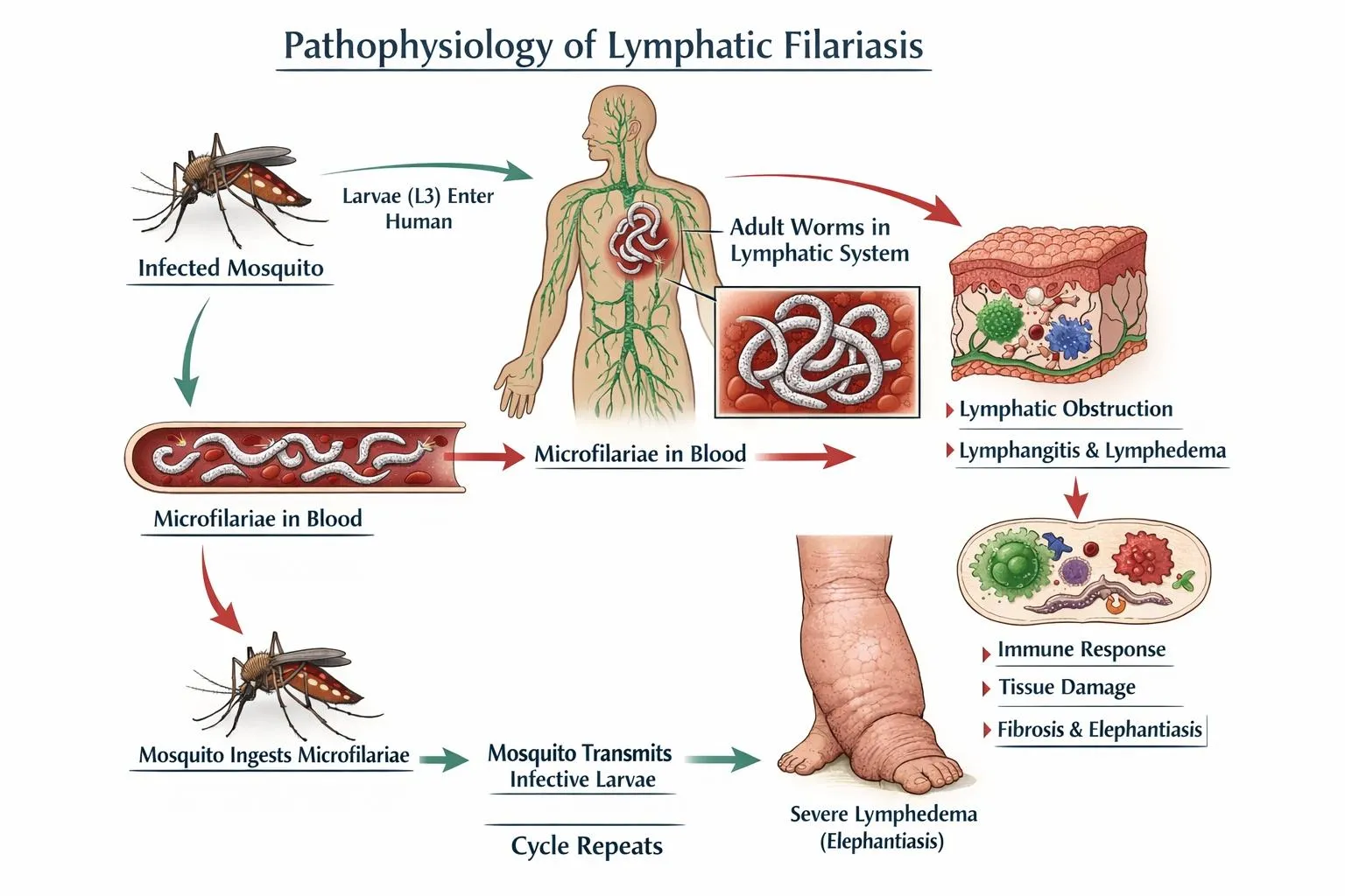
Humans are the only definitive hosts for Wuchereria bancrofti, while Brugia malayi and Brugia timori can also infect domestic and wild animals [29,30]. When an infected mosquito bites a human, it deposits third-stage larvae into the skin. These larvae migrate to the lymphatic system, mature into adults, and reproduce [31]. Female worms release microfilariae with nocturnal periodicity, which circulate in the blood [32]. A mosquito ingests the microfilariae during a subsequent blood meal. Inside the mosquito, microfilariae develop into infectious third-stage larvae, which are transmitted to another human during the mosquito's next feeding [33]. This cycle takes approximately 12 months, with adult worms living up to 15 years and producing microfilariae [34].

Life cycle of LF

The life cycle of lymphatic filariasis (LF) involves a complex interaction between humans and mosquito vectors, facilitating the transmission of the parasitic worms Wuchereria bancrofti, Brugia malayi, and Brugia timori [35,36]. The cycle begins when an infected mosquito bites a human and ingests microfilariae, the immature larvae, present in the person’s bloodstream [35]. These microfilariae are typically active during the night, aligning with the nocturnal feeding habits of mosquito species such as Anopheles, Culex, Aedes, and Mansonia [37].

Within the mosquito, the microfilariae migrate to the midgut, penetrate the midgut wall, and then move to the thoracic muscles, where they develop into first-stage (L1) and second-stage (L2) larvae. Over 10–14 days, these L2 larvae transform into third-stage larvae (L3), the infective form of the parasite, which then migrate to the mosquito’s proboscis [35,38]. When the mosquito bites another human, it deposits the L3 larvae onto the skin, where they enter the body through the bite wound and migrate to the lymphatic system [38].

Inside the lymphatic system, the larvae develop into fourth-stage larvae (L4) and eventually mature into adult worms over several months [35,39]. These adult worms, which can live for 6–8 years, reside in the lymphatic vessels and nodes, causing significant damage and leading to chronic conditions such as lymphedema and hydrocele [39,40]. The adult worms mate and produce thousands of microfilariae daily, which are released into the bloodstream and circulate during the night, ready to be ingested by another mosquito, thereby continuing the cycle [37,40].


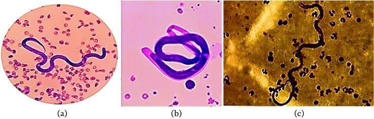
               A. W. bancrofti                                    B. B. malayi                      C. B. timori.

Figure 5. Worms known to cause lymphedema.

Clinical Manifestations of Lymphatic Filarisis

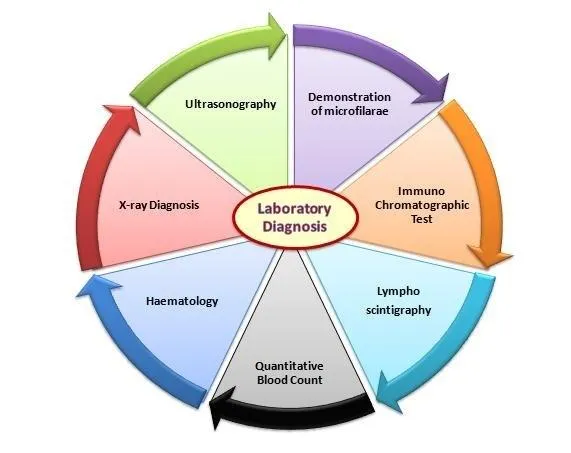
Diagnosis of Lymphatic Filarisis

TREATMENT OF LYMPHATIC FILARIASIS

  1. Confirm Diagnosis
  • Clinical evaluation
  • Night blood smear / Antigen test
  • Ultrasound (filarial dance sign)
  • Antifilarial Drug Therapy
  • Diethylcarbamazine (DEC) – drug of choice
  • Ivermectin
  • Albendazole
  • Combination therapy (DEC + Albendazole)
  • Triple therapy (Ivermectin + DEC + Albendazole) in endemic areas.

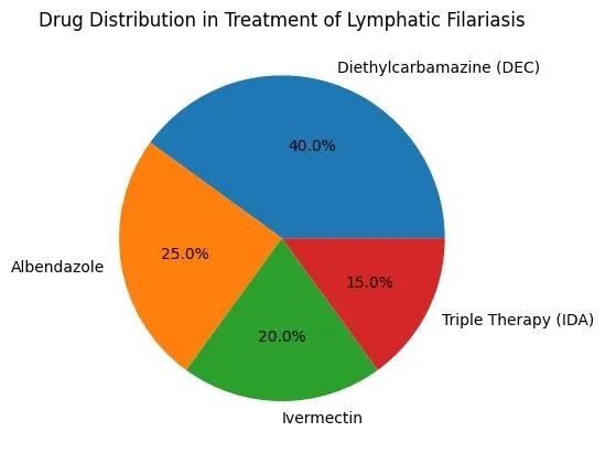
3. Management of Acute Attacks (ADLA)

  • Antibiotics (for secondary bacterial infection)
  • NSAIDs (pain & inflammation)
  • Rest and limb elevation
  • Proper hygiene
  • 4. Management of Chronic Complications
  • Lymphedema care (washing, moisturizing, exercise)
  • Compression bandaging
  • Hydrocele surgery
  • Counseling & community support.

Drug Name

Dose

Duration

Route

Diethylcarbamazine (DEC)

6 mg/kg/day in 3 divided doses

12 days

Oral

Albendazole

400 mg single dose

Single dose (with DEC)

Oral

Ivermectin

150–200 mcg/kg single dose

Single dose

Oral

DEC + Albendazole

DEC 6 mg/kg + Albendazole 400 mg

Single annual dose (MDA program)

Oral

Prevention And Control Of Lymphatic Filariasis

  1. Mass Drug Administration (MDA)
  • Annual single-dose therapy in endemic areas
  • DEC + Albendazole
  • Ivermectin + Albendazole
  • Triple therapy (IDA) where recommended
  • Target: Interrupt transmission

 2. Vector Control (Mosquito Control)

  • Use of insecticide-treated bed nets
  • Indoor residual spraying
  • Elimination of stagnant water
  • Proper sanitation & drainage.

3. Personal Protective Measures

  • Mosquito repellents
  • Wearing full-sleeve clothes
  • Sleeping under bed nets
  • Maintaining personal hygiene.

 4. Morbidity Management & Disability Prevention (MMDP)

  • Limb hygiene & skin care
  • Exercise & elevation of affected limb
  • Treatment of acute attacks
  • Hydrocele surgery

Nationwide MDA Campaign for lymphatic Filarisis Elimination:

The second phase of the Bi-annual Nationwide Mass Drug Administration (MDA) campaign for Lymphatic Filariasis elimination has recently been launched. This phase targets 63 endemic districts in Bihar, Jharkhand, Karnataka, Odisha, Telangana, and Uttar Pradesh. It involves a door-to-door administration of preventive medications in these areas, pushing India closer to achieving its goal of eliminating Lymphatic Filariasis ahead of the global target.

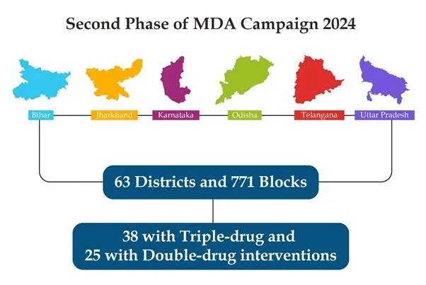
As part of the second phase of the MDA campaign, preventive measures are being implemented across 63 districts (comprising 38 triple-drug and 25 double-drug areas) and 771 blocks in 6 states.

This phase emphasizes not only the distribution of medications but also ensuring their consumption to maximize the campaign's effectiveness and achieve its goals.

Notably, the first phase of the Bi-annual MDA campaign 2024, which covered 96 districts across 11 states, achieved a national coverage rate of 95% of the eligible population.

MDA Campaign

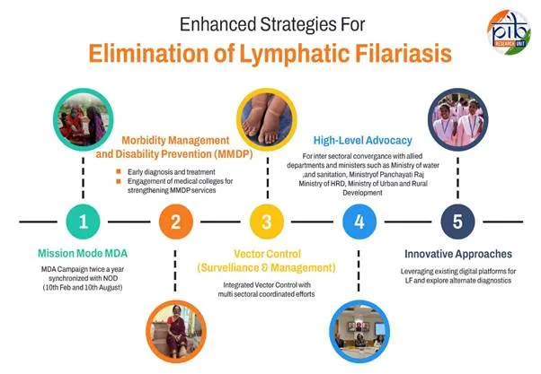
India's efforts to eliminate Lymphatic Filariasis (LF) have made substantial strides with the introduction of the Enhanced Strategy for LF Elimination by 2027. This strategy incorporates a mission-mode Annual Mass Drug Administration (MDA) campaign, held biannually on February 10 and August 10, alongside National Deworming Day (NDD).

India has implemented a comprehensive five-pronged strategy to combat LF, which includes Mission Mode Mass Drug Administration (MDA), Morbidity Management and Disability Prevention (MMDP), Vector Control (Surveillance and Management), High-Level Advocacy, and Innovative Approaches.

Significant Progress in MDA Campaign

  • MDA Coverage: In 2023, MDA campaigns reached 82% coverage nationally across 170 districts in 12 states. The first phase of the 2024 campaign achieved a 95% coverage rate in 96 districts across 11 states.

LF Elimination by 2027: An ambitious target

I In 2023, India’s then Union Minister for Health and Family Welfare, Mansukh Mandaviya, stated that lymphatic filariasis (LF) is not a neglected disease in India but a priority disease targeted for elimination in a time-bound manner (41). The ministry committed to eliminating LF by 2027 through a nationwide Sarva Dawa Sevan (Mass Drug Administration, MDA) campaign, which is three years ahead of the global target set by the World Health Organization (42). During the launch of this initiative, the Additional Secretary and Mission Director (National Health Mission), Roli Singh, highlighted that despite 10–15 rounds of MDA in the past, LF elimination could not be achieved due to suboptimal coverage (43).

The 2027 elimination goal follows a series of missed and extended deadlines, with earlier national targets initially set for 2015 and later extended to 2017 (44,45). At the global level, WHO’s Global Programme to Eliminate Lymphatic Filariasis initially targeted elimination by 2020; however, ongoing challenges have led to a revised global timeline of 2030 (46).

The second phase of the bi-annual nationwide MDA campaign was launched in August, targeting 63 high-risk districts across six states, including Bihar, Jharkhand, Karnataka, Odisha, Telangana, and Uttar Pradesh (47). This included both triple-drug and double-drug districts covering a total of 771 blocks. According to the Ministry of Health and Family Welfare, MDA campaigns achieved 82% national coverage across 170 districts in 2023, while the first phase of 2024 reached a 95% coverage rate in 96 districts across 11 states (47).

Epidemiologically, at least 65% of the total population must receive preventive chemotherapy for effective control of LF. However, even when adult worms are eliminated, complications may persist due to the lack of a fully effective microfilaricidal or adulticidal drug. Ongoing studies are therefore focused on identifying adulticidal agents, which are crucial both for reducing transmission and alleviating disease symptoms in affected patients (46).

India’s familiar challenges and the way ahead

India has over 23 million patients suffering from lifelong disability as a result of lymphatic filariasis (LF) (48), and it inflicts an annual economic loss of about $1 billion in India (49). According to the World Health Organization, indirect losses due to diminished productivity act as a severe drain on local and national economies (50). Acute attacks are estimated to be responsible for losses of $60–85 million per year in India (50).

Earlier, a dedicated budget for filariasis control was not available, and limited human resources resulted in District Malaria Officers managing multiple responsibilities across rural and urban populations. Operational challenges, delayed funding, and interruptions in research activities were also reported, indicating systemic programmatic limitations (51). Previous national plans acknowledged that earlier control efforts had limited impact due to low commitment, implementation gaps, and operational constraints (51).

Experts have emphasized that adequate drug administration coverage and compliance are essential to achieve LF elimination targets. However, lack of awareness among communities regarding the importance of mass drug administration (MDA) leads to gaps in both distribution and consumption (52). Evidence suggests that successful implementation of MDA at the primary healthcare level depends on patient adherence as well as active involvement of healthcare workers in monitoring and motivation (52).

Furthermore, LF is often under-recognized due to its non-fatal nature, leading to low community-level urgency until advanced stages develop. Misconceptions regarding government-provided medication further affect compliance. Programmatic experiences highlight that improvements such as shifting from loose tablets to strip-packaged formulations helped enhance acceptance and adherence within communities (53).”

As part of a branding attempt, a logo and tagline have been developed for the national Filaria Eradication Campaign. The tagline reads “Safe drug, assurance for better health”.

REFERENCES

  1. Ottesen EA. Lymphatic filariasis: treatment, control and elimination. Adv Parasitol. 2006;61:395–441.
  2. Farrar J, Hotez PJ, Junghanss T, Kang G, Lalloo D, White NJ. Lymphatic filariasis. In: Manson’s Tropical Diseases. 23rd ed. Philadelphia: Elsevier; 2014. p. 737–765.
  3. Simonsen PE, Mwakitalu ME. Urban lymphatic filariasis. Parasitol Res. 2013;112(1):35–44.
  4. Ramaiah KD, Ottesen EA. Progress and impact of 13 years of the Global Programme to Eliminate Lymphatic Filariasis. PLoS Negl Trop Dis. 2014;8(10):e3319.
  5. Müller RH, Mäder K, Gohla S. Solid lipid nanoparticles (SLN) for controlled drug delivery – a review of the state of the art. Eur J Pharm Biopharm. 2000;50(1):161–177.
  6. Mehnert W, Mäder K. Solid lipid nanoparticles: production, characterization and applications. Adv Drug Deliv Rev. 2001;47(2–3):165–196.
  7. Pardeike J, Hommoss A, Müller RH. Lipid nanoparticles (SLN, NLC) in pharmaceutical applications. Int J Pharm. 2009;366(1–2):170–184.
  8. Cox FEG. History of human parasitology. Clin Microbiol Rev. 2002;15(4):595–612.
  9. Ramaiah KD, Ottesen EA. Progress and impact of 100 years of lymphatic filariasis control activities. Ann Trop Med Parasitol. 2014;108(5):1–13.
  10. Hoeppli R. Parasitic diseases in ancient Persia and the Near East. J Trop Med Hyg. 1959;62:1–18.
  11. Kiple KF. Disease in ancient Egypt: evidence and interpretation. J Egypt Archaeol. 1993;79:45–58.
  12. Desowitz RS. Who gave Pinta to the Santa Maria? Tropical diseases in a temperate climate. J Trop Med Hyg. 1991;94(5):262–266.
  13. Nutman TB. Insights into the pathogenesis of disease in human lymphatic filariasis. Lymphat Res Biol. 2013;11(3):144–148.
  14. Bockarie MJ, Deb RM. Elimination of lymphatic filariasis: do we have the drugs to complete the job? Curr Opin Infect Dis. 2010;23(6):617–620.
  15. Dreyer G, Norões J, Addiss D. The silent burden of sexual disability associated with lymphatic filariasis. Acta Trop. 1997;63(1):57–60.
  16. Taylor MJ, Hoerauf A, Bockarie M. Lymphatic filariasis and onchocerciasis. Lancet. 2010;376(9747):1175–1185.
  17. Simonsen PE, Fischer PU, Hoerauf A, Weil GJ. The filariases. In: Farrar J, Hotez PJ, Junghanss T, Kang G, Lalloo D, White NJ, editors. Manson’s Tropical Diseases. 23rd ed. Philadelphia: Elsevier; 2014. p. 737–765.
  18. Bockarie MJ, Deb RM. Elimination of lymphatic filariasis: do we have the drugs to complete the job? Curr Opin Infect Dis. 2010;23(6):617–620..
  19. World Health Organization. Lymphatic filariasis: epidemiology. Geneva: WHO; 2023. p. 1–10.
  20. Centers for Disease Control and Prevention (CDC). Lymphatic Filariasis – Epidemiology & Risk Factors. Atlanta: CDC; 2022. p. 1–5.
  21. Simonsen PE, Fischer PU, Hoerauf A, Weil GJ. The filariases. In: Farrar J, Hotez PJ, Junghanss T, et al., editors. Manson’s Tropical Diseases. 23rd ed. Philadelphia: Elsevier; 2014. p. 737–765.
  22. World Health Organization. Global programme to eliminate lymphatic filariasis: progress report. Wkly Epidemiol Rec. 2022;97(41):513–528.
  23. Ottesen EA. Lymphatic filariasis: treatment, control and elimination. Adv Parasitol. 2006;61:395–441.
  24. Gubler DJ. Vector-borne diseases. In: Schaechter M, editor. Encyclopedia of Microbiology. 3rd ed. Elsevier; 2009. p. 549–563.
  25. Uniting to Combat NTDs. London Declaration on Neglected Tropical Diseases. London; 2012. p. 1–12.
  26. World Health Organization. Guideline: Alternative mass drug administration regimens to eliminate lymphatic filariasis. Geneva: WHO; 2017. p. 1–60.
  27. Michael E, Bundy DA. Global mapping of lymphatic filariasis. Parasitol Today. 1997;13(12):472–476.
  28. Ramaiah KD, Ottesen EA. Progress and impact of 13 years of the global programme to eliminate lymphatic filariasis. Infect Dis Poverty. 2014;3:30 (p. 1–7).
  29. Manson’s Tropical Diseases. Farrar J, Hotez PJ, Junghanss T, et al., editors. Philadelphia: Elsevier; 2014. p. 737–765.
  30. World Health Organization. Lymphatic filariasis. Geneva: WHO; 2023. p. 1–12.
  31. Centers for Disease Control and Prevention. Lymphatic Filariasis – Biology. Atlanta: CDC; 2022. p. 1–6.
  32. Ottesen EA. Lymphatic filariasis: treatment, control and elimination. Adv Parasitol. 2006;61:395–441.
  33. Simonsen PE, Fischer PU, Hoerauf A, Weil GJ. Filariases. In: Farrar J, Hotez PJ, Junghanss T, et al., editors. Manson’s Tropical Diseases. 23rd ed. Philadelphia: Elsevier; 2014. p. 737–765.
  34. World Health Organization. Global programme to eliminate lymphatic filariasis: progress report. Wkly Epidemiol Rec. 2022;97(41):513–528.
  35. Simonsen PE, Fischer PU, Hoerauf A, Weil GJ. The filariases. In: Farrar J, Hotez PJ, Junghanss T, Kang G, Lalloo DG, White NJ, editors. Manson’s Tropical Diseases. 23rd ed. Philadelphia: Elsevier; 2014. p. 737–765.
  36. World Health Organization. Lymphatic filariasis. Geneva: WHO; 2023. Available from: https://www.who.int/news-room/fact-sheets/detail/lymphatic-filariasis⁠
  37. Ottesen EA. Lymphatic filariasis: treatment, control and elimination. Adv Parasitol. 2006;61:395–441.
  38. Centers for Disease Control and Prevention. Parasites – Lymphatic Filariasis (Biology). Atlanta: CDC; 2022. Available from: https://www.cdc.gov/parasites/lymphaticfilariasis/biology.html⁠�
  39. Nutman TB. Lymphatic filariasis. In: Goldman L, Schafer AI, editors. Goldman-Cecil Medicine. 26th ed. Philadelphia: Elsevier; 2020. p. 2150–2153.
  40. Ramaiah KD, Ottesen EA. Progress and impact of 13 years of the global programme to eliminate lymphatic filariasis. Infect Dis Poverty. 2014;3:30.
  41. Press Information Bureau (PIB), Government of India. For India, LF is not a neglected disease but a priority disease for elimination in a time-bound manner [Internet]. 2023 [cited 2026 Jun 8]. Available from: https://pib.gov.in/PressReleasePage.aspx?PRID=1890935⁠
  42. World Health Organization. Lymphatic filariasis: reporting continued progress towards elimination as a public health problem [Internet]. 2020 Oct 29 [cited 2026 Jun 8]. Available from: https://www.who.int/news/item/29-10-2020-lymphatic-filariasis-reporting-continued-progress-towards-elimination-as-a-public-health-problem⁠
  43. Press Information Bureau (PIB), Government of India. Launch of Sarva Dawa Sevan (MDA) campaign for elimination of lymphatic filariasis [Internet]. 2023 [cited 2026 Jun 8]. Available from: https://pib.gov.in/PressReleasePage.aspx?PRID=1898092⁠
  44. National Centre for Vector Borne Diseases Control (NCVBDC), Ministry of Health and Family Welfare, Government of India. National programme for elimination of lymphatic filariasis [Internet]. [cited 2026 Jun 8]. Available from: https://ncvbdc.mohfw.gov.in/index4.php?lang=1&level=0&linkid=462&lid=3740⁠
  45. National Centre for Vector Borne Diseases Control (NCVBDC), Ministry of Health and Family Welfare, Government of India. LF elimination programme timeline and updates [Internet].. https://ncvbdc.mohfw.gov.in/index4.php?lang=1&level=0&linkid=461&lid=3739⁠
  46. World Health Organization. Global programme to eliminate lymphatic filariasis [Internet]. Geneva: WHO; [cited 2026 Jun 8]. Available from: https://www.who.int/teams/control-of-neglected-tropical-diseases/lymphatic-filariasis/global-programme-to-eliminate-lymphatic-filariasis⁠
  47. Press Information Bureau (PIB), Government of India. National Mass Drug Administration (MDA) campaign coverage report for lymphatic filariasis elimination [Internet]. 2024 [cited 2026 Jun 8]. Available from: https://pib.gov.in/PressNoteDetails.aspx?NoteId=152023&ModuleId=3&reg=3&lang=1
  48. Sasaoka T, et al. Lymphatic filariasis in India: burden of disease and disability-adjusted life years lost. PLoS Negl Trop Dis [Internet]. 2022 [cited 2026 Jun 8]. Available from: https://pmc.ncbi.nlm.nih.gov/articles/PMC9154644/⁠
  49. National Centre for Vector Borne Diseases Control (NCVBDC), Ministry of Health and Family Welfare, Government of India. Economic burden and control strategies for lymphatic filariasis in India [Internet]. [cited 2026 Jun 8]. Available from: https://ncvbdc.mohfw.gov.in/WriteReadData/l892s/1031567531528881007.pdf
  50. World Health Organization. Lymphatic filariasis: epidemiology and economic impact [Internet]. Geneva: WHO; [cited 2026 Jun 8]. Available from: https://iris.who.int/bitstream/handle/10665/85347/9789241505291_eng.pdf

Reference

  1. Ottesen EA. Lymphatic filariasis: treatment, control and elimination. Adv Parasitol. 2006;61:395–441.
  2. Farrar J, Hotez PJ, Junghanss T, Kang G, Lalloo D, White NJ. Lymphatic filariasis. In: Manson’s Tropical Diseases. 23rd ed. Philadelphia: Elsevier; 2014. p. 737–765.
  3. Simonsen PE, Mwakitalu ME. Urban lymphatic filariasis. Parasitol Res. 2013;112(1):35–44.
  4. Ramaiah KD, Ottesen EA. Progress and impact of 13 years of the Global Programme to Eliminate Lymphatic Filariasis. PLoS Negl Trop Dis. 2014;8(10):e3319.
  5. Müller RH, Mäder K, Gohla S. Solid lipid nanoparticles (SLN) for controlled drug delivery – a review of the state of the art. Eur J Pharm Biopharm. 2000;50(1):161–177.
  6. Mehnert W, Mäder K. Solid lipid nanoparticles: production, characterization and applications. Adv Drug Deliv Rev. 2001;47(2–3):165–196.
  7. Pardeike J, Hommoss A, Müller RH. Lipid nanoparticles (SLN, NLC) in pharmaceutical applications. Int J Pharm. 2009;366(1–2):170–184.
  8. Cox FEG. History of human parasitology. Clin Microbiol Rev. 2002;15(4):595–612.
  9. Ramaiah KD, Ottesen EA. Progress and impact of 100 years of lymphatic filariasis control activities. Ann Trop Med Parasitol. 2014;108(5):1–13.
  10. Hoeppli R. Parasitic diseases in ancient Persia and the Near East. J Trop Med Hyg. 1959;62:1–18.
  11. Kiple KF. Disease in ancient Egypt: evidence and interpretation. J Egypt Archaeol. 1993;79:45–58.
  12. Desowitz RS. Who gave Pinta to the Santa Maria? Tropical diseases in a temperate climate. J Trop Med Hyg. 1991;94(5):262–266.
  13. Nutman TB. Insights into the pathogenesis of disease in human lymphatic filariasis. Lymphat Res Biol. 2013;11(3):144–148.
  14. Bockarie MJ, Deb RM. Elimination of lymphatic filariasis: do we have the drugs to complete the job? Curr Opin Infect Dis. 2010;23(6):617–620.
  15. Dreyer G, Norões J, Addiss D. The silent burden of sexual disability associated with lymphatic filariasis. Acta Trop. 1997;63(1):57–60.
  16. Taylor MJ, Hoerauf A, Bockarie M. Lymphatic filariasis and onchocerciasis. Lancet. 2010;376(9747):1175–1185.
  17. Simonsen PE, Fischer PU, Hoerauf A, Weil GJ. The filariases. In: Farrar J, Hotez PJ, Junghanss T, Kang G, Lalloo D, White NJ, editors. Manson’s Tropical Diseases. 23rd ed. Philadelphia: Elsevier; 2014. p. 737–765.
  18. Bockarie MJ, Deb RM. Elimination of lymphatic filariasis: do we have the drugs to complete the job? Curr Opin Infect Dis. 2010;23(6):617–620..
  19. World Health Organization. Lymphatic filariasis: epidemiology. Geneva: WHO; 2023. p. 1–10.
  20. Centers for Disease Control and Prevention (CDC). Lymphatic Filariasis – Epidemiology & Risk Factors. Atlanta: CDC; 2022. p. 1–5.
  21. Simonsen PE, Fischer PU, Hoerauf A, Weil GJ. The filariases. In: Farrar J, Hotez PJ, Junghanss T, et al., editors. Manson’s Tropical Diseases. 23rd ed. Philadelphia: Elsevier; 2014. p. 737–765.
  22. World Health Organization. Global programme to eliminate lymphatic filariasis: progress report. Wkly Epidemiol Rec. 2022;97(41):513–528.
  23. Ottesen EA. Lymphatic filariasis: treatment, control and elimination. Adv Parasitol. 2006;61:395–441.
  24. Gubler DJ. Vector-borne diseases. In: Schaechter M, editor. Encyclopedia of Microbiology. 3rd ed. Elsevier; 2009. p. 549–563.
  25. Uniting to Combat NTDs. London Declaration on Neglected Tropical Diseases. London; 2012. p. 1–12.
  26. World Health Organization. Guideline: Alternative mass drug administration regimens to eliminate lymphatic filariasis. Geneva: WHO; 2017. p. 1–60.
  27. Michael E, Bundy DA. Global mapping of lymphatic filariasis. Parasitol Today. 1997;13(12):472–476.
  28. Ramaiah KD, Ottesen EA. Progress and impact of 13 years of the global programme to eliminate lymphatic filariasis. Infect Dis Poverty. 2014;3:30 (p. 1–7).
  29. Manson’s Tropical Diseases. Farrar J, Hotez PJ, Junghanss T, et al., editors. Philadelphia: Elsevier; 2014. p. 737–765.
  30. World Health Organization. Lymphatic filariasis. Geneva: WHO; 2023. p. 1–12.
  31. Centers for Disease Control and Prevention. Lymphatic Filariasis – Biology. Atlanta: CDC; 2022. p. 1–6.
  32. Ottesen EA. Lymphatic filariasis: treatment, control and elimination. Adv Parasitol. 2006;61:395–441.
  33. Simonsen PE, Fischer PU, Hoerauf A, Weil GJ. Filariases. In: Farrar J, Hotez PJ, Junghanss T, et al., editors. Manson’s Tropical Diseases. 23rd ed. Philadelphia: Elsevier; 2014. p. 737–765.
  34. World Health Organization. Global programme to eliminate lymphatic filariasis: progress report. Wkly Epidemiol Rec. 2022;97(41):513–528.
  35. Simonsen PE, Fischer PU, Hoerauf A, Weil GJ. The filariases. In: Farrar J, Hotez PJ, Junghanss T, Kang G, Lalloo DG, White NJ, editors. Manson’s Tropical Diseases. 23rd ed. Philadelphia: Elsevier; 2014. p. 737–765.
  36. World Health Organization. Lymphatic filariasis. Geneva: WHO; 2023. Available from: https://www.who.int/news-room/fact-sheets/detail/lymphatic-filariasis?
  37. Ottesen EA. Lymphatic filariasis: treatment, control and elimination. Adv Parasitol. 2006;61:395–441.
  38. Centers for Disease Control and Prevention. Parasites – Lymphatic Filariasis (Biology). Atlanta: CDC; 2022. Available from: https://www.cdc.gov/parasites/lymphaticfilariasis/biology.html??
  39. Nutman TB. Lymphatic filariasis. In: Goldman L, Schafer AI, editors. Goldman-Cecil Medicine. 26th ed. Philadelphia: Elsevier; 2020. p. 2150–2153.
  40. Ramaiah KD, Ottesen EA. Progress and impact of 13 years of the global programme to eliminate lymphatic filariasis. Infect Dis Poverty. 2014;3:30.
  41. Press Information Bureau (PIB), Government of India. For India, LF is not a neglected disease but a priority disease for elimination in a time-bound manner [Internet]. 2023 [cited 2026 Jun 8]. Available from: https://pib.gov.in/PressReleasePage.aspx?PRID=1890935?
  42. World Health Organization. Lymphatic filariasis: reporting continued progress towards elimination as a public health problem [Internet]. 2020 Oct 29 [cited 2026 Jun 8]. Available from: https://www.who.int/news/item/29-10-2020-lymphatic-filariasis-reporting-continued-progress-towards-elimination-as-a-public-health-problem?
  43. Press Information Bureau (PIB), Government of India. Launch of Sarva Dawa Sevan (MDA) campaign for elimination of lymphatic filariasis [Internet]. 2023 [cited 2026 Jun 8]. Available from: https://pib.gov.in/PressReleasePage.aspx?PRID=1898092?
  44. National Centre for Vector Borne Diseases Control (NCVBDC), Ministry of Health and Family Welfare, Government of India. National programme for elimination of lymphatic filariasis [Internet]. [cited 2026 Jun 8]. Available from: https://ncvbdc.mohfw.gov.in/index4.php?lang=1&level=0&linkid=462&lid=3740?
  45. National Centre for Vector Borne Diseases Control (NCVBDC), Ministry of Health and Family Welfare, Government of India. LF elimination programme timeline and updates [Internet].. https://ncvbdc.mohfw.gov.in/index4.php?lang=1&level=0&linkid=461&lid=3739?
  46. World Health Organization. Global programme to eliminate lymphatic filariasis [Internet]. Geneva: WHO; [cited 2026 Jun 8]. Available from: https://www.who.int/teams/control-of-neglected-tropical-diseases/lymphatic-filariasis/global-programme-to-eliminate-lymphatic-filariasis?
  47. Press Information Bureau (PIB), Government of India. National Mass Drug Administration (MDA) campaign coverage report for lymphatic filariasis elimination [Internet]. 2024 [cited 2026 Jun 8]. Available from: https://pib.gov.in/PressNoteDetails.aspx?NoteId=152023&ModuleId=3&reg=3&lang=1
  48. Sasaoka T, et al. Lymphatic filariasis in India: burden of disease and disability-adjusted life years lost. PLoS Negl Trop Dis [Internet]. 2022 [cited 2026 Jun 8]. Available from: https://pmc.ncbi.nlm.nih.gov/articles/PMC9154644/?
  49. National Centre for Vector Borne Diseases Control (NCVBDC), Ministry of Health and Family Welfare, Government of India. Economic burden and control strategies for lymphatic filariasis in India [Internet]. [cited 2026 Jun 8]. Available from: https://ncvbdc.mohfw.gov.in/WriteReadData/l892s/1031567531528881007.pdf
  50. World Health Organization. Lymphatic filariasis: epidemiology and economic impact [Internet]. Geneva: WHO; [cited 2026 Jun 8]. Available from: https://iris.who.int/bitstream/handle/10665/85347/9789241505291_eng.pdf

Photo
Rutuja Shete
Corresponding author

SVPM college of pharmacy Malegaon, Baramati

Photo
Sharmila Shah
Co-author

SVPM college of pharmacy Malegaon, Baramati

Rutuja Shete*, Sharmila Shah, Lymphatic Filariasis: Current Status, Pathogenesis, And Global Elimination Strategies, Int. J. of Pharm. Sci., 2026, Vol 4, Issue 6, 5417-5430. https://doi.org/10.5281/zenodo.20774173

More related articles
Formulation And Evaluation of a Polyherbal Anti-Ac...
Vijay Gadiwan , Ekta Waghmare, Shubham Shirsat , Om Ghuge, Sunil ...
Formulation and Evaluation of Sunscreen For UV Protection ...
Ritik Patel , Shivanand M. Patil, Manisha Rana...
Formulation and Evaluation of Sunscreen For UV Protection ...
Ritik Patel , Shivanand M. Patil, Manisha Rana...
Comparative UV Spectrophotometric Analysis of Different Brands of Paracetamol Ta...
Shivkanya Gadkar, Dr. P. N. Sable, Megha Hange, Anushka Shingade...