

We use cookies to ensure our website works properly and to personalise your experience. Cookies policy
Department of pharmaceutics, Anurag College of Pharmacy, Warthi, Bhandara, Maharashtra.
Microneedle (MN) technology has emerged as a promising transdermal drug delivery platform capable of overcoming the limitations associated with conventional hypodermic injections and topical formulations. Owing to their minimally invasive nature, enhanced patient compliance, and ability to improve drug bioavailability, MNs have attracted significant interest from both academic researchers and the pharmaceutical industry. This review provides a comprehensive overview of microneedle-based drug delivery systems, including their classification into solid, coated, hollow, dissolving, and hydrogel-forming microneedles. Various fabrication approaches and materials employed in microneedle production are discussed, with emphasis on their influence on mechanical strength, drug loading, and release characteristics. Recent applications of microneedles for the delivery of small molecules, peptides, proteins, vaccines, and nucleic acids are highlighted. Furthermore, current challenges related to large-scale manufacturing, regulatory approval, safety, and clinical translation are critically evaluated. Finally, future perspectives and emerging trends are presented, emphasizing the potential of microneedle technology to transform transdermal therapeutics.
Over the past few decades, transdermal drug delivery systems (TDDS) have attracted significant interest as a viable alternative to oral and injectable 'formulations due to their ability to provide sustained drug release, bypass first-pass metabolism, and enhance patient compliance. However, the efficacy of traditional transdermal delivery is greatly hindered by the stratum corneum, the skin's outermost layer, which serves as a highly effective barrier to the penetration of most therapeutic agents.[1] To address this challenge, microneedle (MN) technology has emerged as an innovative and minimally invasive platform for transdermal drug delivery. Microneedles are microscopic, needle-shaped structures measuring 25 to 2000 μm in length that form temporary microchannels through the stratum corneum without stimulating pain receptors or causing significant tissue damage. This distinctive method merges the superior delivery efficiency of hypodermic injections with the convenience and patient-friendly attributes of transdermal patches. Based on their design, microneedles are categorized into solid, coated, hollow, dissolving, and hydrogel-forming systems, each providing unique benefits for delivering diverse therapeutic agents such as small molecules, peptides, proteins, vaccines, and biologics [2].Transdermal drug delivery has developed from old topical treatments to advanced medical devices that allow for controlled and effective delivery of medication through the skin [3].In recent decades, progress in microfabrication and material science has led to the development of microneedle (MN) technologies, marking a significant advancement that addresses the shortcomings of oral and traditional injectable drug delivery methods [4].This technology allows for nearly painless, minimally invasive administration of various drugs and biological agents, which enhances patient adherence and improves treatment results[5] Microneedles (MNs) are composed of arrays of microscopic projections, typically less than 1000 µm in length. Upon application, these projections create temporary microchannels in the stratum corneum, enabling efficient delivery of therapeutic agents either into the skin layers or across the skin for localized or systemic treatment [6].
Microneedles are microscopic solid or hollow needle-like structures designed for minimally invasive drug delivery. They generally range from 50 to 900 µm in length and have an outer diameter of 300 µm or less, enabling them to penetrate the outer layer of the skin while minimizing pain and tissue damage [7].
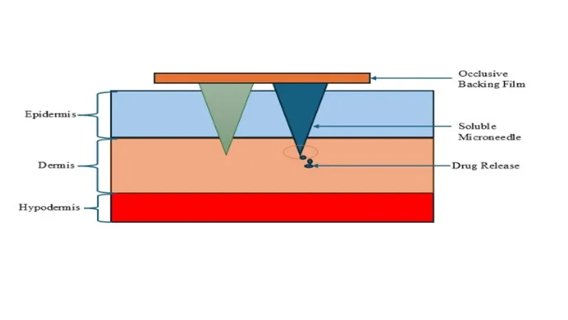
Fig.No.1: - Microneedle Transdermal Drug Delivery System
Table No.1: -Timeline of microneedle development: -
|
Year
|
Researcher(S) |
Development |
Significance |
|
1976 |
Gerstel and place |
Submitted the earliest patent describing the fundamental concept of microneedles for drug delivery. |
Introduced the concept of a novel transdermal drug delivery device. |
|
1996
|
Gross and Kelly |
Development and patented the first hallow microneedle systems. |
Enabled direct drug delivery into the dermal layer of the skin |
|
1997 |
Jang |
Introduced a skin performing device for transdermal drug administration. |
Created microscopic skin channels to enhance drug permeation. |
|
1998 |
- |
Fabricated the first solid silicon microneedle. |
Demonstrated enhanced transdermal delivery of calcine. |
|
2000 |
Zahn |
Designed hollow microneedle for liquid drug injection. |
Improved precision in intradermal drug administration. |
|
2004 |
Cormier |
Developed coated microneedle for transdermal delivery of desmopressin. |
Demonstrated drug-coated microneedles for efficient transdermal administration. |
|
2006 |
Park |
Fabricated dissolving microneedle using PLGA as the matrix material. |
Successfully delivered calcine and bovine serum albumin through the skin. |
|
2012 |
Donnelly |
Introduced hydrogel-forming microneedles. |
Enabled controlled drug diffusion by swelling after insertion without leaving polymer residues in the skin. |
Advantages of Microneedles: -
1)Minimally invasive and painless: Microneedles are extremely small, allowing them to penetrate the outer layers of the skin without reaching deep nerve endings or blood vessels. As a result, they cause little to no pain, minimize bleeding, and provide greater patient comfort compared with conventional hypodermic needles.
2)Improved patient compliance: Because microneedles are less painful and easier to use, they enhance patient acceptance and adherence to treatment, particularly among individuals with needle phobia.
3)Combines the benefits of patches and injections: Microneedle systems integrate the convenience of transdermal patches with the efficient drug delivery capabilities of injectable formulations, offering a simple and effective alternative to traditional administration methods.
4)Enhanced drug bioavailability: Drugs delivered through microneedles bypass the gastrointestinal tract and first-pass liver metabolism, reducing enzymatic degradation and improving systemic absorption, especially for biomolecules such as peptides and proteins.
5)Improved vaccine delivery: Microneedles enable efficient vaccine administration with the potential for dose sparing while eliciting strong immune responses. This approach has shown promise for vaccines against diseases such as influenza, rabies, poliomyelitis, rotavirus, and herpes simplex virus.
6)Stronger immune response: Delivery of vaccines into the skin using microneedles can stimulate both humoral and cellular immune responses more effectively than conventional intramuscular injections, resulting in enhanced protective immunity.
7)Rapid skin recovery: Microneedle application produces only temporary microchannels in the skin. These channels close naturally within a short period after removal, reducing the likelihood of prolonged irritation, infection, or tissue damage.
Disadvantages of Microneedles: -
1)Limited drug loading capacity: Microneedle systems, particularly coated and dissolving types, can deliver only small quantities of drug, making them less suitable for medications that require high therapeutic doses.
2)Mechanical fragility: Some microneedles may not possess sufficient mechanical strength to penetrate the skin effectively. If excessive insertion force is applied, the needles may bend, fracture, or break, potentially affecting drug delivery efficiency.
3)Influence of skin properties: Variations in the viscoelastic characteristics of the skin can alter the depth of microneedle penetration. This effect is more pronounced with shorter or blunt microneedles, leading to inconsistent drug administration.
4)Variability in insertion depth: Differences in skin thickness, elasticity, and mechanical resistance among individuals make it difficult to achieve uniform microneedle penetration and reproducible dosing. Consequently, specialized applicators are often used to ensure reliable insertion.
5)Safety concerns of polymeric materials: In dissolving and biodegradable microneedle systems, the long-term biocompatibility and potential adverse effects of polymer degradation products remaining in the skin require careful evaluation before widespread clinical use [8,9].
Overview: -
Transdermal drug delivery (TDD) is a non-invasive approach in which therapeutic agents are administered through the skin to achieve systemic drug absorption without the need for injections. In this method, a drug-containing formulation is applied to intact skin, allowing the active pharmaceutical ingredient to penetrate the stratum corneum, the primary barrier of the skin, and subsequently diffuse through the epidermal and dermal layers. After reaching the dermis.[2] the drug enters the dermal microvascular network and is transported into the systemic circulation rather than remaining within the skin tissue. Compared with conventional oral and parenteral routes, transdermal delivery offers improved patient comfort by eliminating needle-associated pain and reducing the risk of infection and accidental needle-stick injuries. In addition, the extensive surface area of the skin provides a suitable interface for sustained and controlled drug absorption, making transdermal delivery an attractive option for a wide range of therapeutic applications. Allowing the drug to be released in a controlled and sustained manner over an extended period [10].
Materials Used in Microneedle (MN) Technology: -
Various materials have been employed in the fabrication of microneedles (MNs), each offering distinct advantages and limitations that influence their suitability for transdermal drug delivery applications.
Metal Microneedles: -
Metals are widely used because of their excellent mechanical strength and stiffness, which minimize the risk of needle breakage during skin insertion. They also possess high chemical resistance, making them suitable for repeated or demanding applications. However, metal microneedles may be susceptible to fracture under excessive tensile forces and are generally not biodegradable, which may limit their use in certain biomedical applications [11,12,13].
Ceramic Microneedles: -
Ceramic materials exhibit superior mechanical strength, excellent biocompatibility, and remarkable stability under harsh environmental conditions such as high temperatures and humidity. They provide controlled porosity, facilitate drug loading, and may enhance transdermal drug permeation through electrostatic interactions with the ceramic surface. Despite these advantages, ceramic microneedles are inherently brittle and prone to fracture, requiring careful handling during fabrication and application [14,15,16].
Silica Glass Microneedles: -
Silica glass enables the fabrication of microneedles with a wide variety of sizes and geometries, providing significant design flexibility. Nevertheless, their manufacturing process is complex, time-consuming, and expensive. In addition, concerns regarding biocompatibility, the possibility of needle grooving or fracture, poor thermal tolerance, and the absence of commercially available products have restricted their widespread clinical use [17,18,19].
Carbohydrate Polymer Microneedles: -
Natural carbohydrate polymers such as hyaluronic acid, chitin, chitosan, chondroitin sulfate, cellulose, and starch are increasingly used for dissolving microneedles due to their excellent biocompatibility, biodegradability, low toxicity, and sustainability. These materials offer diverse structural properties and are considered safe and cost-effective for biomedical applications. However, they are susceptible to degradation or contamination during extraction and processing and generally possess lower mechanical strength than metals or ceramics [20,21,22,23].
Polymer Microneedles: -
Synthetic polymers are among the most commonly used materials for microneedle fabrication because they are tougher than glass and ceramics, can be molded into various shapes, and are inexpensive to manufacture. They are highly suitable for large-scale production and disposable applications. However, some polymers exhibit lower mechanical strength than metals, which may affect their skin penetration efficiency depending on the formulation and design [24,25,26].
Mechanism of Microneedle: -
The therapeutic performance of topical drug delivery primarily depends on the diffusion of drug molecules across the skin. In microneedle (MN)-based delivery systems, the stratum corneum is temporarily disrupted, allowing drugs to bypass the skin's major barrier. A typical microneedle patch consists of an array of hundreds to thousands of microscopic needles mounted on a small backing, resembling a conventional transdermal patch. After insertion into the skin, the microneedles create transient microchannels that facilitate drug transport into the epidermal and dermal layers while also permitting the movement of interstitial fluid toward the patch. Hydration of the patch backing by interstitial fluid can further promote drug diffusion and improve delivery efficiency [27,28].
Recent research has focused on polymer-based microneedles designed to provide controlled and sustained drug release instead of rapid drug dissolution. Such systems maintain therapeutic drug concentrations over an extended period, which may reduce dosing frequency, improve patient adherence, and minimize adverse effects. To achieve controlled release, several advanced microneedle designs have been developed, including slow-dissolving, biodegradable, and stimuli-responsive (bioresponsive) microneedles, each employing distinct mechanisms to regulate drug release according to therapeutic requirements [29].
Types of Microneedles: -
1.Solid Microneedles: -
Solid microneedles are commonly fabricated from durable and biocompatible materials, including silicon, stainless steel, titanium, and medical-grade polymers. Their primary function is to create temporary microscopic pores in the stratum corneum, thereby improving the penetration of topically applied drug formulations. This approach, often referred to as the "poke-and-patch" technique, enhances drug transport across the skin while avoiding penetration into deeper tissues where pain receptors are located. Because drug application occurs after microneedle insertion, this method requires two separate steps, which may reduce convenience during self-administration [30].
2. Coated Microneedles: -
Coated microneedles consist of a solid needle coated with a thin layer of drug on its surface. Following insertion into the skin, the coating rapidly dissolves in the interstitial fluid, resulting in quick drug release. These systems are particularly suitable for vaccines, peptides, proteins, and other potent therapeutics that require rapid systemic absorption. However, the limited surface area available for coating restricts the amount of drug that can be loaded onto each microneedle [31].
3. Dissolving Microneedles: -
Dissolving microneedles are manufactured from biodegradable and water-soluble polymers such as carboxymethyl cellulose (CMC) and polyvinylpyrrolidone (PVP). The therapeutic agent is uniformly incorporated within the polymer matrix during fabrication. After insertion into the skin, the microneedles gradually dissolve, releasing the encapsulated drug in a controlled manner. Since the entire structure dissolves after use, no sharp biomedical waste remains, making this technology safer and highly suitable for vaccination and repeated drug administration [30].
4. Hydrogel-Forming Microneedles: -
Hydrogel-forming microneedles are composed of crosslinked hydrophilic polymers that absorb interstitial fluid after insertion and swell to form a hydrogel network. Unlike dissolving microneedles, they do not dissolve but instead create a pathway for drug diffusion from an attached drug reservoir into the skin. Their ability to provide sustained and controlled drug delivery, while accommodating larger drug molecules, makes them promising for long-term therapeutic applications [32].
5. Hollow Microneedles: -
Hollow microneedles are miniature versions of conventional hypodermic needles containing an internal channel through which liquid drug formulations can be delivered directly into the skin. Drug administration is achieved using controlled pressure, allowing accurate dosing and rapid delivery of solutions, suspensions, or biologics. These microneedles are particularly advantageous for high-dose formulations and macromolecular therapeutics. Nevertheless, their widespread application is limited by fabrication complexity, higher production costs, and the possibility of lumen blockage during uses [30,31].
Table No.2: - Types of microneedles (Including their types, material, drug loading method, drug release mechanism, advantages. Limitation, common application)
|
Type |
Material |
Drug Loading Method |
Drug Release Mechanism |
Advantages |
Limitations |
Common Application |
|
Solid
|
Silicon, stainless steel, or medical-grade polymers |
Drug is applied after microneedle insertion |
Drug diffuses through temporary microchannels formed in the skin |
Easy to manufacture, mechanically strong, and highly reproducible |
Limited control over drug release |
Skin pretreatment to improve penetration of topical formulations |
|
Coated |
Metal or silicon |
Drug is coated directly onto the needle surface |
The coating dissolves rapidly after insertion onto the skin |
Delivers drug quickly and is well suited for vaccination |
Limited drug-loading capacity |
accine delivery and administration of potent small molecule drugs |
|
Dissolving |
Biodegradable polymers such as PVP, CMC and hyaluronic acid |
Drug is uniformly incorporated into the polymer matrix |
Drug is released as the polymer gradually dissolves in the skin fluids |
Eliminates sharp waste, biodegradable, and patient friendly |
Lower mechanical strength and restricted drug loading capacity |
Delivery of vaccines, insulin, peptides, proteins, and other biologics |
|
Hollow |
Glass, metal, or polymer |
Liquids drug formulation is filled into the internal channel |
Drug is infused through the hollow bore under applied pressure |
Suitable for large doses and viscous formulations with accurate dosing |
Fabrication is complex and needle blockage may occur |
Delivery of monoclonal antibodies, enzymes, vaccines, and liquid therapeutics |
|
Hydrogel-forming |
Cross-linked hydrogel polymer such as PVA and PEG |
Drug is contained in an external reservoir attached to the microneedle patch |
Swollen hydrogel controls sustained diffusion of the drug |
Enables prolonged and controlled drug release without leaving polymer residues in the skin |
Slower onset of action and may require hydration before use |
Sustained delivery of hormones, biologics, and wearable diagnostic systems |
Fabrication Techniques: -
The performance, mechanical properties, and clinical applicability of microneedles (MNs) are strongly influenced by their fabrication method. Depending on the intended microneedle type such as solid, coated, dissolving, hollow, or hydrogel-forming an appropriate manufacturing technique must be selected to achieve the desired geometry, mechanical strength, drug-loading capacity, reproducibility, and production cost. Advances in microfabrication technologies over the past two decades have enabled the production of highly precise and reproducible microneedle arrays, supporting their transition from laboratory research to commercial biomedical applications [33,34].
A. Photolithography: -
Photolithography is a well-established microfabrication technique widely employed for the production of solid and coated microneedles. The process involves transferring a predefined pattern onto a photosensitive material deposited on silicon or metal substrates, followed by selective etching to create the desired structures. This method provides excellent dimensional accuracy and uniformity, making it highly suitable for research and prototype development. However, its complex processing steps, specialized equipment, and high manufacturing costs limit its large-scale industrial application [34].
B. Micromolding: -
Micromolding is one of the most commonly used techniques for fabricating dissolving and hydrogel-forming microneedles. In this approach, flexible Molds, typically prepared from polydimethylsiloxane (PDMS), are filled with a drug-containing polymer solution. After curing or drying, the formed microneedle arrays are carefully removed from the Mold. This technique offers excellent reproducibility, is cost-effective, and is compatible with biodegradable polymers, making it suitable for both laboratory research and commercial manufacturing [33].
C. Laser Ablation: -
Laser ablation employs a highly focused laser beam to selectively remove material from metal or polymer substrates, thereby creating microneedles with precise geometries. The technique enables rapid design modification and is particularly useful for prototype development because it does not require dedicated Molds or masks. Nevertheless, the high thermal energy generated during processing may produce surface roughness or heat-related defects, often requiring additional finishing procedures to improve surface quality [34].
D. Three-Dimensional (3D) Printing: -
Three-dimensional (3D) printing has significantly expanded the possibilities for microneedle fabrication through layer-by-layer additive manufacturing. This technology allows the production of complex and customized microneedle architectures with minimal material waste. It also supports the fabrication of multi-material systems and the incorporation of functional components such as drug reservoirs or biosensors within the microneedle structure. Although printing resolution differs among available systems, continuous technological improvements are steadily enhancing the precision and quality of printed microneedles [33].
E. Microelectromechanical Systems (MEMS) Fabrication: -
Microelectromechanical systems (MEMS) technology is an advanced microfabrication approach used to manufacture highly accurate and reproducible silicon-based microneedle arrays. The technique is particularly valuable for diagnostic devices and biosensor-integrated microneedles because it enables the integration of mechanical structures with electronic components. MEMS fabrication provides excellent dimensional control, structural reliability, and compatibility with multifunctional biomedical devices, making it an attractive platform for next-generation microneedle systems [32].
Application of microneedle: -
1)Delivery of Low Molecular Weight Drugs: -
Microneedle (MN) technology has been widely explored to improve the transdermal administration of low molecular weight drugs by overcoming the barrier function of the stratum corneum. Arshad and co-workers demonstrated the successful delivery of the hydrophilic drug cetirizine hydrochloride using dissolving microneedle patches prepared from chitosan and sodium alginate through centrifugation and vacuum micromoulding techniques. In vitro permeation studies using rat skin showed approximately a two-fold increase in drug permeation compared with patches lacking microneedles. Histological analysis further confirmed temporary disruption of the stratum corneum, indicating the effectiveness of the microneedle system in enhancing drug transport across the skin [35].
Similarly, enhanced transdermal delivery of lornoxicam was achieved using a cellulosic microsponge gel applied after microneedle pretreatment. In this approach, the skin was micropunctured with a dermaroller before application of the gel containing the permeation enhancer Transcutol® P. Evaluation in a rat paw oedema model demonstrated significantly greater drug permeation through microneedle-treated skin than untreated skin, resulting in approximately 72% reduction in inflammation within four hours [36].
2)Protein and Peptide Delivery: -
Microneedle (MN) technology has shown significant potential for the transdermal delivery of therapeutic proteins and peptides, particularly for localized treatment with minimal systemic exposure. One important application involves the targeted delivery of peptides that modulate calcitonin gene-related peptide (CGRP), a neuropeptide associated with the development and maintenance of neuropathic pain.
Xie et al. developed a dissolving microneedle patch using sodium carboxymethyl cellulose (SCMC) as the matrix material and incorporated CGRP8–37, a peptide antagonist of CGRP. The therapeutic performance of the formulation was evaluated in rat models of diabetes-induced neuropathy, peripheral nerve injury, and neuropathic pain. The microneedle patch produced localized analgesic effects while preserving normal pain sensation, demonstrating its potential as a targeted treatment strategy for neuropathic pain [37].
In another study, Liu et al. employed polyethylene glycol diacrylate (PEGDA) as the microneedle matrix to investigate peptide/protein delivery, further highlighting the versatility of microneedle platforms for the controlled transdermal administration of biomacromolecules [38].
3) Delivery of Vaccines: -
Microneedle (MN)-based systems have gained considerable attention as an alternative approach for vaccine administration because they enable minimally invasive, painless, and efficient delivery through the skin. Unlike conventional injectable vaccines, which are generally administered by hypodermic needles and may reduce patient acceptance, microneedles offer improved compliance and the possibility of self-administration. These advantages make them particularly valuable for large-scale vaccination campaigns and emergency situations such as the COVID-19 pandemic, especially in resource-limited settings [39].
Arshad et al. demonstrated successful transdermal delivery of the Bacillus Calmette–Guérin (BCG) vaccine using dissolving sodium alginate-based microneedle patches. Immunization studies in rats showed elevated IgG antibody levels after nine weeks, confirming that the microneedle system effectively elicited an immune response [40].
In another investigation, microneedle patches prepared from chitosan, polyvinyl alcohol (PVA), and polyvinylpyrrolidone (PVP) were used to deliver ovalbumin as a model antigen. The formulation provided sustained antigen release for approximately 28 days and maintained high antibody levels in immunized rats for up to 18 weeks. Compared with administration of free antigen or chitosan solution, the ovalbumin-loaded microneedle patches produced a stronger and more prolonged immune response, highlighting the potential of microneedle technology for enhanced vaccine delivery [41].
4) Cancer Therapy: -
Microneedle-based drug delivery systems have emerged as a promising platform for localized combination chemotherapy. One innovative approach involved the development of a three-dimensional, Christmas tree-shaped microneedle array designed to deliver the multi-drug chemotherapy regimen FOLFIRINOX, which consists of fluorouracil, leucovorin, irinotecan, and oxaliplatin. The microneedle arrays were produced using 3D printing technology, with oxaliplatin and leucovorin incorporated into the upper layer, while fluorouracil and irinotecan were loaded into the lower layer. This layered configuration enabled controlled, site-specific release of multiple anticancer agents from a single device. The fabricated microneedles also exhibited strong skin adhesion, demonstrating their potential as an effective transdermal platform for combination therapy in the treatment of pancreatic cancer [42].
Current Challenges in Large-Scale Manufacturing of Microneedle-Based Transdermal Drug Delivery Systems: -
1. Consistent Manufacturing Quality: -
2. Limited Scalability of Fabrication Techniques: -
3. Uniform Drug Loading
4. Material Selection
5. Sterility and Sterilization
Safety Considerations and Clinical Translation Challenges of Microneedle-Based Transdermal Drug Delivery Systems: -
The advancement of microneedle (MN)-based transdermal drug delivery systems has opened new possibilities for painless, self-administered, and patient-friendly drug administration. Despite remarkable progress in laboratory and preclinical investigations, successful clinical translation requires comprehensive evaluation of safety, reliability, regulatory compliance, and long-term therapeutic performance. Several critical factors continue to influence the transition of microneedle technologies from experimental platforms to clinically approved products [46,47].
1. Skin Compatibility and Local Tissue Response: -
Since microneedles overcome the skin barrier by creating temporary microchannels within the stratum corneum, evaluation of skin safety is a fundamental requirement. Short-term effects such as mild redness, swelling, itching, or localized irritation have been reported following application; however, these responses are generally reversible and disappear within a short duration. For repeated administration, further studies are required to assess possible long-term effects, including chronic inflammation, sensitization, and alterations in skin integrity [46,48].
2 Biomaterial Safety and Biocompatibility; -
The selection of appropriate microneedle materials plays a crucial role in ensuring patient safety. Various materials, including silicon, metals, ceramics, and biodegradable polymers, have been investigated for microneedle fabrication. Among these, polymeric microneedles have gained considerable attention due to their biodegradability, reduced risk of sharp waste generation, and suitability for controlled drug release. Nevertheless, comprehensive evaluation of material toxicity, degradation products, and immune responses is essential before clinical application [47,49].
3. Mechanical Stability and Application Reliability: -
For effective and safe drug delivery, microneedles must possess sufficient mechanical strength to penetrate the skin without bending, breaking, or losing structural integrity. Mechanical failure may result in incomplete drug administration or retention of fragments within the skin, which could compromise patient safety. Therefore, optimization of needle geometry, polymer composition, fabrication processes, and mechanical testing methods is necessary to ensure consistent clinical performance [46,48].
4. Sterility and Risk of Microbial Infection: -
Although microneedle-induced microchannels typically close rapidly after application, temporary disruption of the skin barrier may create a potential pathway for microbial entry. Maintaining sterility throughout manufacturing, storage, and administration is therefore essential. The development of suitable sterilization techniques that preserve both drug stability and microneedle structure remains an important challenge, particularly for sensitive biological molecules such as vaccines, proteins, and peptides [47,50].
5. Limited Clinical Evidence and Long-Term Safety Assessment: -
Although numerous microneedle systems have demonstrated promising results in animal studies and early clinical investigations, extensive human trials are still limited for many applications. Large-scale, well-controlled clinical studies are required to establish long-term safety, therapeutic effectiveness, dose consistency, and performance among diverse patient populations before widespread clinical adoption [46,48].
6. Regulatory Pathway and Product Approval Challenges: -
The regulatory approval of microneedle-based products remains complex because these systems often represent a combination of pharmaceutical formulations and medical devices. Differences in regulatory classification between countries create challenges related to quality requirements, manufacturing standards, clinical evaluation, and approval procedures. Establishing internationally accepted regulatory frameworks will be essential to accelerate commercialization of microneedle technologies [47,48].
7. Patient Acceptance and Practical Usability: -
One of the major advantages of microneedle technology is improved patient acceptance compared with conventional injections. Reduced pain, minimal invasiveness, and potential for self-administration make microneedle patches attractive for chronic therapies and vaccination programs. However, variations in skin properties, application technique, and user handling may influence drug delivery efficiency. Development of simple application devices and standardized administration procedures will improve reliability in clinical settings [46,49].
8. Economic and Commercial Considerations: -
The transition of microneedle systems into healthcare practice also depends on economic feasibility. High costs associated with precision manufacturing, sterilization, packaging, and quality control may limit large-scale adoption. Advances in scalable manufacturing technologies and cost-efficient production strategies are required to make microneedle-based products commercially competitive with conventional drug delivery approaches [47,48].
Future Perspectives and Emerging Trends: -
Microneedle (MN) technology is anticipated to progress from a conventional transdermal drug delivery platform to an integrated system capable of drug administration, biomarker sensing, and real-time health monitoring. The convergence of microneedles with wearable biosensors and digital healthcare technologies is expected to facilitate continuous analysis of interstitial fluid biomarkers, enabling early disease detection, personalized therapy, and point-of-care diagnostics [51,52].
Recent research has highlighted the growing interest in stimuli-responsive microneedles that can modulate drug release in response to physiological cues such as glucose levels, pH, temperature, enzyme activity, or inflammatory mediators. These smart systems have the potential to enhance therapeutic precision while minimizing systemic adverse effects, making them particularly promising for the treatment of diabetes, cancer, and chronic inflammatory disorders [51].
The incorporation of artificial intelligence (AI) and machine learning (ML) into microneedle technology is emerging as an important area of research. AI-based computational tools can support the optimization of microneedle geometry, material selection, drug-loading capacity, and release behavior. Furthermore, AI-driven analysis of biosensor data may enable automated decision-making and personalized, closed-loop drug delivery systems [53,54].
Another significant trend is the development of microelectronic and wearable microneedle platforms that integrate sensing components, wireless communication, and controlled drug delivery within a single device. Such multifunctional systems enable continuous physiological monitoring, remote patient management, and improved treatment adherence, thereby supporting next-generation digital healthcare [55,56].
Although remarkable progress has been achieved, several obstacles continue to limit widespread clinical application. Key challenges include large-scale and cost-effective manufacturing, reproducible fabrication, sterilization, long-term mechanical and biological safety, quality assurance, regulatory approval, and commercialization. Addressing these issues will require coordinated efforts among researchers, clinicians, industry partners, and regulatory authorities to ensure successful clinical translation [51,57].
Overall, continuous advances in biomaterials, nanotechnology, wearable electronics, biosensing, and artificial intelligence are expected to expand the capabilities of microneedle systems beyond drug delivery. These innovations are likely to play a crucial role in personalized medicine, disease diagnosis, vaccination, and precision therapeutics in the coming years [51,53].
CONCLUSION
Microneedle technology has emerged as one of the most promising approaches for transdermal drug delivery by overcoming the limitations of conventional oral, topical, and injectable dosage forms. Its minimally invasive nature, improved patient compliance, and ability to deliver a wide range of therapeutic agents, including small molecules, peptides, proteins, vaccines, and nucleic acids, make it an attractive platform for modern healthcare. Continuous advancements in biomaterials, fabrication techniques, and drug-loading strategies have significantly enhanced the safety, efficiency, and versatility of microneedle systems.espite these advantages, several challenges remain before microneedle-based products can achieve widespread clinical use. Large-scale manufacturing, cost-effective production, sterilization, long-term safety, regulatory approval, and quality standardization continue to be major barriers to commercialization. Addressing these challenges will require close collaboration among researchers, clinicians, manufacturers, and regulatory authorities.Overall, the future of microneedle technology extends beyond drug delivery alone. The integration of microneedles with nanotechnology, wearable biosensors, artificial intelligence, and personalized medicine is expected to transform disease diagnosis, continuous health monitoring, and precision therapy. With ongoing scientific and technological progress, microneedle-based systems have the potential to become an important component of next-generation healthcare and significantly improve patient outcomes.
REFERENCES
DOI: 10.1007/s00604-024-06458-2.
DOI: 10.1007/s00604-024-06458-2.




Jatin Rahangdale, Dinesh Bawankar, Ankita Yadav, Dr. Sachin Lohe, Microneedle-Based Transdermal Drug Delivery Systems: Current Challenges and Future Perspectives: A Review, Int. J. of Pharm. Sci., 2026, Vol 4, Issue 8, 3784-3800, https://doi.org/10.5281/zenodo.22076213
 10.5281/zenodo.22076213