

We use cookies to ensure our website works properly and to personalise your experience. Cookies policy
Department of Chemistry, University College of Engineering (Autonomous), Jawaharlal Nehru Technological University, Kakinada, 533003, Andhra Pradesh, India.
A new series of aryl amide derivatives of Imidazo[1,5-a]pyridine-1,2,4-thiadiazoles 1a-j were designed and screening the molecular docking simulations against EGFR target. The derivative 1f showed binding energy against EGFR with -8.5 kcal/mol
The nitrogenous fused hetero-aromatic scaffolds are found broadly in biological and natural products motifs, these are considered as significant core blocks in medicinal and material field.1-3 In particularly, imidazo[1,5-a]pyridine are most unique class of nitrogen atom having fused bicyclic heterocyclic molecules and are played a vital role in synthetic and medicinal chemistry.4-6 They possessed a broad spectrum of biological activities including antibacterial,7 thromboxane synthetase inhibitors,8 inhibitors of aromatase,9 antiviral,10 positive inotropic agents,11 anti-tuberculosis,12 antifungal,13 antiprotozoal,14 anti-inflammatory,15 AChE and BChE inhibition,16 anticandidosic activity,17 anti-plasmodial,18 protein-kinase inhibitor,19 and vascular endothelial growth factor (VEGF)-receptor20 activities.
RESULTS AND DISCUSSION:
Based on the above literature, we designed the following aryl amide derivatives of Imidazo [1,5-a]pyridine-1,2,4-thiadiazoles.
Figure 1: Structure of aryl amide derivatives of Imidazo[1,5-a]pyridine-1,2,4-thiadiazoles
In this current docking study, we focused on key cancer-related proteins that are implicated in the progression of various cancer types. We chose EGFR, for molecular docking due to their pivotal roles in cancer pathology including tumor growth, metastasis, and angiogenesis. This protein are well-established therapeutic targets in cancer treatment, making them ideal candidates for exploring potential therapeutic compounds.
Binidng energy and interaction summary of compounds with human EGFR TKD
Molecular interaction summary of top compounds with Human EGFR TKD was shown in table 1. The 2D NMR structures of all compounds were shown in figure 2. Protein ligand complexes representing the binding modes of 1a, 1b, 1c, 1f, 1j compounds and and Erlotinib and interacting residues in the active site in human human EGFR TKD B) Pharmacophore features of compounds along with Erlotinib were shown in figure 3.
Table 1. Molecular interaction summary of top compounds with Human EGFR TKD
|
Compounds |
Binding Energy (K.cal/mol) |
Interacting Amino acids |
Nature of interactions |
|
1a |
-7.8 |
THR830, PRO770, LYS721, MET742, LEU694, LEU820, ALA719, LEU820, VAL702, CYS773, ASP776, PHE771, THR766, ILE720, ILE765, LEU764, LEU753, LEU738, ASP831, MET769, LEU768, HIS781, TYR777, GLU780, GLY772, LYS704 |
H-bond, π-sigma, π-sulfur, alkyl, π-alkyl, π-cation, π-donor hydrogen bond, carbon hydrogen bond, van der waals
|
|
1b |
-8 |
MET769, PRO770, GLU780, ASP831, LEU694, LEU820, VAL702, LYS721, AAL719, PHE771, HIS781, TYR777, ASP776, CYS773, GLY772, LYS704, PHE699, THR830, GLN767, LEU768 |
H-bond, π-anion, π-sigma, π-alkyl, carbon hydrogen bond, van der waal s
|
|
1c |
-8 |
THR830, MET742, LYS721, LEU820, LEU694, MET769, PRO770, VAL702, ALA719, LEU694, THR766, ILE720, ILE765, LEU753, GLU738, ASP831, PHE771, LEU768, LYS692, LYS704, GLY772, CYS773 |
H-bond, π-cation, π-donor hydrogen bond, π-sigma, π-sulfur, π-alkyl, carbon hydrogen bond, van der waals |
|
1f |
-8.5 |
THR830, PRO770, MET742, LEU694, LEU820, ALA719, LYS721, VAL702, THR766, PHE771, HIS781, TYR777, GLU780, CYS773, GLY772, ILE720, ILE765, LEU764, LEU753, GLU738, ASP831, MET769, LEU768 |
H-bond, π-sigma, π-sulfur, π-alkyl, π-donor hydrogen bond, van der waals |
|
1j |
-8.3 |
THR830, PRO770, LEU820, PHE771, VAL702, THR766, ALA719, TYR777, LEU694, HIS781, MET769, ASP831, GLU738, LEU753, LEU764, ILE765, ILE720, LYS704, GLU780, ASP776, GLY772, CYS773, LEU768 |
H-bond, π-sigma, π-sulfur, amide- π stacked, π-alkyl, π-donor hydrogen bond, π-cation, carbon hydrogen bond, van der waals |
|
Erlotinib |
-6.9 |
MET769, LEU820, LEU694, ALA719, LEU764, LYS721, GLN767, THR830, THR766, MET742, GLU738, ASP831, VAL702, GLY695, PRO770, PHE771, GLY772, LEU768 |
H-bond, alkyl, π-alkyl, π-sigma, π-donor hydrogen bond, carbon hydrogen bond, van der waals
|
H-bond forming residues coloured in green
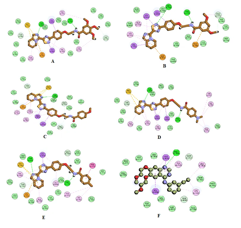
Figure 2: 2D molecular representation of interactions of compounds A) 1a, B) 1b, C) 1c, D) 1f, E) 1j F) Erlotinib with the active site residues of the EGFR. Interactions were displayed as color coded dashed lines, green lines indicated the H–bonds.
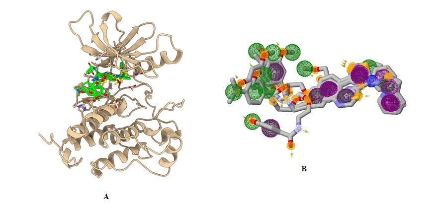
Figure 3: Protein ligand complexes representing the binding modes of 1a, 1b, 1c, 1f, 1j compounds and and Erlotinib and interacting residues in the active site in human human EGFR TKD B) Pharmacophore features of compounds along with Erlotinib (Pharmacophore features color coding include: Purple spheres-Aromatic, Green spheres-Hydrophobic, Orange spheres-HBD and White spheres-HBA).
Molecular interaction profile of compounds with EGFR TKD
Erlotinib forms a hydrogen bond with MET769 through the nitrogen atom of its quinazoline ring. LEU694 and LEU820 engage in pi-sigma interactions with the central scaffold, while ALA719 and LYS721 are involved in pi-alkyl interactions. Additionally, the alkynyl group on the phenyl ring forms alkyl interactions with LEU764, and LEU820 and MET769 also participate in alkyl interactions. THR830 and GLN767 contribute to the interaction profile with carbon-hydrogen bonds and pi-donor hydrogen bonds.
Compound 1a displayed two hydrogen bond interactions: one between the imidazole nitrogen and THR830, and another involving the amide nitrogen with PRO770. The imidazopyridine ring formed a π-sulfur interaction with MET742 and a π-cation interaction with LYS721. Additionally, LEU820 exhibited π-σ interactions with the thiadiazine ring, while LEU694 engaged in π-σ interactions with the phenoxy ring. ALA719 and CYS773 showed π-alkyl interactions, and VAL702 and PHE771 participated in alkyl interactions. Furthermore, THR766 formed a π-donor hydrogen bond interaction, and ASP776 established a carbon-hydrogen bond interaction.
Compound 1b exhibited two hydrogen bond interactions: one between the amide nitrogen and PRO770, and another involving the thiadiazine ring nitrogen with MET769. The dimethoxy phenyl ring showed π-anion interactions with GLU780, while the imidazopyridine ring engaged in π-cation interactions with ASP831. Additionally, LEU820 formed a π-σ interaction with the thiadiazine ring, and ALA719 displayed π-alkyl interactions with it. VAL702 showed both π-σ and π-alkyl interactions with the imidazopyridine ring, and LEU694 contributed π-σ interactions with the phenoxy ring. Finally, PHE771 exhibited a carbon-hydrogen bond interaction.
Compound 1c had a hydrogen bond interaction with THR830 via the imidazopyridine ring. The imidazopyridine ring also engaged in π-sulfur interactions with MET742 and π-cation interactions with LYS721. Additionally, LEU820 and LEU694 displayed π-σ interactions, while MET769 and PRO770 contributed carbon-hydrogen bond interactions. ALA719, VAL702, and LEU764 showed π-alkyl interactions, and THR766 formed π-donor hydrogen bond interactions.
Compound 1f displayed a hydrogen bond interaction profile similar to compound 19a, with interactions involving THR830 and PRO770 residues. MET742 engaged in π-sulfur interactions, while LEU820 and LEU694 formed π-σ interactions. ALA719, LYS721, and VAL702 showed π-alkyl interactions, and PHE771, HIS781, and TYR777 participated in alkyl interactions with the dimethylamino groups. Additionally, THR766 formed π-donor hydrogen bond interactions.
Compound 1j exhibited a hydrogen bond and hydrophobic interaction profile similar to other compounds but featured a distinct π-cation interaction with LYS721. PHE771 formed an amide-π stacked interaction, MET769 contributed a carbon-hydrogen bond interaction, and LEU694 showed π-alkyl interactions with the phenoxy ring in the scaffold structure.
The residues involved in hydrogen bond interactions with these compounds, such as LYS721 and THR830, also participate in critical interactions with Erlotinib. Compound 1b, in particular, demonstrates a key hydrogen bond interaction with MET769, similar to Erlotinib. Additionally, LEU694 and LEU820 exhibit π-σ interactions with most of the compounds, including Erlotinib. These common interaction profiles suggest that the compounds have the potential to modulate the target in a manner similar to Erlotinib, contributing to their promising anti-cancer activity.
Determination of ADMET profile, Lipinski rule, and pharmacokinetics
ADMET properties (absorption, distribution, metabolism, excretion, and toxicity) are critical for further sucessful progression in the drug discovery were shown in table 2.
Table 2: Physico-chemical properties and drug-likeness prediction of compounds with better binding energy and interaction profile using SWISS ADME
|
Parameters |
1a |
1b |
1c |
1f |
1j |
|
Molecular Weight (g/mol) |
531.58 |
501.56 |
471.53 |
484.57 |
455.53 |
|
Log P o/w |
3.83 |
3.90 |
3.89 |
3.94 |
4.28 |
|
No. of. H-bond Donors |
1 |
1 |
1 |
1 |
1 |
|
No. of H-bond Acceptors |
8 |
7 |
6 |
5 |
5 |
|
Solubility |
Poor |
Poor |
Poor |
Poor |
Poor |
|
TPSA(Å2) |
137.34 |
128.11 |
118.88 |
112.89 |
109.65 |
|
GI absorption |
Low |
Low |
High |
High |
High |
|
BBB permeation |
No |
No |
No |
No |
No |
|
P-gp substrate |
Yes |
Yes |
Yes |
Yes |
Yes |
|
Drug likeness (Lipinski) |
Yes; 1 violation MW>500 |
Yes; 1 violation MW>500 |
Yes |
Yes |
Yes |
|
CYP450 isoforms inhibition |
CYP2C9, CYP2C19, CYP2D6 CYP3A4 |
CYP2C9, CYP2C19, CYP2D6 CYP3A4 |
CYP2C9, CYP2C19, CYP2D6 CYP3A4 |
CYP2C9, CYP2C19, CYP2D6 CYP3A4 |
CYP2C9, CYP2D6 CYP3A4 |
|
Bioavailability score |
0.55 |
0.55 |
0.55 |
0.55 |
0.55 |
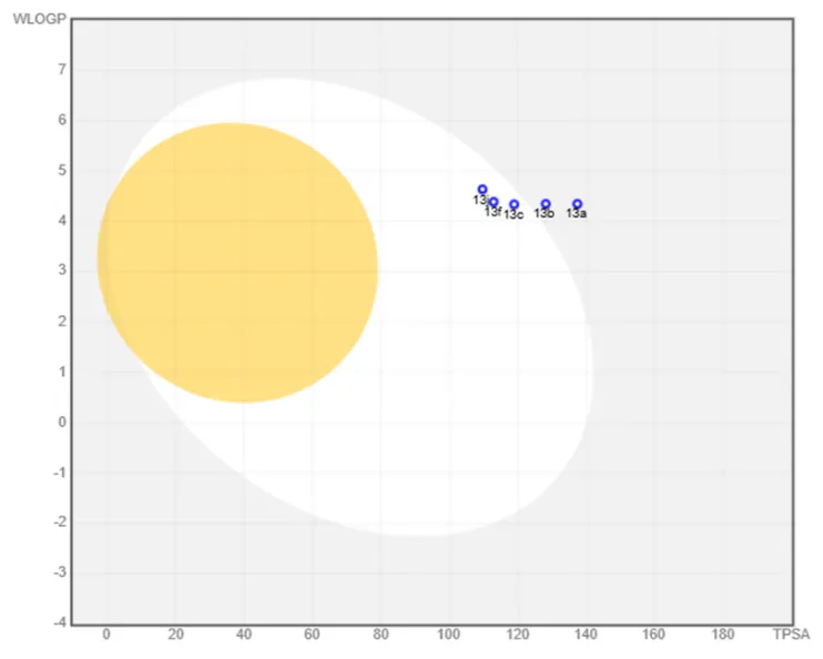
Figure 4: The Brain Or Intestinal Estimated Permeation (BOILED-Egg) method illustrates the absorption, blood-brain barrier (BBB) permeation, and substrate selectivity for P-glycoprotein (PGP) of compounds 1a, 1b, 1c, 1f and 1j. Blue indicates substrate of P-glycoprotein (PGP), while red represents non-substrate of PGP.
The ADME predictions highlight the need for optimization of the compounds, particularly due to the significant challenge posed by the poor solubility of all the compounds, along with the low gastrointestinal absorption observed for compounds 1a and 1b.
Compounds 1c, 1f, and 1j demonstrate high gastrointestinal (GI) absorption, while all compounds were predicted not to cross the blood-brain barrier (BBB). According to the BOILED-Egg model in Figure 4, all the compounds are identified as substrates of P-glycoprotein (P-gp). This characteristic may affect their bioavailability and distribution, as P-gp actively transports substances out of cells, potentially reducing their intracellular concentrations.
Compounds 1c, 1f, and 1j adhere to Lipinski's Rule of Five, while compounds 1a and 1b slightly deviate due to their molecular weight exceeding 500. Additionally, these compounds are predicted to inhibit several CYP isoforms, including CYP2C19, CYP2C9, CYP3A4, and CYP2D6.
Toxicity assessment using the pkCSM webserver predicts that all the compounds under investigation are hERG II inhibitors, but they do not show signs of hepatotoxicity, mutagenicity, or skin sensitization. hERG II inhibition suggests a potential risk of cardiac issues, such as QT prolongation and arrhythmias. To mitigate these risks, it is crucial to optimize the compounds to enhance their therapeutic efficacy and safety profile.
CONCLUSION
In conclusion, these compounds also demonstrated stronger binding affinities with the EGFR target compared to the co-crystallized inhibitor Erlotinib, which has a binding energy of -6.9 kcal/mol. All compounds exhibited higher binding energies in the range of -7.8 to -8.5 kcal/mol, with compound 19f showing the highest affinity at -8.5 kcal/mol, followed closely by compound 19j at -8.3 kcal/mol.
REFERENCES
https://doi.org/10.3390/molecules24213839
imidazo[1,2-a]pyrimidine derivatives. Eur J Med Chem 26:13-18. https://doi.org/10.1016/0223-5234(91)90208-5
Veikkola T, Karkkainen M, Claesson-Welsh L, Alitalo K (2000) Regulation of angiogenesis via vascular endothelial growth factor receptors. Cancer Res 60(2):203-212.https://pubmed.ncbi.nlm.nih.gov/10667560
https://doi.org/10.3390/molecules24213839
imidazo[1,2-a]pyrimidine derivatives. Eur J Med Chem 26:13-18. https://doi.org/10.1016/0223-5234(91)90208-5

Paila Suresh, Gadupudi Purna Chandra Rao, Molecular docking simulations of various aryl amide derivatives of Imidazo[1,5-a]pyridine-1,2,4-thiadiazoles against EGFR protein, Int. J. of Pharm. Sci., 2026, Vol 4, Issue 7, 972-880, https://doi.org/10.5281/zenodo.21186004
 10.5281/zenodo.21186004