

We use cookies to ensure our website works properly and to personalise your experience. Cookies policy
Department of Pharmaceutics, Karnataka College of Pharmacy.
Impetigo is a highly contagious superficial bacterial skin infection caused primarily by Staphylococcus aureus and Streptococcus pyogenes, and it remains one of the most common dermatological infections in children worldwide. Topical antibiotics such as mupirocin, fusidic acid, retapamulin, and ozenoxacin are widely used, but their clinical effectiveness is often constrained by poor skin penetration, short residence time at the infection site, frequent dosing requirements, and the steady emergence of antimicrobial resistance. These limitations have driven interest in advanced topical delivery systems capable of improving local drug bioavailability and therapeutic outcomes.Among the nanotechnology-based approaches under investigation, nano-emulgels have emerged as a particularly promising carrier system. By combining the superior skin-permeation characteristics of nanoemulsions with the favourable retention, spreadability, and patient acceptability of gel systems, nano-emulgels address several of the practical shortcomings of liquid nanoemulsions on their own.This review provides a comprehensive overview of impetigo, covering its epidemiology, pathogenesis, clinical presentation, diagnosis, and current treatment strategies, before turning to the design principles, formulation components, preparation methods, characterisation techniques, and evaluation parameters that define nano-emulgel technology. Recent advances in antibiotic-loaded and herbal nano-emulgel formulations, along with relevant patents, are discussed in relation to their potential to overcome the limitations of conventional therapy. The review also considers the practical challenges of large-scale manufacturing, formulation stability, and regulatory approval, and closes with a view toward future research directions.Taken together, the evidence indicates that nano-emulgel technology represents a credible strategy for improving topical antibiotic delivery and enhancing therapeutic efficacy in impetigo management, while minimising systemic exposure and adverse effects.
Impetigo is a highly contagious bacterial skin infection that mainly affects infants and young children, though adults living in humid or overcrowded conditions can also be affected. It is most often caused by Staphylococcus aureus and Streptococcus pyogenes, with a growing involvement of methicillin-resistant Staphylococcus aureus (MRSA) strains. Clinically, impetigo is characterised by erythematous lesions, vesicles, pustules, and honey-coloured crusts; if left untreated, it may lead to complications such as cellulitis and post-streptococcal glomerulonephritis.1,2
Conventional management relies mainly on topical antibiotics — mupirocin, fusidic acid, retapamulin, and ozenoxacin — while systemic antibiotics are reserved for severe or widespread infections.3 Topical therapy is generally preferred for its localised action and reduced systemic adverse effects, but conventional creams and ointments often suffer from poor skin penetration, short residence time at the infection site, frequent dosing requirements, and rising bacterial resistance. Resistance to commonly used topical antibiotics — particularly mupirocin and fusidic acid — has been widely reported, compromising therapeutic efficacy and contributing to treatment failure.4,5
The growing challenge of antimicrobial resistance, especially in MRSA-associated superficial skin and soft-tissue infections, has underscored the need for advanced topical delivery systems that can raise local drug concentration and therapeutic effectiveness while limiting systemic exposure.6 Nanotechnology-based topical systems have emerged as a promising response. Nanocarriers such as nanoemulsions, liposomes, solid lipid nanoparticles, nanostructured lipid carriers, and polymeric nanoparticles have each demonstrated improved drug solubility, enhanced penetration through the stratum corneum, sustained drug release, and better interaction with bacterial cells and biofilms.7,8
Among these, nano-emulgel technology has attracted particular attention for topical delivery. Nano-emulgels bring together the nano-sized droplets and improved skin permeation of nanoemulsions with the practicality and stability of a gel base, addressing the principal drawback of liquid nanoemulsions by enhancing viscosity, spreadability, skin retention, and overall patient compliance. Several studies have shown that nano-emulgels markedly increase drug deposition in the epidermal and dermal layers, prolong drug residence time, and offer better antibacterial efficacy than conventional topical formulations.9
Given the superficial nature of impetigo and the rising incidence of antibiotic-resistant pathogens, nano-emulgel-based delivery offers a logical and effective strategy for disease management. This review presents a comprehensive overview of nano-emulgel technology — its design principles, formulation approaches, and advantages — and considers its potential to improve therapeutic outcomes in impetigo treatment.
2. Clinical Overview of Impetigo
Impetigo is a highly contagious, superficial bacterial skin infection that most often affects children aged 2–5 years. It typically involves only the outermost layer of the skin and is clinically recognised by small pustules that rupture to form characteristic honey-coloured crusted erosions, often called “school sores.” The infection spreads mainly through direct skin-to-skin contact and is more common where hygiene is poor, conditions are overcrowded, socioeconomic status is low, or an underlying skin condition such as scabies is present. The primary causative organisms are Staphylococcus aureus and Streptococcus pyogenes (Group A β-haemolytic streptococci), with S. aureus now the most frequently isolated pathogen. Impetigo is usually self-limiting, but appropriate treatment remains important for reducing transmission and preventing complications.10
2.1 Classification of Impetigo
Impetigo is classified into three clinical forms based on lesion morphology, depth of skin involvement, and causative organism:
i. Non-bullous impetigo
Non-bullous impetigo, also known as impetigo contagiosa, is the most prevalent form and is mainly caused by Staphylococcus aureus and Streptococcus pyogenes. It presents as erythematous macules that progress into vesicles or pustules and subsequently rupture to form the characteristic honey-coloured crusted lesions.
ii. Bullous impetigo
Bullous impetigo is a less common form, caused exclusively by toxin-producing strains of Staphylococcus aureus. The exfoliative toxins produced by the bacteria induce intraepidermal cleavage, leading to large, flaccid bullae filled with clear or yellow fluid. These bullae rupture easily, leaving superficial erosions with minimal crusting.
iii. Ecthyma
Ecthyma represents a deeper variant of impetigo in which the infection extends into the dermis. It is predominantly caused by Streptococcus pyogenes and occasionally by Staphylococcus aureus. Clinically, ecthyma is characterised by ulcerative lesions covered with thick, adherent crusts and is more likely to result in scarring. It is commonly associated with poor hygiene, malnutrition, and immunocompromised states.11,12
Impetigo is a highly prevalent superficial bacterial skin infection that predominantly affects infants and young children. Population-based studies estimate that approximately 12% of children are affected globally at any given time, with more than 162 million children suffering from impetigo worldwide.1 The burden of disease is disproportionately higher in low- and middle-income countries, particularly in tropical and resource-limited regions, where overcrowding, poor hygiene, and limited access to healthcare contribute to increased transmission. Although impetigo is most common in paediatric populations, it can also occur in adults, especially following skin trauma or in immunocompromised conditions.2
This high prevalence — particularly in paediatric populations — underscores the need for effective, patient-friendly topical drug delivery systems.
Impetigo commonly presents as erythematous papules, vesicles, pustules, and superficial erosions that subsequently develop the characteristic honey-coloured crust. Non-bullous impetigo predominantly affects the face and extremities, whereas bullous impetigo is marked by fluid-filled bullae caused by the exfoliative toxins of Staphylococcus aureus. Patients may experience itching, mild pain, and local discomfort, while systemic symptoms are generally uncommon except in severe infections. The principal clinical features are summarised below.13
Impetigo is mainly caused by Staphylococcus aureus and occasionally by Streptococcus pyogenes. Both the bullous and non-bullous forms are predominantly associated with S. aureus. Overall incidence is similar between males and females across all age groups, although adult men are affected somewhat more frequently. The disease occurs most commonly in children but may develop at any age, with peak occurrence during the summer and autumn months. Bullous impetigo is seen mainly in infants, with nearly 90% of cases occurring in children under two years of age.
Host defence mechanisms — including intact skin barrier function, acidic skin pH, sebum secretion containing fatty acids such as oleic acid, production of antimicrobial peptides like lysozyme and defensins, and adequate nutritional status — all play an important role in protecting against infection. Most bacteria grow optimally at a neutral pH and a temperature of approximately 37 °C. Several factors increase susceptibility, including:
Impetigo occurs when bacteria enter through minor skin injuries such as cuts, insect bites, or abrasions. The infection begins in the superficial epidermis, where bacterial toxins cause proteolytic cleavage of desmoglein-1, a protein responsible for cell-to-cell adhesion between keratinocytes in the granular layer. This disruption causes loss of cohesion between epidermal cells, resulting in vesicle or blister formation. Based on clinical presentation and toxin activity, impetigo is classified into non-bullous and bullous types, summarised alongside ecthyma in Figure 1.15
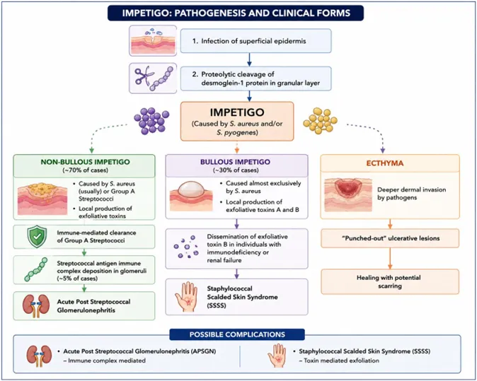
Figure 1. Pathogenesis of impetigo and its principal clinical forms — non-bullous impetigo, bullous impetigo, and ecthyma — together with their associated complications, acute post-streptococcal glomerulonephritis (APSGN) and staphylococcal scalded skin syndrome (SSSS).
I. Non-bullous impetigo
Non-bullous impetigo accounts for approximately 70% of cases and is mainly caused by Staphylococcus aureus, with occasional involvement of Group A Streptococcus. It begins as an erythematous macule that progresses to a vesicle or pustule and ruptures to form the characteristic honey-coloured crust. Exfoliative toxins contribute to superficial epidermal damage, and in rare streptococcal cases, immune-complex deposition may lead to acute post-streptococcal glomerulonephritis.
II. Bullous impetigo
Bullous impetigo, accounting for roughly 30% of cases, is caused almost exclusively by Staphylococcus aureus producing exfoliative toxins A and B. It presents as flaccid bullae on an erythematous base, followed by rupture and collarette scaling. In vulnerable individuals, systemic toxin spread may result in staphylococcal scalded skin syndrome.
III. Ecthyma
When infection penetrates deeper into the epidermis and dermis, it leads to ecthyma — a more severe form of impetigo presenting as punched-out ulcerative lesions that may heal with scarring.
Because impetigo primarily affects the superficial layers of the epidermis, topical drug delivery systems capable of maintaining adequate drug concentrations at the site of infection are particularly advantageous.
Impetigo is diagnosed mainly on clinical grounds and presents in two principal forms — non-bullous and bullous impetigo.
Non-bullous impetigo begins as a red macule or papule that rapidly develops into a vesicle, which ruptures to form shallow erosions covered by honey-coloured crusts. Lesions are often pruritic and spread by auto-inoculation, commonly affecting the face and extremities. The condition usually resolves spontaneously without scarring within a few weeks if untreated.16
A secondary form, known as common (impetiginous) impetigo, may complicate systemic diseases such as diabetes mellitus and acquired immunodeficiency syndrome, or occur at sites of skin injury caused by insect bites or viral infections. Its clinical appearance closely resembles primary non-bullous impetigo.17
Bullous impetigo, caused by toxin-producing Staphylococcus aureus, is more common in neonates but can also affect older children and adults. It presents as flaccid bullae with well-defined margins and minimal surrounding erythema; ruptured bullae leave yellow crusts, often with a characteristic collarette of scale.16 Lesions favour moist intertriginous areas, systemic symptoms are uncommon, and most cases resolve without scarring. Bullous impetigo is generally less contagious than the non-bullous form.18
Patients with impetigo should maintain good hygiene, and lesions should be gently cleaned with soap and warm water to remove crusts and secretions before antimicrobial therapy begins.14 Although impetigo is often self-limiting, appropriate treatment is recommended to shorten disease duration, prevent spread of infection, and avoid complications.14,3
Topical antibiotics are considered first-line treatment for localised impetigo. Strong evidence indicates that topical antibiotics are superior, or at least equivalent, to oral antibiotics for localised disease, while being associated with fewer systemic adverse effects.3 For localised, uncomplicated non-bullous or bullous impetigo, topical therapy alone is therefore recommended, with crusts removed beforehand to enhance drug penetration.19
2.7.1 Topical Therapy
The most commonly recommended topical antibiotics are mupirocin 2%, retapamulin 1%, and fusidic acid.20
Mupirocin acts by inhibiting bacterial protein synthesis through binding to isoleucyl-tRNA synthetase, and is highly effective against Staphylococcus aureus and Streptococcus pyogenes with minimal effect on normal skin flora.19 Retapamulin, a pleuromutilin antibiotic, interferes with bacterial protein synthesis at multiple binding sites and shows a low potential for resistance development.20 Fusidic acid demonstrates good skin penetration and achieves high local concentrations at the site of infection, though its availability varies by region.19
Ozenoxacin, a newer, non-fluorinated topical quinolone, has recently been approved in the United States for the treatment of impetigo and in several European countries for non-bullous impetigo in patients aged six months and older. Studies have shown that it exhibits strong bactericidal activity against Staphylococcus aureus strains resistant to methicillin, mupirocin, and ciprofloxacin, while demonstrating minimal systemic absorption. Given the increasing resistance to conventional therapies, ozenoxacin represents a promising topical alternative for impetigo management — although further clinical studies are needed to better establish its safety profile, therapeutic benefits, and comparative efficacy against currently recommended treatments.21
2.7.2 Systemic Therapy
Systemic antibiotics are reserved for extensive disease, multiple lesions, deeper tissue involvement, systemic symptoms, or situations where topical therapy is impractical.14 Recommended oral agents include first-generation cephalosporins and penicillinase-resistant penicillins, typically given for seven days.22,20 Trimethoprim-sulfamethoxazole may be considered in MRSA-suspected cases.20
Overall, topical therapy remains preferred whenever feasible, while systemic antibiotics should be used judiciously — both to minimise adverse effects and to limit the further development of antimicrobial resistance.
Taken together, these limitations point to the need for better topical drug delivery approaches that can maintain effective drug levels at the site of infection while also improving ease of use in impetigo management.
Table 1. Commonly used topical antibiotics for impetigo.
|
Drug (Generic) |
Brand / Description |
Action / Notes |
|
Mupirocin |
Bactroban® cream / ointment |
Widely used topical antibiotic for localised impetigo; effective against S. aureus and S. pyogenes; low systemic absorption. |
|
Retapamulin |
Altabax® ointment |
Newer topical agent used for localised impetigo treatment. |
|
Fusidic acid |
Fucidin® cream / ointment |
Topical antibiotic option commonly used, especially in certain regions. |
|
Ozenoxacin |
Xepi™ 1% cream |
Newer quinolone antibiotic with bactericidal activity against S. aureus, including some resistant strains; approved for impetigo in adults and children. |
Source: refs. 23
Table 2. Oral antibiotics used in the management of impetigo.
|
Oral Antibiotic |
Indication / Use |
Notes |
|
Dicloxacillin |
Empiric therapy for presumed MSSA impetigo |
Typical adult dose 250 mg QID for 7 days. |
|
Cephalexin |
Empiric therapy for MSSA impetigo |
Common first-line oral agent; paediatric dosing 25–50 mg/kg/day in divided doses. |
|
Amoxicillin–Clavulanate |
Empiric therapy covering S. aureus |
Useful when broad-spectrum coverage is desired. |
|
Oral Penicillin |
When culture confirms streptococcal infection only |
Not preferred empirically, owing to poor S. aureus coverage. |
|
Clindamycin |
Suspected or confirmed MRSA |
Alternative to TMP-SMX; good soft-tissue penetration. |
|
Doxycycline |
Suspected or confirmed MRSA |
Not recommended in young children. |
|
Trimethoprim–Sulfamethoxazole |
Suspected or confirmed MRSA |
Should be combined with an anti-streptococcal agent if streptococcus is suspected. |
Source: refs. 3
3. Limitations of Conventional Therapy
Conventional topical antibiotics used in impetigo management present several limitations that reduce their clinical effectiveness. Poor penetration into the deeper layers of the epidermis — especially in crusted or inflamed lesions — can compromise antibacterial activity. These formulations often require frequent application and prolonged treatment duration, which may translate into poor patient compliance, particularly among paediatric patients.
The increasing prevalence of antibiotic resistance, notably to commonly used agents such as mupirocin and fusidic acid, has further reduced treatment success. Local adverse reactions, including irritation and sensitisation, may occur with topical therapy, while systemic antibiotics carry their own systemic side effects and disrupt normal microbial flora. Collectively, these limitations restrict the overall effectiveness of conventional approaches to impetigo management.21
4. Need for Advanced Topical Drug Delivery Systems
The limitations associated with conventional creams, ointments, and gels underscore the need for advanced topical drug delivery systems in the effective treatment of impetigo. The stratum corneum acts as a major barrier to drug permeation, often resulting in inadequate drug concentration at the site of infection and suboptimal therapeutic outcomes.24 Improved delivery approaches are therefore required to enhance drug bioavailability at the infection site, prolong residence time, and reduce dosing frequency.
In this context, nanotechnology-based carriers — liposomes, polymeric nanoparticles, nanoemulsions, and nano-emulgels — have emerged as promising alternatives. These systems are designed to improve skin penetration, enable controlled drug release, and enhance formulation stability, with the potential to reduce both local and systemic side effects while improving antibacterial efficacy against resistant pathogens.25,26
Table 3. Comparison of nanocarrier-based topical delivery systems considered for impetigo management.
|
Nanocarrier System |
Advantages |
Limitations |
|
Liposomes |
Biocompatible; enhance skin penetration; suitable for both hydrophilic and lipophilic drugs. |
Physical instability; drug leakage during storage. |
|
Solid Lipid Nanoparticles (SLNs) |
Controlled release; improved stability; skin occlusion effect. |
Limited drug loading; possible drug expulsion during storage. |
|
Nanostructured Lipid Carriers (NLCs) |
Higher drug loading; improved stability; sustained release. |
Complex formulation process. |
|
Polymeric Nanoparticles |
Controlled release; protects drug from degradation. |
Higher production cost; possible polymer toxicity concerns. |
|
Nanoemulsions |
Excellent drug solubilisation; enhanced skin permeation; ease of preparation. |
Low viscosity; poor retention at the application site. |
|
Nano-emulgels |
Enhanced permeation; prolonged skin retention; controlled release; improved patient compliance. |
Scale-up and long-term stability challenges. |
Source: refs. 27, 26, 28
Among these carriers, nano-emulgels offer specific advantages. While nanoemulsions enhance skin permeation, their low viscosity often results in poor retention at the application site. Liposomes and solid lipid nanoparticles may be limited by low drug loading, physical instability, or structural disruption on skin contact. Nano-emulgels, by contrast, combine the nanoscale penetration efficiency of nanoemulsions with the prolonged residence time, spreadability, and patient acceptability of gel systems — supporting enhanced epidermal drug deposition, sustained local drug release, and improved therapeutic performance in superficial infections such as impetigo.29,30
5. Advances and Novel Approaches for Impetigo Treatment
Recent advances in impetigo management have focused on novel topical strategies that address increasing antibiotic resistance, particularly involving methicillin-resistant Staphylococcus aureus (MRSA). The reduced effectiveness of conventional topical and oral antibiotics has highlighted the need for alternative approaches that ensure effective local drug delivery while minimising systemic exposure.26
Nanotechnology-based topical systems — including nanoemulsions, nano-emulgels, liposomes, solid lipid nanoparticles, and polymeric nanoparticles — have gained attention for their ability to enhance drug solubility, skin penetration, and retention in superficial epidermal layers. These properties make them well suited to impetigo, which primarily affects the outer epidermis, while also supporting sustained drug release and improved activity against resistant pathogens.8
Bacterial biofilms further contribute to persistent staphylococcal infections by limiting antibiotic penetration and protecting bacteria from host defences. Nanocarrier-based systems can improve drug diffusion within biofilms and maintain prolonged local drug levels, thereby enhancing bacterial eradication. Emerging adjunct approaches — antimicrobial peptides, bacteriophage therapy, photodynamic therapy, and antiseptic-based formulations — are also being explored to disrupt biofilms and reduce reliance on conventional antibiotics.31,32
6. Recent Patents on Impetigo Treatment
A number of recent patents and patent applications reflect continued commercial and academic interest in topical antibacterial formulations relevant to impetigo. A representative selection is summarised in Table 4.
Table 4. Recent patents relevant to topical impetigo treatment.
|
Publication No. / Year |
Title of Invention |
Summary of Invention |
Ref. |
|
US11795192B2 (2023) |
Antimicrobial compounds and compositions and uses thereof |
Discloses novel antimicrobial compounds and pharmaceutical compositions for treating bacterial infections, with topical administration for skin infections — including impetigo — targeting S. aureus and S. pyogenes claimed specifically. |
33 |
|
US20250127836A (2025) |
Compositions for treatment of bacterial skin conditions |
A patent application describing topical compositions that combine antimicrobial, moisturising, and barrier-repair agents, addressing superficial bacterial skin diseases including impetigo with improved skin healing and reduced irritation. |
34 |
|
US12440529B1 (2025) |
Topical compositions for treating bacterial colonization and infection of the skin |
Covers broad-spectrum topical formulations aimed at bacterial infections associated with skin disorders; impetigo is included as a target condition, especially secondary infections in compromised skin. |
35 |
|
Ozenoxacin patent family (2014–present) |
Topical quinolone antibacterial compositions |
Covers ozenoxacin-based topical formulations approved for impetigo treatment, with claims relating to formulation stability, antimicrobial efficacy, and reduced resistance development. |
36 |
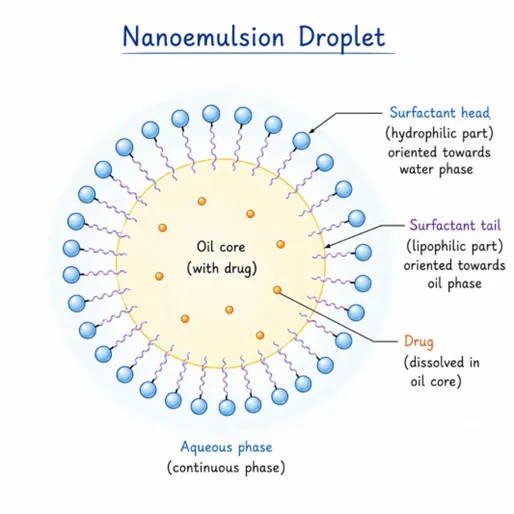
Figure 2. Structure of a nanoemulsion droplet, showing the oil core carrying the dissolved drug, the surrounding surfactant layer, and the continuous aqueous phase.
A nano-emulgel is an advanced topical drug delivery system developed to combine the high penetration ability of nanoemulsions with the convenience and retention of gels. In simple terms, it is a formulation in which extremely small, drug-carrying oil droplets are uniformly distributed within a soft gel base. This design allows the drug to remain on the skin surface for longer while simultaneously penetrating the deeper layers more efficiently.
Nanoemulsions consist of oil and water phases stabilised by surfactants and co-surfactants, with droplet sizes typically in the nanometre range. These nanosized droplets provide a large surface area, which improves drug solubility and facilitates closer contact with the skin. However, nanoemulsions are liquid in nature and may spread uncontrollably or run off the skin during application. Converting them into a gel system resolves this issue by offering a semi-solid, easy-to-apply formulation — making the nano-emulgel highly suitable for topical therapy, especially in superficial skin infections such as impetigo.37,38
7.2 Rationale Behind Nano-Emulgel Design
Conventional topical formulations such as creams, ointments, and gels often show limited effectiveness due to poor skin penetration, low drug retention, and the need for frequent application. In bacterial skin infections, maintaining an effective drug concentration at the site of infection is crucial for complete eradication of pathogens and prevention of resistance.
The nano-emulgel system is designed to overcome these limitations by:
For impetigo, where treatment relies mainly on topical antibiotics, the nano-emulgel offers a promising approach by ensuring prolonged drug contact with infected skin and better therapeutic outcomes.38,39
7.3 Structural Design of Nano-Emulgel
The design of a nano-emulgel involves two integrated systems: a nanoemulsion that carries the drug, and a gel matrix that houses and stabilises it.
7.3.1 Nanoemulsion System
The nanoemulsion acts as the drug-carrying unit and serves as the core delivery component of the nano-emulgel. It is a thermodynamically or kinetically stable colloidal dispersion consisting of nano-sized oil droplets dispersed in an aqueous phase with the aid of surfactants and co-surfactants. Droplet size generally ranges between 20–200 nm, providing a large surface area that improves drug solubilisation, skin penetration, and bioavailability. Owing to their extremely small size, nanoemulsion droplets can interact closely with the stratum corneum and enhance the permeation of both hydrophilic and lipophilic drugs across the skin barrier. The system is composed of four principal elements:
1. Oil Phase
The oil phase acts as the reservoir for lipophilic drugs and plays an important role in enhancing skin permeation. It solubilises poorly water-soluble drugs and improves drug-loading capacity, and can also fluidise the lipid bilayers of the stratum corneum, thereby increasing transdermal permeation. Commonly used oils include caprylic/capric triglycerides, isopropyl myristate, oleic acid, Capryol 90, and medium-chain triglycerides. Selection of the oil phase is based mainly on drug solubility, compatibility, and permeation-enhancing ability.
2. Surfactants
Surfactants are surface-active agents that reduce interfacial tension between the oil and aqueous phases, thereby stabilising the nano-sized droplets. They form a protective interfacial film around the oil droplets and prevent coalescence. Non-ionic surfactants such as Tween 80, Tween 20, Cremophor RH40, and Span 80 are widely preferred for topical formulations because they exhibit low irritation potential, good biocompatibility, and favourable safety profiles. The concentration and hydrophilic–lipophilic balance (HLB) value of the surfactant greatly influence droplet size, stability, and drug release behaviour.
3. Co-surfactants
Co-surfactants are incorporated to further reduce interfacial tension and improve the flexibility of the surfactant film surrounding the droplets. They assist in the spontaneous formation of nanoemulsions and improve formulation stability by preventing droplet aggregation, while also enhancing drug solubilisation and skin permeation. Common examples include propylene glycol, polyethylene glycol (PEG 400), ethanol, and Transcutol HP. The combination of surfactant and co-surfactant — often referred to as Smix — plays a critical role in determining the stability region of the nanoemulsion.
4. Aqueous Phase
The aqueous phase generally consists of purified water and forms the continuous external phase in oil-in-water (O/W) nanoemulsions. It helps maintain the fluidity and dispersion of the nano-sized droplets, and contributes to skin hydration, which can further improve drug permeation through the stratum corneum. In some formulations, the aqueous phase may also contain preservatives, buffers, or humectants to improve stability and patient acceptability.
The synergistic interaction of oil, surfactant, co-surfactant, and aqueous phase results in stable nano-sized droplets with improved drug delivery performance — providing enhanced surface contact with the skin, improved penetration, controlled drug release, and better therapeutic efficacy compared with conventional emulsions.37,40
7.3.2 Gel Matrix
The gel matrix is the structural component that transforms the liquid nanoemulsion into a semi-solid nano-emulgel suitable for topical application. Incorporating the nanoemulsion into a gel base improves viscosity, spreadability, patient compliance, and retention time at the site of application; it also provides a cooling effect, ease of application, and a non-greasy texture, making the formulation cosmetically acceptable.
The gel matrix forms a three-dimensional polymeric network that entraps the nanoemulsion droplets uniformly without affecting their nanoscale size or stability. This network helps maintain prolonged contact of the formulation with the skin surface, thereby enhancing drug absorption and therapeutic activity. Its principal functions are summarised below.
7.4 Key Design Parameters in Nano-Emulgel Development
Successful nano-emulgel design depends on optimising several critical parameters:
7.5 Mechanism of Drug Delivery from Nano-Emulgel
Once applied to the skin, the gel matrix ensures prolonged contact at the site of application. The nanoemulsion droplets slowly release the drug, which then penetrates the skin through intercellular, transcellular, and appendageal pathways via:
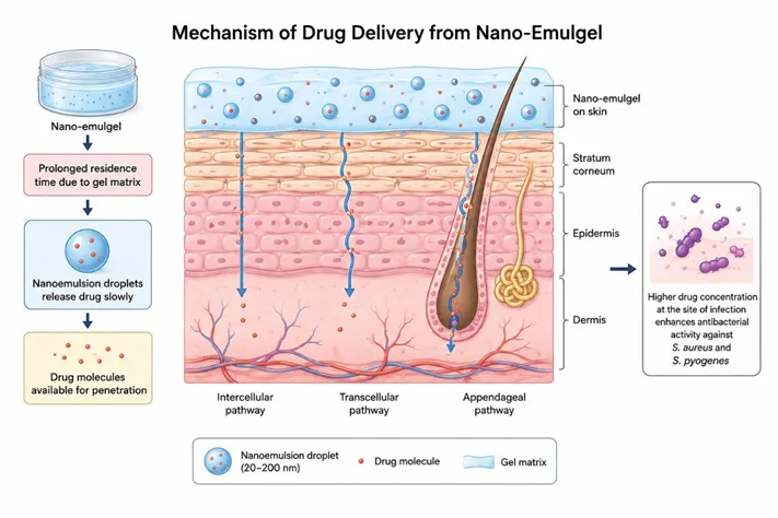
Figure 3. Mechanism of drug delivery from a nano-emulgel through the skin. The gel matrix prolongs surface residence time while nanoemulsion droplets release drug through intercellular, transcellular, and appendageal pathways, raising local drug concentration at the infection site.
This dual mechanism — retention by the gel and penetration by the nanoemulsion — is the key design advantage of nano-emulgel systems.
7.6 Advantages of Nano-Emulgel Design
8. Preparation of Nano-Emulgel
Nano-emulgels can be prepared by various methods, distinguished mainly by the sequence in which the oil and aqueous phases are combined. In this process, the active pharmaceutical ingredient (API) is initially solubilised and stabilised in the oil phase (Phase I), while the gelling agents are separately dispersed in the aqueous phase to form the gel base (Phase II). The oil phase containing the drug is then gradually incorporated into the gel phase under continuous stirring, followed by homogenisation to obtain a uniform and stable emulsion.
Overall, the preparation of a nano-emulgel involves two main steps: first, formulation of the nanoemulsion; second, incorporation of the prepared nanoemulsion into the gelling agent system to yield the final nano-emulgel, as outlined in Figure 4.
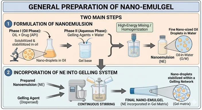
Figure 4. General preparation scheme for a nano-emulgel, showing (1) formulation of the nanoemulsion from an oil phase and aqueous gel base, and (2) incorporation of the nanoemulsion into the gelling system to yield the final product.
8.1 Methods of Preparation of Nanoemulsions for Nano-Emulgel Systems
1. High-Pressure Homogenization (HPH)
High-pressure homogenization is one of the most widely used high-energy techniques for preparing pharmaceutical nanoemulsions intended for nano-emulgel systems. A coarse emulsion of oil phase, aqueous phase, surfactant, and co-surfactant is first prepared by mechanical stirring. This pre-emulsion is then passed through a high-pressure homogenizer at pressures typically ranging from 500 to 20,000 psi for several cycles. The intense shear forces, cavitation, and turbulence generated during homogenization break the larger emulsion droplets down into nanosized droplets, usually below 200 nm.
This method is particularly well suited to topical nano-emulgels, producing a uniform droplet size distribution, improved physical stability, and enhanced drug solubilisation — all of which are essential for efficient skin permeation and gel incorporation. It is, however, energy-intensive and may not suit thermolabile drugs.41
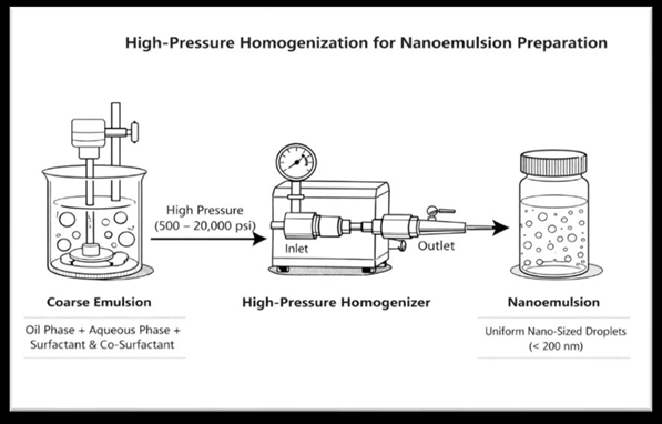
Figure 5. High-pressure homogenization for nanoemulsion preparation: a coarse pre-emulsion is forced through a high-pressure homogenizer, breaking droplets down to a uniform nanoscale size.
2. Ultrasonication Method
Ultrasonication is another commonly used high-energy method, especially at laboratory scale. A coarse emulsion is first prepared by mixing oil, surfactant, co-surfactant, and aqueous phase using magnetic stirring; the resulting emulsion is then subjected to ultrasonic waves via a probe sonicator. The acoustic cavitation generated leads to the formation and collapse of microbubbles, producing strong shear forces that reduce droplet size into the nanometre range.
Ultrasonication is valued for its simplicity, shorter processing time, and ability to produce fine nanoemulsions suitable for gel incorporation. Prolonged sonication, however, may cause drug degradation and a rise in temperature, both of which must be carefully controlled.41,42
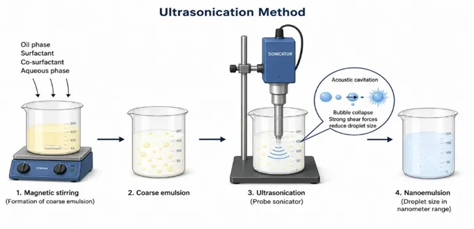
Figure 6. Ultrasonication method: a coarse emulsion is exposed to acoustic cavitation via a probe sonicator, collapsing microbubbles to generate the shear forces that reduce droplet size into the nanometre range.
3. Phase Inversion Temperature (PIT) Method
The phase inversion temperature method is a low-energy emulsification technique based on the temperature-dependent solubility of non-ionic surfactants. The oil phase, aqueous phase, and non-ionic surfactant are mixed and heated above the PIT, at which point the surfactant has equal affinity for both oil and water phases, triggering phase inversion. On controlled cooling, the system inverts from water-in-oil (w/o) to oil-in-water (o/w) nanoemulsion with very small droplet sizes.
PIT-based nanoemulsions are thermodynamically favourable and exhibit a narrow size distribution, making them well suited to topical nano-emulgel formulations — though the method is limited to temperature-stable drugs and requires precise temperature control.42,43
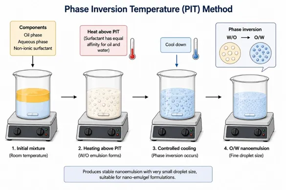
Figure 7. Phase Inversion Temperature (PIT) method: heating above the PIT inverts a water-in-oil emulsion to oil-in-water on controlled cooling, yielding a fine, stable nanoemulsion.
4. Phase Inversion Composition (PIC) Method
The phase inversion composition method is a low-energy emulsification technique that relies on gradual changes in composition rather than temperature. The oil phase, containing drug and surfactant, is titrated slowly with the aqueous phase under continuous stirring; as water content increases, the system undergoes phase inversion, leading to spontaneous formation of nanoemulsion droplets.
The PIC method is particularly advantageous for nano-emulgel development because it requires no specialised equipment, reduces energy input, and produces stable nanoemulsions with fine droplet size. It is extensively reported for topical formulations owing to its simplicity, reproducibility, and compatibility with the polymers used in gel formation.41,44
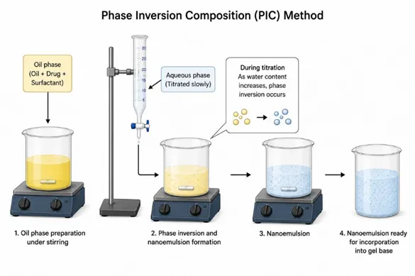
Figure 8. Phase Inversion Composition (PIC) method: slow titration of the aqueous phase into the oil phase drives spontaneous phase inversion and nanoemulsion formation as water content increases.
5. Solvent Displacement (Spontaneous Emulsification) Method
Solvent displacement, also known as spontaneous emulsification, is widely used for preparing nanoemulsions containing poorly water-soluble drugs. The oil phase and drug are dissolved in a water-miscible organic solvent such as ethanol or acetone; this organic phase is then injected into an aqueous phase containing surfactant under continuous stirring. Rapid diffusion of the solvent into the aqueous phase drives spontaneous formation of nano-sized oil droplets, after which the organic solvent is removed by evaporation.
This method suits nano-emulgel systems particularly well, producing transparent, stable nanoemulsions with enhanced drug solubility and uniform droplet size that facilitate homogeneous gel incorporation.44,42
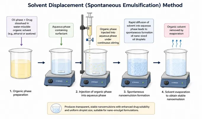
Figure 9. Solvent displacement (spontaneous emulsification) method: injecting a drug-loaded organic phase into an aqueous surfactant phase drives spontaneous nanodroplet formation as the solvent diffuses out and evaporates.
6. Microfluidization
Microfluidization is an advanced high-energy emulsification technique in which the pre-emulsion is forced through microchannels at very high velocities, producing intense shear and impact forces. The collision of fluid streams within the microfluidizer efficiently disrupts droplets, yielding nanoemulsions with extremely small and uniform droplet sizes.
The method is highly reproducible and well suited to scale-up, making it advantageous for industrial nano-emulgel production; the uniform droplet size enhances physical stability and drug-release characteristics in topical applications. Its main drawback is the high cost of equipment, which limits routine laboratory use.40,45
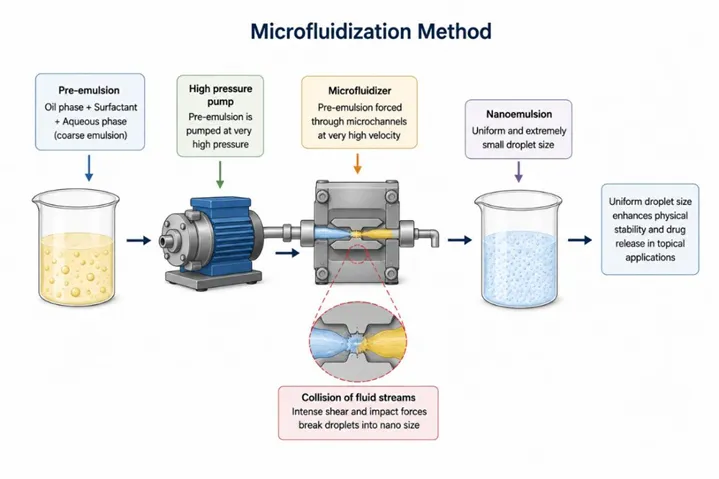
Figure 10. Microfluidization method: a pre-emulsion is forced through microchannels at high velocity, where colliding fluid streams generate the intense shear needed for a uniform, extremely fine nanoemulsion.
8.2 Incorporation of Nanoemulsion into the Gel Base
After successful preparation of a stable nanoemulsion by a suitable high- or low-energy technique, the nanoemulsion is incorporated into a gel base to obtain the final nano-emulgel formulation.9,47 This step is essential for transforming the low-viscosity nanoemulsion into a patient-acceptable semisolid dosage form with enhanced retention at the site of application.
The gel base is typically prepared separately by dispersing gelling agents — such as Carbopol 934 or 940, hydroxypropyl methylcellulose (HPMC), or poloxamer 407 — in purified water, followed by adequate hydration and neutralisation using agents like triethanolamine to achieve the desired pH and viscosity.9,48 The prepared nanoemulsion is then slowly incorporated into the hydrated gel base under gentle, continuous stirring to ensure uniform distribution of the nanosized droplets without causing phase separation or droplet coalescence.47,25 The result is a homogeneous nano-emulgel system in which the nanoemulsion droplets are uniformly entrapped within the three-dimensional polymeric gel network.
Several studies have reported that nano-emulgels prepared this way exhibit improved physical stability, better spreadability, enhanced skin permeation, prolonged drug residence time, and controlled drug release compared with conventional gels or emulsions.9,25,49 Owing to these advantages, nano-emulgels have gained significant attention for the topical treatment of superficial skin infections such as impetigo, where localised drug delivery with minimal systemic exposure is desirable.47,49
9. Characterization Studies of Nano-Emulgel
Characterization of nano-emulgels is essential to ensure the quality, stability, and performance of the formulation. Physicochemical characterization helps determine the structural integrity of the nanoemulsion droplets, their interaction with excipients, and their suitability for topical administration. Parameters such as droplet size, polydispersity index, zeta potential, morphology, thermal behaviour, and crystallinity are commonly evaluated to predict formulation stability and drug delivery efficiency. The major characterization techniques are summarised in Table 5.
Table 5. Characterization studies commonly applied to nano-emulgel formulations.
|
Characterization Parameter |
Purpose |
Method / Instrument |
Ref. |
|
Droplet / globule size |
Determines stability and penetration ability |
Dynamic Light Scattering (DLS) |
50,45 |
|
Polydispersity Index (PDI) |
Indicates uniformity of size distribution |
DLS (Zetasizer) |
50,45 |
|
Zeta potential |
Predicts physical stability of the formulation |
Zeta potential analyzer |
50,45 |
|
Morphology |
Confirms shape and surface characteristics |
SEM / TEM |
45 |
|
Drug–excipient compatibility |
Detects possible interactions |
FTIR spectroscopy |
45 |
|
Thermal behaviour |
Assesses thermal stability of the drug |
DSC |
45 |
|
Crystallinity |
Determines amorphous or crystalline nature |
XRD |
45 |
10. Evaluation Studies of Nano-Emulgel
Evaluation studies assess the topical applicability, drug release characteristics, and overall performance of nano-emulgel formulations. Parameters such as pH, viscosity, spreadability, drug content, in vitro drug release, ex vivo skin permeation, and stability provide valuable information on formulation quality, patient acceptability, and therapeutic potential. The commonly reported evaluation parameters are presented in Table 6.
Table 6. Evaluation studies commonly applied to nano-emulgel formulations.
|
Evaluation Parameter |
Purpose |
Method / Instrument |
Ref. |
|
Physical appearance |
Ensures uniformity and absence of phase separation |
Visual inspection |
50 |
|
Homogeneity |
Confirms uniform drug distribution |
Visual inspection |
50 |
|
pH |
Ensures skin compatibility |
Digital pH meter |
50 |
|
Viscosity / rheology |
Determines flow behaviour and consistency |
Brookfield viscometer |
50,45 |
|
Spreadability |
Measures ease of application |
Glass slide method |
50,45 |
|
Drug content uniformity |
Ensures uniform drug distribution |
UV–Visible spectrophotometry |
50 |
|
In vitro drug release |
Evaluates the release profile |
Franz diffusion cell |
50,51 |
|
Ex vivo skin permeation |
Assesses drug permeation through skin |
Franz diffusion cell |
51 |
|
Stability studies |
Evaluates formulation stability |
ICH guidelines |
50 |
11. Recent Research on Nano-Emulgels for Impetigo and Related Bacterial Infections
Recent research on nano-emulgels for impetigo and related bacterial skin infections has focused on improving topical antibiotic delivery, skin retention, and therapeutic efficacy. A QbD-optimized ozenoxacin-loaded nano-emulgel demonstrated suitable physicochemical characteristics for topical administration, enhanced in vitro drug release, improved ex vivo skin permeation and retention, and superior antibacterial efficacy in an impetigo mouse model compared with a marketed ozenoxacin cream. The formulation also showed satisfactory physical and chemical stability under accelerated storage conditions.52
Similarly, a mupirocin-loaded nano-emulgel prepared using a Carbopol-based gel matrix showed a homogeneous appearance, pseudoplastic rheological behaviour, and enhanced drug deposition within skin layers, suggesting improved local drug retention and sustained antibacterial activity.53
Beyond antibiotic-loaded formulations, herbal nano-emulgels have also shown promising antimicrobial potential. A turmeric- and neem-based nano-emulgel exhibited significant antibacterial activity against multiple microbial strains compared with conventional hydrogel formulations, indicating potential applicability in managing superficial bacterial skin infections.54
Collectively, these findings highlight the potential of nano-emulgel systems to improve local drug delivery, enhance skin retention, promote sustained drug release, and increase antibacterial efficacy. Such advantages support the continued development of nano-emulgel formulations as advanced topical therapeutic systems for the effective management of impetigo and other superficial bacterial skin infections.52,53
Owing to the limited number of studies specifically targeting impetigo, recent investigations involving nano-emulgels for topical antibacterial therapy and related skin infections are also included here to provide a broader perspective on the therapeutic potential of this delivery system.
CHALLENGES, REGULATORY ASPECTS, AND FUTURE PERSPECTIVES
Although nano-emulgels show promising laboratory-scale performance, large-scale production remains challenging. High-energy techniques such as ultrasonication and high-pressure homogenization may introduce batch-to-batch variability, difficulty in maintaining a consistent droplet size distribution, and increased production costs during scale-up.55,56
Nano-emulgels are also inherently prone to physical and chemical instability, including phase separation, droplet aggregation, and viscosity changes during storage. Long-term stability depends heavily on the selection of surfactants, gelling agents, and storage conditions, making stability optimisation a critical step in formulation development.55,57
From a regulatory standpoint, nano-emulgels face additional challenges due to the absence of well-defined, harmonised guidelines specific to nanostructured topical drug delivery systems. Regulatory agencies require extensive characterization and safety evaluation — and in some cases nanotoxicity assessment — which can delay product approval and increase development complexity.58,59
Despite these limitations, nano-emulgels hold considerable potential for clinical translation, particularly for localised skin infections such as impetigo, where enhanced skin retention and reduced systemic exposure are advantageous. Future research should focus on scalable manufacturing strategies, robust stability enhancement, and well-designed clinical studies to facilitate regulatory approval and commercialisation.56,59
Impetigo continues to be a frequent and easily transmissible skin infection, especially in children, and its treatment is often limited by the poor skin penetration and short residence time of conventional topical formulations.
As this review has set out, nano-emulgels offer a practical and promising approach to overcoming these limitations by combining the penetration efficiency of nanoemulsions with the convenience and prolonged retention of gel systems. Recent studies involving antibiotics such as ozenoxacin and mupirocin show clearly that nano-emulgels can enhance local drug delivery, improve skin retention, and achieve better antibacterial outcomes without increasing systemic exposure.
Challenges related to large-scale production, long-term stability, and regulatory approval still remain, but ongoing formulation advances and increasing regulatory clarity are gradually addressing these concerns. Overall, nano-emulgels represent a meaningful step forward in the topical management of impetigo, with real potential to improve treatment effectiveness and patient compliance in everyday clinical settings.

Bhuvana, Beny Baby, Pallavi A, Tejashree K C, Navya N, Nano-Emulgels as Advanced Topical Drug Delivery Systems for Impetigo A Comprehensive Review, Int. J. of Pharm. Sci., 2026, Vol 4, Issue 9, 2392-2416, https://doi.org/10.5281/zenodo.22869413
 10.5281/zenodo.22869413