

We use cookies to ensure our website works properly and to personalise your experience. Cookies policy
Department of pharmaceutics, Rajiv Gandhi Institute of Pharmaceutical Sciences and Research, Kasargod-671310, Kerala, India.
Rheumatoid arthritis (RA) is a chronic, systemic autoimmune inflammatory disorder that primarily affects synovial joints, causing pain, swelling, stiffness, and progressive joint destruction. It is characterized by persistent synovitis, cartilage degradation, and bone erosion, leading to functional disability and reduced quality of life. The disease arises from a dysregulated immune response with excessive production of pro-inflammatory cytokines, including tumour necrosis factor-? and interleukins, and activation of the JAK–STAT signaling pathway, which sustains inflammation and tissue damage. Globally, 17.6–17.9 million people are affected, with prevalence increasing by about 14% since 1990, and projections estimate cases may reach 31.7 million by 2050.currently oral preparations are widely administered to control the disease, but it has many adverse effects. Topical application of novel drug delivery systems like nanoparticles, liposomes, hyalurosomes, solid lipid nanoparticles, ethosomes, transferosomes, and dendrimers can increase the therapeutic effect and reduces the systemic side effects. Natural polymer based nanoparticle preparations are capable of site specific delivery and it has biocompatibility. Hydrogels are non sticky, biocompatible preparation that provide controlled drug release and reduce systemic side effects. Their soft, non-greasy nature makes them particularly suitable for topical administration in chronic inflammatory conditions. Therefore, formulating tofacitinib nanoparticle loaded hydrogels provide targeted anti-inflammatory effects at affected joints, minimize systemic toxicity.
Rheumatoid arthritis is a chronic, systemic autoimmune inflammatory disease that mostly affects synovial joints, rheumatoid arthritis (RA) causes pain, swelling, stiffness, and gradual joint deterioration(1) . Persistent synovitis, bone erosion, and cartilage deterioration are its hallmarks, which result in functional impairment and a lower quality of life. The illness is caused by a dysregulated immune response that produces excessive amounts of pro-inflammatory cytokines, such as interleukins and tumor necrosis factor-α, and activates the JAK–STAT signaling pathway, which perpetuates tissue damage and inflammation(2). Non-inflammatory arthritis (osteo-arthritis) and inflammatory arthritis, which are brought on by crystal deposition (pseudogout, basic calcium phosphate disease, gout), bacterial and viral infections (Staphylococcus aureus, Neisseria gonorrhoea, Lyme disease complications, Parvovirus, Enterovirus), or auto-immune processes, have been identified through research and documentation.
RA is a prevalent form of chronic inflammatory arthritis that significantly affects patient and public health. It affects 0.5–1% of adults and is two–three times more common in women than in men. About 5 out of 1000 persons have RA, which can cause severe joint damage and disability. In individuals between the ages of 20 and 54, RA has been recognized as a global health concern. The incidence rate rose from 11.66 to 13.48 per lakh, whereas the fatality rate decreased from 0.09 to 0.06 per lakh(3). Over 14 million individuals worldwide suffer from RA, with an estimated prevalence rate ranging from 0.24 to 1%.
Numerous signal transduction pathways have been implicated in the development of RA, while the exact causes of the disease are still being investigated. Janus kinases (JAK) mediate the intracellular signalling transduction of lymphocytes. JAK1, JAK2, JAK3, and tyrosine kinase 2 (TYK) are the four protein tyrosine kinases that make up the JAK family. Numerous cytokines can trigger the JAK/signal transducers and activators of transcription (STAT) pathway, an intracellular signalling system that leads to the production of additional pro- and anti-inflammatory cytokines as well as locally harmful enzymes. The JAK/STAT pathway either directly or indirectly activates the many cytokines. The JAK/STAT pathway directly activates the cytokines IL-6, IL-1, L-10, and (IFN)-γ, while it indirectly activates TNF-α and interleukin (IL)-1(4).
Tofacitinib, an oral Janus kinase ( JAK) inhibitor, has become an effective treatment for moderate-to -severe rheumatoid arthritis by blocking cytokine-driven inflammatory signaling through selective inhibition of JAK1 and JAK3(5). However, long-term oral use is often linked to systemic side effects such as infections, gastrointestinal issues, liver toxicity, abnormal lipid levels, and cardiovascular problems, which restrict its long-term safety and reduce patient compliance(6). In recent days, efforts have been made to address these issues through the topical delivery of TF using methods such as liquid crystal nanoparticles, proposomes, and microneedles for treating various skin disorders(4). Applying the nanoparticulate drug delivery system ( NDDS) topically to the skin can: improve drug solubility, deliver drugs precisely to their site of action, create a localized reservoir of the drug, and provide controlled drug release (7).
Hydrogels three-dimensional, water-loving polymer networks that can hold substantial amounts of waterprovide benefits including biocompatibility, simple application, controlled drug delivery, and higher local drug concentration with less systemic exposure(8). Their soft, non-greasy texture makes them especially appropriate for topical use in chronic inflammatory conditions. Thus, developing hydrogels loaded with tofacitinib could deliver targeted anti-inflammatory action directly to affected joints, reduce systemic toxicity, and enhance treatment effectiveness and patient adherence.
RHEUMATOID ARTHRITIS:
PATHOPHYSIOLOGY AND CURRENT TREATMENT STRATEGIES
Pathophysiology
Rheumatoid arthritis ( RA) typically presents as a condition that impacts several joints, often developing slowly over time. However, some people might develop symptoms abruptly, which either fluctuate or shift between joints, or they may exhibit the condition limited to a single joint. The symptoms of the condition can significantly restrict patients’ ability to perform everyday tasks, including walking, climbing stairs, dressing, using the restroom, standing up from a chair, opening containers, typing, and their professional abilities. In up to a third of patients who suddenly develop polyarthritis, systemic symptoms including severe muscle pain, fatigue, low-grade fever, weight loss, and depression may also appear. In some cases, patients may also show symptoms beyond the joints, such as nodules or eye inflammation(9).
The development of RA entails intricate immune system interactions that result in persistent inflammation and joint deterioration. RA is regarded as an autoimmune condition, in which the immune system erroneously attacks the body’s own tissues, especially the synovium the lining of the joints. While the precise cause of this autoimmune reaction remains unclear, it is thought to result from a mix of genetic vulnerability and environmental influences. Research indicates that immunological processes referred to as the ‘ pre RA phase’ can occur years prior to the onset of visible joint inflammation. Peptide arginine deiminases carry out a post-translational modification called citrullination on proteins that contain arginine residues, transforming them into citrulline. Additionally, joint abnormalities can provoke cytokine release, leading to joint inflammation and altered self antigens, such as synovial hyperplasia or synovial infections.Citrullinated proteins (vimentin, type II collagen, histones, fibrin, fibro nectin, Epstein‑Barr nuclear antigen 1 and α‑enolase) are no longer recognised by the immune system as self‑structures because of the susceptibility of genes HLA‑DR1 and HLA‑DR4. Antigen‑ presenting cells are activated dendritic cells that take up antigens in order to start immune response.The entire complex moves to the lymph node, where CD4+ helper T‑cell activation occurs. Moreover, B‑cells in the germinal centre the lymph node are stimulated by T‑cells in a reciprocating and sequential signalling mechanism known as co‑stimulation(10) .
Infectious agents, for example, EpsteinBarr virus, cytomegalovirus, Proteus species, and Escherichia coli, along with their products (e.g., Heat-shock proteins) have been associated with rheumatoid arthritis for a long time; while a unifying mechanism has not yet been identified, some form of molecular mimicry is believed to be involved. During infection, immune complex formation may lead to the production of rheumatoid factor a high-affinity autoantibody targeting the Fc region of immunoglobulinthat has historically been used as a diagnostic indicator of rheumatoid arthritis and is also thought to contribute to the disease’s development. Moreover, rheumatoid arthritis seems to be linked with periodontal disease(11).
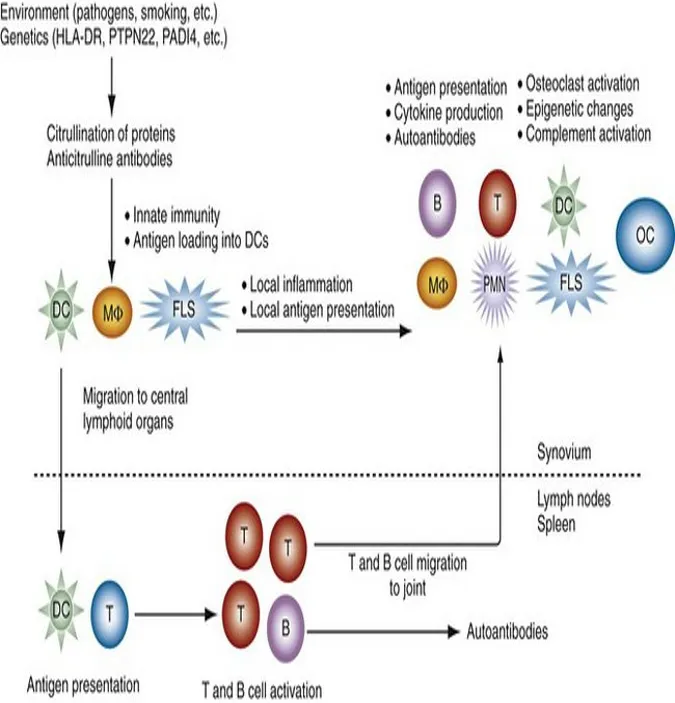
Fig 1:Pathogenesis of rheumatoid arthritis
Current treatment strategies
Non-steroidal anti-inflammatory medications (NSAIDs), glucocorticoids (GCs), disease-modifying antirheumatic drugs (DMARDs), and biological agents are the primary categories of pharmacological therapy for rheumatoid arthritis.
The main objective of RA treatment is to help each patient reach a target of low disease activity (LDA) or persistent clinical remission, which is usually only achievable with the aid of disease-modifying anti-rheumatic medications (DMARDs). DMARDs can regulate synovitis, prevent or lessen joint damage, maintain joint integrity and function, and slow or halt the radiographic development. Currently, RA can be treated with a variety of DMARDs, such as targeted synthetic DMARDs, biological DMARDs (bDMARDs), and conventional synthetic DMARDs (csD MARDs). Methotrexate (MTX; 4-amino-10-methyl folate acid) is currently regarded as the "anchor drug" for the treatment of RA among the csDMARDs. Actually, up to 70% of RA patients receive a prescription for it(12).
Leflunomide, sulfasalazine, and hydroxychloroquine alone or in combination (e.g., triple therapy) are choices for patients who cannot use MTX or do not respond well to it. In order to rapidly suppress synovitis, short-course or low-dose GCs are still frequently employed as bridge therapy. Analgesics and NSAIDs help with symptoms, but they don't stop the condition from getting worse.For patients who have not responded well to the best csDMARD treatment, biologic DMARDs (bDMARDs), categorized by their unique mechanism, are immunomodulatory medicines to take into consideration. First-line treatment is probably going to involve tumor-necrosis factor inhibitors (adalimumab, etanercept, infliximab, certoli zumab, and golimumab). It has been demonstrated that oral targeted synthetic DMARDs, specifically the Janus kinase inhibitors (tofacitinib, baricitinib, upadacitinib, and filgotinib), are convenient to take orally and have comparable clinical efficacy to biologics on the majority of endpoints(13).
Table No.1 Advances in RA therapy(14)
|
Type of DMARDs |
Category |
Mechanism of action |
Drugs |
|
Traditional DMARDs
|
1. Methotrexate and sulfasalazine
2.Hydroxychloroquine and leflunomide |
MTX mediates the resolution of inflammation by upregulating adenosine and inhibiting methylation reactions. Sulfasalazine (SSZ) exerting multiple anti-inflammatory effects in vitro. SSZ is often used as one of the combination therapies for RA. HCQ is weakly alkaline, can change the pH of lysosomes and ER, leading to decreased protein digestion and antigen presentation ability of monocytes/macrophages. Leflunomide inhibits the activation and proliferation of T lymphocytes by targeting dihydroorotate dehydrogenase (DHODH). Alsoinhibit tyrosine kinase phosphorylation |
Methotrexate Sulfasalazine
Leflunomide |
|
BiologicDMARDs |
1.TNF-α inhibitors
2. IL-6 inhibitors |
Decrease inflammatory mediators such as IL-1, IL-6, IL-8, MMPs, to downregulate VEGF for endothelial cell-specific angiogenesis, and to reduce the migration of lymphocytes and macrophages into the joints. Can effectively inhibit series of reactions induced by IL-6. |
Etanercept,Infliximab, Adalimumab, Golimumab, Certolizumab Tocilizumab, Sarilumab, Sirukumab |
|
Small molecule DMARDs |
1.JAK inhibitor
2.Phosphodiesterase-4 inhibitors |
Inhibit JAK–STAT signaling pathway in inflammatory responses. Phosphodiesterase-4 (PDE4) is an enzyme responsible for degradation of intracellular cyclic adenosine monophos phate (cAMP). PDE4 inhibitors can significantly increase the level of cAMP, |
Tofacitinib Baricitinib
Ibudilas |
|
Emerging therapies |
1.IL-23/Th17 pathway 2.Sphingosine-1-phosphate receptor Modulators |
Inhibit IL-23/Th17 pathway Targeting the S1P/S1PR signalling axis reduces the trafficking of auto reactive lymphocytes and the Th17/Treg ratio, there by controlling autoimmunity and inflammatory responses. |
Secukinumab Ixekizumab NIBR-0213 LX2932
|
ROLE OF TOFACITINIB IN RA MANAGEMENT
JAK–STAT pathway
First, it is discovered that a highly activated JAK-STAT pathway is intimately linked to the development of RA. JAK2 is the most extensively researched protein of the four members of the JAK family(15). Tyrosine kinase 2 (TYK), JAK1, JAK2, and JAK3 are the four protein tyrosine kinases that make up the JAK family.Numerous cytokines can stimulate the JAK/signal transducers and activators of transcription (STAT) pathway, an intracellular signaling mechanism that leads to the production of additional pro- and anti-inflammatory cytokines as well as locally harmful enzymes. The JAK/STAT pathway either directly or indirectly activates the many cytokines. The JAK/STAT pathway directly activates IL-6, IL-1, L-10, and (IFN)-γ, while it indirectly activates TNF α and interleukin (IL)-1(16).
The United States Food and Drug Administration (USFDA) approved tofacitinib (TF), a strong inhibitor of JAK 1 and JAK 3, in 2012 to treat moderate to severe arthritis. A novel small-molecule inhibitor of multiple JAK subtypes, including JAK3 and JAK1, is tofacitinib. Tofacitinib blocks the function of JAK in the synovial response to treat RA by acting on synovial JAK/STAT targets via JAK-mediated IFN and IL-6 signaling pathways(16-17). Many nations, including the USA, Latin America, Asia, and certain European nations, have approved tofacitinib(18).
As of right now, tofacitinib is solely available orally and comes in tablet and solution form. Tofacitinib topical preparation has been suggested as a treatment for autoimmune disorders. Serious side effects, including an increase in cholesterol, a decrease in neutrophil count (after three months of continuous use), gastrointestinal perforations, systemic effects, and immunological suppression, have been seen with long-term oral TF treatment. As a result, the topical route may be the best way to improve patient compliance through a non-invasive method while avoiding the negative effects of oral TF administration. Combining tofacitinib with transdermal preparations may be a helpful way to minimize systemic adverse effects while especially addressing the treatment of RA(16-19).
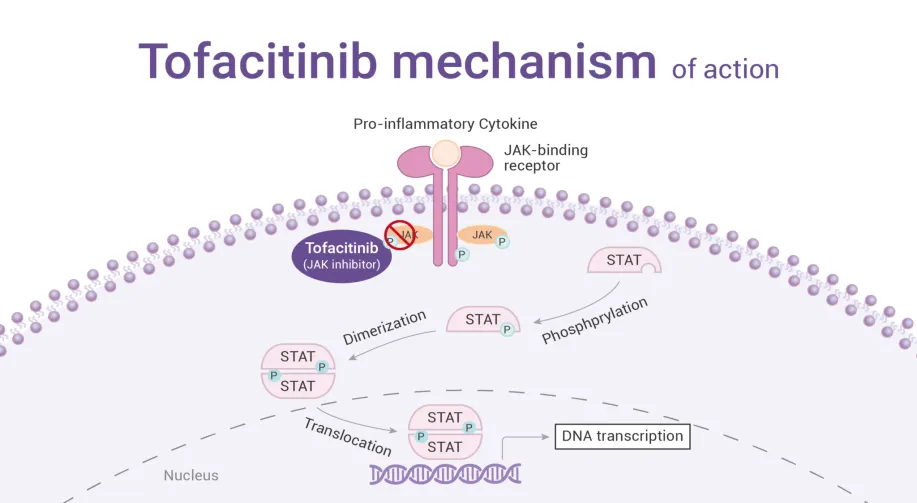
Fig 2: Mechanism of action of tofacitinib
NANOPARTICLE-BASED DRUG DELIVERY SYSTEMS FOR RHEUMATOID ARTHRITIS
Targeted, controlled, and sustained release of anti-rheumatic therapy directly to inflamed synovial tissues is made possible by new advanced drug delivery techniques, such as nanotechnology-based carriers (liposomes, polymeric nanoparticles, gold nanoparticles) that minimize systemic toxicity and maximize the therapeutic index. Preparing bioactive substances in biocompatible nanosystems, such as nanoparticles (NPs), is necessary for nano-sized medication delivery. The sizes of NPs range from 1 to 100 nm. There are various medical uses for nanoparticles (NPs). By delivering anti-inflammatory or immunomodulatory medicines while concentrating them in an inflamed joint, nanotechnology aims to improve the therapeutic index of RA drugs. In rheumatoid arthritis models, a variety of nanocarriers, such as liposomes, hyalurosomes, solid lipid nanoparticles, ethosomes, transferosomes, dendrimers, gold nanoparticles, nanotubes, metal-organic frameworks, and quantum dots, have demonstrated encouraging therapeutic results(20).
Advantages of nanoparticles based drug delivery systems
Classification of nanoparticles(20)
1. Organic particles
Well-known examples include ferritin, liposomes, dendrimers, and various organic nanoparticles or polymers. Some of these nanoparticles, such as micelles and liposomes, contain a hollow core and are susceptible to electromagnetic and thermal radiation, including heat, but they are all non-toxic and biodegradable.
2. Nanoparticles that are inorganic
Inorganic nanoparticles are particles made of materials other than carbon. Metal and metal oxide-based nanoparticles are commonly referred to as inorganic nanoparticles.such as metal nanoparticles and nanoparticles based on metal oxides.
3. Nanoparticles based on carbon
Fullerenes and carbon nanotubes (CNTs) are the two primary types of carbon-based nanoparticles. Allotropic forms of carbon and other nanomaterials made of globular hollow cages are found in fullerenes. They have demonstrated a strong interest in business.
Polymeric nanoparticles for effective targeting of RA
By controlling the immune system, preventing the inflammatory response, and encouraging tissue regeneration, polymers can treat a variety of illnesses, including rheumatoid arthritis. The intrinsic biochemistry and functional integration of polysaccharides, particularly hyaluronic acid (HA) and chitosan (or their chemically modified derivatives), make them a distinct class of biomaterials for IA drug delivery. Chitosan and its water-soluble derivatives, such as carboxymethyl-chitosan, provide mucoadhesive interactions, pH/enzymatic responsiveness, and simple chemical handles for conjugation. Chitosan coatings or matrices may enhance cartilage penetration, encourage nanoparticle retention at the synovial surface, and enable stimulus-triggered release of the payload in acidic/proteolytic microenvironments(3).
Table no 2. Advantages and limitations of natural polymers in RA.
|
Polymer |
Advantages |
Limitations |
|
Hyaluronic acid |
1.Long-term IA drug habitation versus free drug(21) 2.decrese synovial inflammation and systemic toxicity |
1. The impact of native HA is inconsistent and low. 2. High safety issues |
|
Chitosan |
1.Create sustained- release in situ de pots. Strong adherence pre vents inflammatory diffusion and enhances retention(22). 2.Minimise joint injury, discomfort, and inflammation |
1. Irritation and complement activation may result from a high cationic charge. |
|
Chondroitin Sulfate |
1.Deep penetration and effective cartilage targeting. Extended retention(23). 2. Improved cartilage repair and cytokine decrease. |
1.Possible immunogenicity issues |
|
Dextran |
1.Increased joint accumulation and effectiveness compared to the free drug(24). 2.Reduced toxicity to the system |
1. High doses carry a risk of immunogenic or anticoagulant consequences. |
|
Pullulan |
1. Increases the drug's solubility and bioavailability. 2. Lowers inflammation and damage to bone and cartilage(25) |
1.Modification is required for targeting. |
CHITOSAN NANOPARTICLE
Chitosan is a partially deacetylated cationic polymer derived from chitin. Its structure is made up of N-acetyl D-glucosamine and (1→4) connected D glucosamine units. In intra-articular procedures, it has been shown that chitosan-based nanoparticles have a good binding ability with the cartilage tissue and are readily absorbed by synovial macrophages. This makes it possible for anti-inflammatory proteins, nucleic acids, and tiny molecules to be released locally and specifically in arthritic joints. Because of their high accessibility, huge surface area, and molecular links to biological processes, they exhibit increased biological activity. One limitation on the use of chitosan is its solubility. Only in acidic settings can it disintegrate. The small size of chitosan nanoparticles often improves the surface area where they can interact with the substrate, increasing activity. One of the most significant features of chitosan nanoparticles is their ability to eradicate free radicals, which are unstable, reactive substances that can cause cellular stress and damage (3-26).
Preparation of chitosan nanoparticles through ionic gelation method
After dissolving the chitosan in a 0.35% (w/v) acetic acid solution, the mixture was left at room temperature for the whole night. The pH of the solution was then lowered to 5.5 using 0.5 M NaOH. A 0.25% (w/v) TPP (sodium tripolyphosphate) solution was prepared in deionized water. The material was made by combining the chitosan solution with the 0.25% TPP solution at a chitosan: TPP ratio of 6:1 (w/w) and using a magnetic stirrer to agitate at 900 g for 60 minutes at room temperature. After centrifuging the solutions at 5000 g to remove white precipitates, the chitosan nanoparticles were recovered using a freeze dryer. The nanoparticles were then stored at 5 ◦ 2 C in glass tubes(26).
Characterization of Nanoparticles
Using sophisticated microscopic methods like atomic force microscopy, transmission electron microscopy (TEM), and scanning electron microscopy (SEM), nanoparticles are often described by their size, shape, and surface charge. The physical stability and in vivo distribution of the nanoparticles are influenced by their average particle diameter, size distribution, and charge. The general shape of polymeric nanoparticles, may impact their toxicity, can be determined with the help of electron microscopy techniques. The polymer dispersion's physical stability, redispersibility, and in vivo performance are impacted by the nanoparticles' surface charge(27)
Size of particles:
The two most crucial aspects of characterizing nanoparticles are their shape and size distribution. Electron microscopy is used to measure size and morphology. Drug targeting and release are the main uses of nanoparticles. Particle size has been demonstrated to have an impact on medication release. Greater surface area is provided by smaller particles. Fast drug release will occur from the majority of the drug placed onto them being exposed to the particle surface.
DLS, or dynamic light scattering:
Nowadays, dynamic light scattering (DLS) or photon-correlation spectroscopy (PCS) are the fastest and most widely used techniques for detecting particle size. Brownian nanoparticles in colloidal suspensions in the nano and submicron ranges are frequently measured using DLS.
Scanning electron microscopy:
Scanning electron microscopy (SEM) provides direct viewing for morphological analysis. Electron microscopy-based methods have a number of benefits for morphological and sizing studies, but they don't reveal much about the size distribution and actual population average. The initial step in SEM characterization is to turn the nanoparticle solution into a dry powder, mount it on a sample holder, and then use a sputter coater to coat it with a conducting metal, like gold. After that, a concentrated, fine electron beam is employed to scan the sample.
Transmission electron microscope:
Although TEM and SEM operate on separate principles, they frequently provide the same kind of data. Because the sample must be extremely thin for electron transmission, TEM sample preparation is difficult and time-consuming. The dispersion of nanoparticles is applied on films or support grids. Nanoparticles are fixed using either plastic embedding or a negative staining substance, such as phosphotungstic acid or derivatives, uranyl acetate, etc., to make them resistant to the instrument vacuum and easier to handle.released from the sample's surface provide information about its surface properties.
Atomic force microscopy:
By physically scanning samples at the sub-micron level with an atomic-scale probe tip, atomic force microscopy (AFM) enables ultra-high resolution in particle size assessment. The instrument creates a topographical map of the sample based on forces between the tip and the sample surface. Depending on their characteristics, samples are often scanned in either contact or noncontact mode.
Surface Charge:
The type and strength of a nanoparticle's surface charge is crucial because it affects how it interacts with the biological environment and how it interacts electrostatically with bioactive substances. The zeta potential of nanoparticles is used to investigate colloidal stability. The surface charge is indirectly measured by this potential. It is equivalent to the potential difference between the shear surface and the outer Helmholtz plane. Predictions regarding the storage stability of colloidal dispersion are made possible by the measurement of the zeta potential. To guarantee stability and prevent particle aggregation, high zeta potential values—whether positive or negative—should be attained. The zeta potential readings can then be used to forecast the degree of surface hydrophobicity.
Future opportunities and challenges
Drug delivery methods using nanoparticles have already been successfully implemented. One of the key instruments in nanomedicine, nanoparticles offers enormous benefits in terms of medication targeting, transport, and the possibility to integrate diagnosis and therapy. The sizes of NPs range from 1 to 100 nm. There are various medical uses for nanoparticles (NPs). The improved solubility of medications that are poorly soluble in water is one major benefit. By increasing surface area and surface contacts, drug nanoparticles with low water solubility might enhance dissolution rates and alter pharmacokinetic characteristics. Preclinical research demonstrates that polysaccharide-based nanoplatforms prolong their stay in the joint, enable regulated and stimulus-responsive drug release, and improve cellular uptake, all of which ultimately result in the suppression of pro-inflammatory cytokines, modification of macrophage phenotypes, and preservation of cartilage and bone. Furthermore, localized distribution reduces the medications' exposure to the rest of the body, which reduces the likelihood of side effects, which are a significant drawback of both conventional and biologic therapy(3-27)
The following methods are difficult to create technically:Virus-like systems for intracellular systems, biomimetic polymer architecture, sensitive drug control, functions (of active drug targeting, bioresponsive triggered systems, systems interacting with body smart delivery), nanochips for the release of nanoparticles, and carriers for advanced polymers for the delivery of therapeutic peptides and proteins. To administer or regulate the quantity and pace, drug delivery methods were developed. The majority of significant and well-established internal drug delivery research programs use formulations and dispersions with nanoscale components(27).
NANOPARTICLE LOADED HYDROGEL FOR MANAGEMENT OF RHEUMATOID ARTHRITIS
A symmetric, long-term autoimmune condition is rheumatoid arthritis (RA). Joint deformities and associated systemic symptoms cause RA patients to lose joint function, become disabled, and have a shorter life expectancy as the disease progresses. An alternate approach to treating RA is the delivery of medications using biomaterials. Too far, numerous drug delivery technologies including liposomes , carbon nanotubes , noble metal nanoparticles , and so on have been developed to increase the safety and bioavailability of drugs(28). There are essentially two types of hydrogels for the treatment of rheumatoid arthritis. One is intra-articular injection hydrogel, and the other is transdermal hydrogel. These two forms of hydrogels have their distinct features.
Transdermal administration can circumvent nonlinear pharmacokinetics and the gastrointestinal tract, so as to decrease the gastrointestinal side effects. At the same time, transdermal administration has an extended duration of action and can also minimize the pain of subcutaneous injection, minimizing the risk of related problems and enhancing the patient compliance. The transdermal hydrogels for the treatment of rheumatoid arthritis should have the following biological features. First of all, transdermal hydrogels must be biocompatible, and they should have little irritation and toxicity to the skin. Secondly, hydrogels must enhance their permeability to break past the skin barrier and enter the joints to play a role(29).
Properties of hydrogel materials required for treatment of rheumatoid arthritis
Persistent synovitis is the primary feature of rheumatoid arthritis. Histologically, it displays angiogenesis, inflammatory leukocyte inflow, cellular hyperplasia, and altered expression of proteinases, many cytokines, cell-surface adhesion molecules, and proteinase inhibitors. In addition to avoiding the discomfort of subcutaneous injection, transdermal administration provides a longer duration of action, which lowers the risk of associated problems and increases patient compliance. Transdermal hydrogels need to be biocompatible and cause very little skin irritation or toxicity. In order to play a part, hydrogels must also increase their permeability in order to penetrate the skin barrier and reach the joints. Lastly, in order to sustain the necessary concentration for a long time, they should have a high drug encapsulation rate(29).
There are two primary forms of hydrogels used in the treatment of rheumatoid arthritis. One type can carry therapeutic medicines. The drugs themselves are chemically cross linked or self-assembled into a hydrogel through the interactions of noncovalent supramolecules in the other hydrogels, which are made from therapeutic agents and are self-assembled or chemically cross linked into the materials for the preparation of hydrogels.
Release mechanism of hydrogel(30)
Temperature-Responsive Type
A class of thermoreactive molecularly imprinted hydrogels known as temperature-responsive hydrogels regulates drug release through dynamic molecular binding sites that undergo conformational changes in response to temperature changes. Drug leakage is reduced below the transition temperature and increases above it. They are therefore perfect for temperature-triggered medication administration.
pH-Responsive
pH-sensitive linkages enable pH-responsive hydrogels to expand, shrink, or degrade in response to changes in the pH of the surrounding environment, hence controlling drug release. For example, in mildly acidic environments (pH 5.0), Schiff bases and borate ester linkages hydrolyze, facilitating medication release. Additionally, in both acidic and alkaline environments, acylhydrazone linkages can cause structural alterations that impact drug diffusion. Similar to temperature-responsive hydrogels, the network structures of these hydrogels expand or contract in response to changes in pH. In alkaline conditions, expansion increases, pore size increases, and drug diffusion is enhanced; in acidic environments (such as pH 3.0 or 5.4), release accelerates to satisfy microenvironmental requirements.
ROS-Responsive
By cleaving ROS-sensitive chemical bonds in the polymer structure, ROS-responsive hydrogels are intended to release medicinal medicines. For example, reactive oxygen species (ROS), which are overexpressed at diseased areas like inflamed or wounded tissues, cause thioether-based bonds to undergo oxidative scission. This technique minimizes off-target effects by enabling highly localized and on-demand medication release. Furthermore, with external stimuli such as ultrasonic irradiation, nanocarriers functionalized with thioether-containing materials can produce ROS. The ensuing oxidation process damages the network integrity of the nanocarrier, causing fast structural disintegration that speeds up the release of medications that have been enclosed.
Light-Responsive Type
In light-responsive hydrogels, photolysis is a common release mechanism in which certain light wavelengths, such as ultraviolet (UV) or near-infrared (NIR), cleave photosensitive chemical bonds within the hydrogel. NIR irradiation causes coumarin ester groups in NIR-responsive hydrogels to dissolve, dissociating the hydrogel network and releasing medications like doxorubicin (DOX). This method allows for precise spatiotemporal drug release and is widely used in anticancer therapy.
Enzyme-Responsive Type
Enzymatic hydrolysis of internal chemical bonds in enzyme-responsive hydrogels causes the cross-linked network structure to break down or collapse, releasing therapeutic chemicals. The activity of β-lactamase, which selectively cleaves β-lactam cross-linking agents incorporated within the hydrogel matrix, serves as an example. The hydrogel's mass and structural integrity are significantly altered by this enzymatic cleavage, which makes it easier to release encapsulated nanoparticles under regulated conditions. Because the degradation process only starts when target enzymes are present, these systems are particularly selective in their responsiveness, which enhances the accuracy and localization of drug delivery.
Site specific drug delivery
In order to improve local medication delivery, NP-gels use nanoparticles incorporated in three-dimensional polymer matrices. Drug localization is handled by the hydrogel network, which enables the design of nanoparticles to be concentrated on regulating drug release. By separating the temporal and spatial components of drug delivery material design, this hybrid paradigm provides a modular approach for highly functional drug targeting applications. By adding targeting ligands to hydrogels that can bind specifically to disease locations, site-specific therapeutic targeting can also be accomplished. For instance, hyaluronic acid (HA), a naturally occurring glycosaminoglycan, is a major component of synovial fluid, which is essential for cartilage maintenance and joint lubrication.A promising method for enhancing targeted drug delivery to inflamed joints is to take advantage of the CD44 receptor-HA interaction. This will increase the drug's local concentration and decrease systemic side effects. Therefore, functionalizing transethosomes with HA opens the door to CD44-mediated drug transport to the synovial membrane, which could transform RA treatment(31-32). Novel bioadhesive hydrogels that operate exceptionally well in a variety of physiological settings are now developing quickly.It is anticipated that combining these hydrogels with drug-loaded nanoparticles will produce new NP-gels with improved drug delivery effectiveness.
CONCLUSION
An autoimmune condition called rheumatoid arthritis (RA) is typified by synovitis. It greatly affects patients' quality of life and is quite prevalent worldwide. The therapy of this illness still faces many challenges and issues.RA has a complicated etiology that involves both environmental and genetic variables. While environmental factors like smoking and infection can cause the start of disease, genetic factors raise the chance of disease.
Hydrogel loaded with nanoparticles offers a biologically sound way to improve the efficacy of intra-articular therapy for rheumatoid arthritis. They benefit from receptor-mediated interactions of polysaccharides like chitosan and hyaluronic acid, as well as biocompatibility and biodegradability. According to the research, polysaccharide-based nanotechnologies prolong joint retention, enable regulated and stimulus-responsive drug release, and increase cellular absorption, which occasionally results in the suppression of pro-inflammatory cytokines, changes in macrophage phenotypes, and protection of bone and cartilage.
There for nanoparticle loaded hydrogel approach will reduce systemic side effects caused by oral therapy, increase drug concentration at effected joints and enhance therapeutic outcome. By formulating as nanoparticles, it can easily penetrate through the skin barrirers and reach into the affected joints; therefore site specific drug delivery can be achieved.
REFERENCES


Arya Rathnakaran, Santhosh Kumar Chinnayyan, Anjana T V, Devika K V, Malavika P R, Prathuish T, Nanoparticle Loaded Hydrogels For Targeted Delivery Of Tofacitinib In Rheumatoid Arthritis: Current Advances And Future Perspectives, Int. J. of Pharm. Sci., 2026, Vol 4, Issue 8, 4140-4154, https://doi.org/10.5281/zenodo.22095314
 10.5281/zenodo.22095314