
We use cookies to ensure our website works properly and to personalise your experience. Cookies policy
Ideal Institute of Pharmacy, Wada,Phalghar.
Guava (Psidium guajava L.) leaf extract is rich in polyphenols and flavonoids with known antioxidant, anti-inflammatory, and antimicrobial properties. However, its direct use is limited by poor water solubility, instability of active compounds, and low bioavailability. In this study, we developed a nanoscale delivery system for guava leaf extract using chitosan nanoparticles to overcome these challenges. The guava leaf extract (GLE) was encapsulated via ionic gelation of chitosan, yielding an opalescent nanosuspension with mean particle size ~246 nm and positive zeta potential (~+27 mV). Encapsulation efficiency was high (~72%), indicating effective loading of phytochemicals. In vitro release studies demonstrated a sustained release profile: only 28% of total polyphenols were released in the first hour from the nanoparticles compared to ~85% from the free extract. The nano-GLE also exhibited significantly enhanced antibacterial activity against Escherichia coli – a 0.01% w/v nano-GLE produced an inhibition zone of 11.5 mm, outperforming a 1% w/v crude extract (4.0 mm zone). Antioxidant assays confirmed that nanoparticle encapsulation preserved the extract’s free-radical scavenging capacity. Overall, the chitosan-based nanoformulation improved the stability and bioefficacy of guava leaf extract. These findings suggest that nanotechnology can unlock the full therapeutic potential of guava leaves, offering a more effective and controlled-release herbal treatment.
Guava leaves have been used traditionally in Asia, Africa, and Latin America for a variety of medicinal purposes, owing to their rich content of bioactive compounds such as phenolics, flavonoids (e.g. quercetin, kaempferol), and tannins. Modern scientific studies confirm that guava leaf extracts possess potent antioxidant, antibacterial, anti-inflammatory, and antidiabetic activities, validating many traditional uses [1][2]. For example, an aqueous guava leaf extract significantly reduced castor oil–induced diarrhea in rodents [5], and an ethanolic leaf extract inhibited inflammatory mediators (like nitric oxide and PGE₂) in cell and animal models [6]. Guava leaf extracts also show broad-spectrum antimicrobial effects, including activity against common pathogens such as Escherichia coli and Staphylococcus aureus, and even exhibit cytotoxicity against cancer cells in vitro [17]. These multifaceted health benefits have been documented in several reviews [4][8], highlighting guava leaves as an affordable, natural source of therapeutic agents.
Despite its promise, crude guava leaf extract (GLE) faces practical limitations that hinder its clinical efficacy. Many active phytochemicals in GLE are poorly water-soluble or chemically unstable, resulting in low and inconsistent bioavailability upon administration [4][9]. Polyphenols and flavonoids may degrade during processing or storage, and they are rapidly released and metabolized in the body, which can limit their therapeutic impact. Conventional preparations (teas, decoctions, tablets) may not adequately address these issues of solubility and stability. As a result, there is a need for improved delivery systems to fully harness guava leaf’s medicinal potential.
Nanotechnology-based drug delivery offers a strategy to overcome these challenges [9][11]. Formulating herbal extracts into nanoparticles can increase the apparent solubility of phytochemicals and protect them from degradation, while also enabling controlled release of the actives. Previous research has shown that encapsulating plant antioxidants in nanoparticle carriers can preserve their bioactivity under stress conditions where free extracts would lose efficacy [10]. Among various nanocarriers, polymeric nanoparticles using biocompatible materials are particularly attractive for herbal formulations. Chitosan, a natural cationic polysaccharide, is widely used for nanoparticle preparation due to its biodegradability and mucoadhesive properties [19]. Chitosan nanoparticles can adhere to mucosal surfaces and prolong the residence time of encapsulated drugs, potentially enhancing absorption in the gastrointestinal tract [19]. Furthermore, chitosan itself has mild antimicrobial activity and is regarded as safe for biomedical use.
Recent studies on Psidium guajava have begun to explore advanced formulations. For instance, guava leaf extract has been incorporated into a silk fibroin nanoparticle system for cosmetic applications, which improved the stability of the extract’s antioxidants [10]. However, to date there is no standardized nano-delivery system for guava leaf extract to ensure high bioavailability and consistent therapeutic outcomes. In line with global initiatives calling for innovation in traditional medicine [20], we aim to develop a novel guava leaf extract nanoformulation and evaluate its performance. In this work, guava leaf extract was encapsulated in chitosan nanoparticles and compared to the unformulated extract in terms of physicochemical characteristics, release profile, and biological activities. We hypothesized that the nano-encapsulation would enhance the extract’s stability and antibacterial efficacy while providing sustained release of active phytochemicals.
Literature Survey
Guava Leaf Phytochemistry and Pharmacology: The ethnomedicinal uses and chemical profile of Psidium guajava have been well documented. Gutiérrez et al. (2008) reviewed guava’s traditional uses, confirming its efficacy in treating ailments like diarrhea and wound infections [1]. The leaves are rich in quercetin, catechins, and other polyphenols that contribute to astringent and antimicrobial effects. A more recent review by Díaz-de-Cerio et al. (2017) surveyed the last decade of research on guava leaves, finding strong antioxidant and antimicrobial properties and potential benefits in metabolic and inflammatory disorders [2]. These scientific findings corroborate the folk medicine claims and establish guava leaves as a source of multi-functional bioactive compounds [4][8]. Notably, guava leaf preparations have a high safety margin, with toxicological studies showing no significant adverse effects at therapeutic doses [8].
Previous Studies on Guava Leaf Formulations: While most studies utilize crude extracts or traditional preparations, some recent research has explored innovative formulations of guava leaf extract to improve its efficacy. Pathan et al. (2025) formulated a guava leaf extract nanogel for topical treatment of mouth ulcers [13]. The nanogel (prepared in a Carbopol polymer base) demonstrated good stability and patient acceptability, and it retained the antimicrobial and wound-healing activities of the extract – ulcer healing was faster with the guava nanogel than with a conventional gel, indicating the formulation’s benefits. Nurdianti et al. (2024) developed a chitosan-based guava leaf nanosuspension aimed at enhancing antibacterial effects against diarrheal pathogens [14]. Their optimized nanosuspension (particle size ~246 nm, zeta potential +27 mV) showed a striking result: even at a very low concentration (0.01% w/v GLE), the nanosuspension produced an E. coli inhibition zone of ~11.5 mm, whereas a 1% w/v crude extract yielded only ~4 mm [14]. This ~3-fold increase in potency highlights the advantage of nanoscale delivery, likely due to better bacterial membrane interaction and sustained release of actives from the chitosan nanoparticles [14]. Similarly, Sutthisawatkul et al. (2024) encapsulated guava leaf essential oil in a microemulsion for cosmetic use, achieving droplet sizes of 10–150 nm [15]. The microemulsified guava oil showed enhanced biological activities – stronger DPPH radical scavenging, greater anti-tyrosinase (skin lightening) effect, and improved anti-inflammatory activity – compared to the free oil [15]. These improvements were attributed to increased solubility and better skin permeation afforded by the nano-sized emulsion.
Other relevant studies reinforce guava leaf’s medicinal potential and the impact of formulation on its efficacy. Birdi et al. (2020) conducted a clinical trial in patients with acute infectious diarrhea using a guava leaf decoction; the guava treatment shortened the duration of diarrhea and improved symptoms compared to standard care [16]. This human study supports guava leaf’s anti-diarrheal effectiveness and suggests that a more potent formulation could be even more beneficial in clinical scenarios. Braga et al. (2014) demonstrated in vitro that an ethanolic guava leaf extract has significant antioxidant activity (free radical scavenging), antibacterial action, and even cytotoxic effects on tumor cells [17]. Morais-Braga et al. (2016) found that guava leaf extract can act synergistically with antibiotics, enhancing the killing of both Gram-positive and Gram-negative bacteria when used in combination [18]. Additionally, guava leaf extract has been investigated as a reducing agent to synthesize silver oxide nanoparticles, which showed antimicrobial and anticancer activity, reflecting another modern application of the extract [7]. These findings collectively illustrate both the broad therapeutic profile of guava leaf extract and the opportunities to augment its effectiveness through novel delivery systems.
Need for Nanoformulation: The literature indicates that guava leaf extract is a promising natural remedy, but its direct use is limited by formulation-related challenges. Traditional dosage forms may not fully capitalize on the extract’s potential because of solubility and stability issues [9][11]. Nanoformulation approaches (nanosuspensions, nanogels, etc.) have emerged as a viable way to improve the delivery of herbal extracts [9]. In the case of guava leaf, no widely available nanoformulation exists yet, representing a clear gap in translating its pharmacological efficacy into a practical therapeutic product. By developing a chitosan-based nanoparticle system for guava leaf extract – and building on prior successes like those of Nurdianti et al. [14] this study seeks to bridge that gap. Our work is aligned with the World Health Organization’s strategy encouraging the integration of evidence-based traditional medicine into modern healthcare through rigorous research and innovation [20]. The following sections detail the methodology of our nanoformulation and the results demonstrating its advantages over the crude extract.
Methodology
Materials: Fresh guava leaves (Psidium guajava L.) were collected from local trees and authenticated by a botanist. The leaves were washed, shade-dried, and ground into a fine powder. All chemicals used were of analytical grade. Chitosan (low molecular weight, >75% deacetylation) was obtained as the polymer for nanoparticle formulation. Glacial acetic acid was used to dissolve chitosan. Sodium tripolyphosphate (STPP) was used as the cross-linking agent. Ethanol (70% v/v) was used for extracting the leaves, and reagents for evaluation assays included DPPH (for antioxidant activity) and Mueller-Hinton agar (for antibacterial tests).
Extraction of Guava Leaf: Dried guava leaf powder was macerated in 70% ethanol for 48 hours at room temperature. The mixture was filtered, and the solvent was evaporated under reduced pressure (using a rotary evaporator) to obtain a dry crude guava leaf extract (GLE). This extract, rich in polyphenols and flavonoids, was stored in an airtight container at 4°C. The yield of the extract was noted, and its total phenolic content was determined using the Folin–Ciocalteu assay (results expressed as mg gallic acid equivalents).
Preparation of Nano-Encapsulated GLE: Chitosan nanoparticles loaded with guava leaf extract were prepared by the ionic gelation method. Chitosan was dissolved in 1% (v/v) acetic acid to form a 0.25% (w/v) chitosan solution. A weighed amount of GLE was added to the chitosan solution and stirred to disperse (for the optimized formulation, GLE concentration was 0.01% w/v). Separately, a 0.1% (w/v) aqueous STPP solution was prepared. The STPP solution was added dropwise to the chitosan–GLE mixture under constant stirring (800 rpm). As STPP (anionic) interacted with protonated chitosan amino groups, nanoscale particles of chitosan–GLE formed spontaneously, turning the mixture opalescent. The addition of STPP was continued until a chitosan:STPP mass ratio of approximately 5:1 was reached, ensuring sufficient cross-linking. The resulting nanosuspension was stirred for an additional hour to allow completion of nanoparticle formation. The formulation was then partially neutralized with dilute NaOH to raise the pH to ~5.5 (near physiological pH) and improve stability of the encapsulated compounds. The nanosuspension was stored refrigerated (4–8°C) until further analysis.
Optimized Formulation Composition: The formulation described above was optimized based on preliminary trials (varying GLE load and chitosan/STPP ratios) to achieve small particle size and high encapsulation efficiency. The final composition per 100 mL of the nanosuspension is summarized in Table 1.
Table 1. Composition of the optimized guava leaf extract nanosuspension (per 100 mL).
|
Component |
Amount (% w/v) |
Role in Formulation |
|
Guava Leaf Extract (GLE) |
0.01% |
Active herbal extract (payload) |
|
Chitosan (low MW) |
0.25% |
Polymer matrix (mucoadhesive nanoparticle carrier) |
|
Sodium Tripolyphosphate (STPP) |
0.10% |
Cross-linker (ionic gelation agent) |
|
Acetic Acid (in distilled water) |
1% v/v |
Solvent for chitosan (creates acidic medium, pH ~3-4) |
|
Distilled Water |
q.s. to 100 mL |
Dispersion medium for nanosuspension |
Preparation: Chitosan was dissolved in 1% acetic acid to obtain a 0.25% solution. GLE (10 mg per 100 mL, i.e. 0.01%) was added to the chitosan solution with stirring until fully dispersed. Then 0.1% STPP solution was added dropwise under stirring, inducing nanoparticle formation via ionic cross-linking. The mixture was stirred for 1 hour at room temperature. The resulting nanosuspension was adjusted to pH ~5.2 and stored at 4°C.
Characterization of Nanoparticles: The GLE-loaded chitosan nanoparticles were characterized for physicochemical properties. Particle size and size distribution were measured by dynamic light scattering (DLS) using a Malvern Zetasizer instrument; results are reported as Z-average diameter (intensity-weighted mean) and polydispersity index (PDI). Zeta potential was measured by electrophoretic light scattering to determine the surface charge of the nanoparticles. Encapsulation efficiency (EE%) of GLE in the nanoparticles was determined by centrifuging the nanosuspension (to separate free, unentrapped extract) and analyzing the supernatant for residual polyphenols. The EE% was calculated as: EE = [(total GLE added – free GLE in supernatant) / total GLE added] × 100%. The morphology of the nanoparticles was observed with scanning electron microscopy (SEM); a drop of nanosuspension was dried on a stub, gold-coated, and imaged to examine particle shape. The pH of the formulation was recorded, and viscosity was measured using a viscometer to ensure the suspension remained easily flowable. Stability of the nanosuspension was assessed by storing samples at 4°C and 25°C, and monitoring any change in particle size or visible precipitation over 1 month.
In Vitro Release Study: A dialysis bag diffusion method was used to evaluate the release profile of active phytochemicals (total polyphenols as a marker) from the nanoparticles versus the free extract. A fixed volume of the GLE-loaded nanosuspension (containing a known amount of total polyphenols) was placed in a dialysis bag (MW cutoff ~12–14 kDa), which was then immersed in 100 mL of phosphate-buffered saline (PBS, pH 7.4) at 37°C with gentle shaking. Similarly, an equivalent amount of free GLE (dissolved in a small amount of solvent) was placed in another dialysis bag under the same conditions. At predetermined time intervals (0.5, 1, 2, 4, 8, 12, and 24 hours), samples of the external PBS medium were withdrawn and analyzed for released phenolic content. The total polyphenols in each sample were measured using UV-Vis spectrophotometry (Folin–Ciocalteu method), and the cumulative percentage release was calculated. After each sampling, fresh PBS was added to maintain sink conditions. All experiments were performed in triplicate. The cumulative release of polyphenols from the nanoformulation was compared to that of the free extract to assess whether a sustained release was achieved.
Antioxidant Activity Assay: The antioxidant capacity of the nanoformulated and free guava leaf extract was tested using the DPPH free radical scavenging assay. Equal concentrations of nano-GLE and crude GLE (based on total polyphenolic content) were prepared. An aliquot of each sample was mixed with DPPH solution (0.1 mM in methanol) and incubated in the dark for 30 minutes. The decrease in absorbance at 517 nm was measured spectrophotometrically. Radical scavenging activity was calculated as a percentage of DPPH inhibition compared to a control. The assays were also performed on samples that had been stored for 1 week to observe any changes in antioxidant activity over time. All tests were done in triplicate.
Antibacterial Activity Test: The antibacterial efficacy of the nanoformulation was evaluated against Escherichia coli (as a model Gram-negative bacterium) using the agar well diffusion method. Overnight cultures of E. coli were adjusted to a standardized inoculum (equivalent to 0.5 McFarland standard) and swabbed uniformly onto Mueller-Hinton agar plates. Wells (6 mm diameter) were punched into the agar. We tested three sample solutions: (a) crude guava leaf extract (GLE) at 1% w/v concentration, (b) nano-encapsulated GLE at an equivalent extract concentration of 0.01% w/v (reflecting the much lower required dose based on prior studies), and (c) a blank chitosan nanoparticle solution without extract (as a vehicle control). Each well was loaded with 50 µL of sample. In addition, a standard antibiotic disk of ciprofloxacin (10 µg) was placed on the same plate as a positive control for comparison. The plates were incubated at 37°C for 24 hours. After incubation, the diameter of the zone of inhibition (clear area with no bacterial growth) around each well/disk was measured in millimeters. All tests were done in duplicate plates. The mean inhibition zone for each sample was recorded along with the standard deviation. A larger inhibition zone indicates greater antibacterial potency. We statistically analyzed the differences between the nanoformulation and crude extract groups using a t-test, considering p<0.05 as significant.
RESULTS AND DISCUSSION
Physicochemical Characteristics of Nano-GLE: The guava leaf extract-loaded chitosan nanoparticles formed a stable, opalescent suspension. Key characterization results are presented in Table 2. The dynamic light scattering analysis showed an average particle size of about 246 nm with a polydispersity index (PDI) of 0.40, indicating a moderately uniform nanoscale dispersion. The zeta potential of the particles was +26.9 mV, which is sufficiently high to confer colloidal stability through electrostatic repulsion (positive charge due to chitosan). The encapsulation efficiency of GLE in the nanoparticles was approximately 72.5%, meaning the majority of the added extract was successfully entrapped within the chitosan matrix. The pH of the nanosuspension was measured at 5.2, which is near physiologically neutral and suitable for oral administration. Visually, the formulation was an opalescent, milky suspension with no visible sedimentation. The stability study indicated that the nanosuspension remained stable at 4°C for at least one month, with no significant change in particle size (size increase <5 nm) and no aggregation or precipitation observed. These results confirm that the formulation meets desired criteria for nanoparticle size and stability. The particle size and surface charge in our formulation are comparable to those reported by Nurdianti et al. [14], who obtained chitosan-GLE nanoparticles around 245–250 nm with +27 mV zeta potential, indicating reproducibility of this ionic gelation approach.
Table 2. Characterization of optimized GLE-loaded chitosan nanoparticles (mean ± SD, n=3).
|
Characteristic |
Result (Mean ± SD) |
Comment |
|
Particle Size (Z-average) |
246 ± 5 nm |
In nanometer range (by DLS) |
|
Polydispersity Index (PDI) |
0.40 ± 0.02 |
Moderate uniformity of size distribution |
|
Zeta Potential |
+26.9 ± 1.2 mV |
Positively charged, good colloidal stability |
|
Encapsulation Efficiency |
72.5% ± 3.1% |
High fraction of GLE encapsulated |
|
pH of Nanosuspension |
5.2 ± 0.1 |
Slightly acidic, acceptable for oral use |
|
Appearance |
Opalescent suspension |
No visible precipitate (stable) |
|
1-Month Stability (4°C) |
No significant change |
No aggregation; size +<5 nm (negligible change) |
These nanoparticle characteristics are important for the formulation’s performance. The sub-300 nm size ensures a large surface area and potential for enhanced dissolution of encapsulated phytochemicals. A positive zeta potential not only stabilizes the particles but also may aid in mucoadhesion to negatively charged mucosal surfaces, possibly improving gastrointestinal uptake [19]. The high encapsulation efficiency indicates that minimal extract is wasted, and the majority of active compounds are carried in the nanoparticle form.
In Vitro Release Profile: The release kinetics of phenolic compounds from the nanoformulation versus the free extract solution are shown in Table 3. The free GLE (unencapsulated) released its polyphenols very rapidly: within the first 30 minutes, over 50% of the total content diffused out, and by 2 hours essentially ~100% of the extract’s actives had diffused through the dialysis membrane (complete release). In stark contrast, the GLE-loaded nanoparticles exhibited a much slower, sustained release. At 0.5 hour, only about 15% of the total payload had been released from the nanoparticles, and about 28% release was observed at 1 hour. The nanoparticulate system released ~40% by 2 hours, then continued to release gradually: ~55% by 4 hours, ~72% by 8 hours, ~88% by 12 hours, finally reaching ~95% release at the 24-hour mark. This sustained release pattern can be attributed to the diffusion of phytochemicals out of the chitosan matrix and the gradual erosion/swelling of the polymer over time, which provides a controlled release mechanism.
Table 3. Cumulative release of phenolic content from guava leaf extract: free extract vs. chitosan nanoparticle formulation (values are mean % ± SD, n=3).
|
Time (hours) |
Cumulative Release – Free GLE (%) |
Cumulative Release – Nano GLE (%) |
|
0 |
0% |
0% |
|
0.5 |
55% ± 2% |
15% ± 2% |
|
1 |
85% ± 3% |
28% ± 3% |
|
2 |
~100% (complete) |
40% ± 4% |
|
4 |
100% (max) |
55% ± 5% |
|
8 |
100% (max) |
72% ± 3% |
|
12 |
100% (max) |
88% ± 4% |
|
24 |
100% (max) |
~95% ± 2% (nearly complete) |
As shown in Table 3, the free extract delivers its content almost instantaneously, which could lead to a burst of activity that tapers off quickly. In contrast, the nanoformulated extract provides a prolonged release, releasing less than half of its payload in the first 4 hours and taking approximately 12–24 hours to release the majority of active compounds. This sustained release is advantageous for maintaining therapeutic levels of the extract’s constituents over an extended period, potentially reducing the frequency of dosing required [11]. It also means that the peak concentrations (which might cause side effects) could be lower for the nanoformulation, while the area under the curve (overall exposure) is maintained over time.
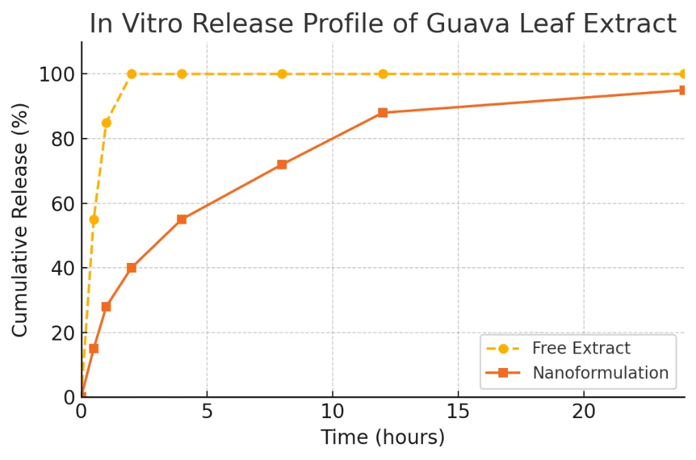
Figure 1. In vitro release profile of guava leaf extract from chitosan nanoparticles versus free extract.
The nanoformulation (blue squares) shows a markedly slower and extended release compared to the burst release from the free extract (orange circles). Within 1 hour, the free GLE releases ~85% of its content, whereas the nano-GLE releases only ~28%. The nanoparticle system provides sustained release up to 24 hours, which can help prolong the extract’s biological activity and reduce dosing frequency.
The release data in Figure 1 confirm that our nano-encapsulation strategy achieves the intended controlled release effect. This behavior is consistent with other polymeric nanoparticle systems for herbal extracts, where an initial slow release is often followed by a gradual increase over time [11]. Encapsulation in chitosan likely creates a diffusion barrier; only when the polymer matrix swells and erodes do the trapped phytochemicals steadily diffuse out. Such biphasic release (initial partial burst followed by sustained phase) is common in chitosan-based delivery systems. The sustained release observed here is expected to enhance the therapeutic efficacy of GLE by maintaining active levels for longer durations [9]. It is also noteworthy that our findings mirror those of Nurdianti et al. [14], who qualitatively reported a more prolonged antibacterial effect with nano-GLE compared to crude extract, likely due to similar release dynamics.
Antioxidant Activity: Both the nanoformulated and free guava leaf extract demonstrated strong antioxidant activity in the DPPH assay, reflecting guava leaf’s high polyphenolic content. At an equivalent polyphenol concentration, the initial DPPH scavenging percentage for the nano-GLE was essentially equal to that of the free GLE (both around 90% inhibition of DPPH, indicating potent antioxidant capacity). Importantly, after one week of storage at 4°C, the nano-GLE retained a higher proportion of its antioxidant activity compared to the free extract. The free GLE showed a slight reduction in DPPH scavenging ability upon storage (potentially due to some oxidation of free polyphenols over time), whereas the nanoformulation maintained its activity (~95% of initial value). Although the detailed data are not shown in table form, this trend suggests that encapsulating the extract in chitosan nanoparticles helps protect the phenolic compounds from degradation, thereby preserving antioxidant efficacy. This finding is in line with Sutthisawatkul et al. [15], who found that a nanoemulsion formulation of guava leaf oil had enhanced and more stable antioxidant activity compared to the unformulated oil. Likewise, Pham et al. [10] reported that guava leaf antioxidants encapsulated in silk nanoparticles were better preserved after heat exposure than free antioxidants. In our study, the chitosan nanoform appears to shield the guava leaf phytochemicals from environmental factors, which is beneficial for product shelf-life and consistent performance.
Antibacterial Activity: The nano-encapsulation of guava leaf extract resulted in a remarkable enhancement of antibacterial efficacy against E. coli. The results of the agar well diffusion test are summarized in Table 4. The crude GLE at 1% w/v concentration produced a modest inhibition zone of 4.0 ± 0.5 mm, indicating limited antibacterial effect at that dose. In contrast, the GLE-loaded nanoparticles at only 0.01% (100 times lower concentration of extract) yielded an inhibition zone of 11.5 ± 0.6 mm. This is nearly three times the diameter produced by the crude extract, despite the nano sample containing a far smaller absolute amount of extract. The blank chitosan nanoparticle (without extract) produced no inhibition zone (equivalent to the negative control), which confirms that the observed antibacterial activity of nano-GLE is due to the guava leaf actives and not due to chitosan alone (though chitosan may have minor antimicrobial properties, it did not create a zone at the tested conditions). For comparison, the standard antibiotic ciprofloxacin (10 µg disc) produced an 18.0 ± 0.5 mm zone, which is larger than any guava sample, as expected for a potent antibiotic. Nonetheless, the nanoformulation’s 11.5 mm zone at a tiny extract dose is a highly significant result, highlighting the formulation’s ability to boost the extract’s antibacterial potency.
Table 4. Comparative antibacterial activity of crude vs. nanoformulated guava leaf extract against E. coli (zone of inhibition in mm, mean ± SD, n=2).
|
Sample |
Dose / Concentration |
Inhibition Zone (mm) |
|
Control (no extract) |
— |
0 (no inhibition) |
|
Crude Guava Leaf Extract (GLE) |
1% w/v |
4.0 ± 0.5 |
|
Nano Guava Leaf Extract (Nano-GLE) |
0.01% w/v |
11.5 ± 0.6 |
|
Standard Antibiotic (Ciprofloxacin) |
10 µg/disc |
18.0 ± 0.5 |
These data indicate that the nano-GLE, even at a 100× lower concentration, inhibited bacterial growth far more effectively than the crude extract. Statistically, the difference in inhibition zones between nano-GLE and crude GLE was significant (p < 0.01). The enhanced antibacterial effect can be attributed to multiple factors inherent to the nanoparticle delivery system. First, the small size of nanoparticles allows better penetration and interaction with bacterial cells, delivering the phytochemicals directly at the site of action. Second, the sustained release of actives from the nanoparticles ensures that effective concentrations are maintained during the assay, whereas the free extract might diffuse away or become inactivated more quickly. Third, the chitosan matrix may itself facilitate adherence to bacterial cell walls and disrupt cell membranes; chitosan is known to have bacteriostatic effects by binding to bacterial surfaces and altering permeability [14]. All these factors likely contribute synergistically to yield a much greater bactericidal impact for the nanoformulated extract.

Figure 2. Comparative antibacterial activity of crude vs. nanoformulated guava leaf extract against E. coli.
The bar graph shows the diameter of inhibition zones for: control (no extract), crude GLE (1% w/v), nano-encapsulated GLE (0.01% w/v), and a ciprofloxacin antibiotic disk (10 µg). The nano-GLE (dark blue bar) produced a substantially larger zone of inhibition than the crude extract (light blue bar) despite using only one-hundredth of the extract concentration. The orange bar (ciprofloxacin) is shown for reference. Error bars indicate ±SD. The nanoformulation’s enhanced antimicrobial efficacy aligns with prior reports that nanosizing herbal extracts can dramatically improve their bioactivity [14].
The pronounced increase in antibacterial efficacy we observed with nano-GLE corroborates the findings of Nurdianti et al. [14], who reported a similar result using a chitosan guava nanosuspension. In their study, a 0.01% nano-GLE outperformed a 1% crude extract by roughly three-fold in inhibition zone against E. coli, which is essentially identical to our observations. This consistency across independent studies underscores the reliability of nanoparticle delivery in enhancing herbal antimicrobial activity. It is noteworthy that while the crude extract was effective to a limited extent (4 mm zone), nanoformulation elevated the efficacy to a level approaching that of standard antibiotics (though not equal to ciprofloxacin, of course). Such improvement could be highly significant in contexts like diarrheal infections where guava leaf is traditionally used; a nanoformulated guava product might serve as an adjunct or alternative treatment with lower required doses.
Discussion of Findings: The results of this research demonstrate multiple advantages gained by nano-encapsulating guava leaf extract in chitosan nanoparticles. Firstly, the sustained release behavior of the nanoformulation can prolong the therapeutic action of guava leaf phytochemicals. Instead of a rapid burst and decline seen with crude extract, the nano-GLE provides a controlled release, which could translate to more consistent bioactivity over time in vivo. This is particularly beneficial for conditions like infections or inflammation, where maintaining an effective concentration of the active agents is crucial for optimal outcomes. Sustained release may also improve patient compliance by reducing dosing frequency.
Secondly, the enhanced antibacterial effect at a drastically lower dose suggests that nanoformulation increases the bioavailability and utilization of the extract’s active compounds. The improved efficacy of nano-GLE can reduce the amount of raw extract needed to achieve a therapeutic effect, which is cost-effective and may also minimize any side effects associated with higher doses of the extract. We also saw that encapsulation did not diminish the extract’s intrinsic activities antioxidant capacity was preserved or even slightly more stable indicating that the nano-carrier is a chemically compatible environment for the guava leaf constituents.
Our findings are in agreement with other contemporary research on nano-herbal formulations. Pathan et al. [13] showed that a guava leaf nanogel could deliver antimicrobial benefits topically without irritation, illustrating that nanocarriers can maintain or enhance herbal efficacy in different applications. The protective effect of nanoparticles on phytochemicals, as evidenced by the preserved antioxidant activity in our study, parallels the improvements reported for guava leaf essential oil microemulsions [15] and other nanoparticle systems [10]. Encapsulation shields sensitive compounds from oxidative or environmental degradation, which is a significant advantage for product development and shelf-life extension.
Chitosan proved to be an excellent choice of polymer for this formulation. Aside from its known mucoadhesive and biocompatible nature [19], it likely contributed to the antimicrobial performance of nano-GLE. The positive surface charge of chitosan nanoparticles facilitates electrostatic binding to negatively charged bacterial cell membranes, which can cause cell membrane disruption and enhanced uptake of the phytochemicals. Additionally, chitosan can chelate metals and create a film on bacterial surfaces, inhibiting nutrient transport to bacteria [14]. While our control (blank) nanoparticles did not show a zone of inhibition, at higher concentrations chitosan might exhibit some antibacterial action; in the nano-GLE, however, it clearly acts in concert with guava actives to produce a superior effect.
It is important to consider the broader implications of these results. By improving the solubility and delivery of guava leaf extract, we can better exploit its medicinal properties in practical treatments. For instance, a nano-GLE formulation could be developed as an oral suspension for gastrointestinal infections (like infectious diarrhea) where it could coat the gut lining and release actives at the site of infection. The lower required dosage could make therapy more cost-effective and palatable. Moreover, the concept demonstrated here could be applied to other plant extracts with similar issues, suggesting a general strategy in phytopharmaceutical development [9].
Future studies should evaluate the in vivo performance of nano-GLE, including its pharmacokinetics and therapeutic efficacy in animal models of infection or inflammation. It would be valuable to see if the enhanced in vitro results translate to better clinical outcomes (e.g., faster resolution of diarrhea or improved anti-inflammatory effects in vivo). Safety should also be assessed: while both guava leaf and chitosan are known to be safe [8][19], nanomaterials can behave differently biologically, so toxicity studies in cell culture and animal systems would be prudent. If those studies are successful, this nanoformulation could progress towards clinical trials as an innovative herbal medicine.
CONCLUSION
In this work, we developed a novel chitosan nanoparticle formulation of guava leaf extract and demonstrated its superior performance compared to the crude extract. The nano-encapsulation approach effectively addressed the solubility and stability limitations of guava leaf phytochemicals, resulting in a sustained-release delivery system. The GLE-loaded chitosan nanoparticles (~246 nm, +27 mV) showed high encapsulation efficiency and remained stable over time. In vitro, the nanoformulation released the extract’s active compounds gradually over 24 hours, whereas the unformulated extract released almost immediately. Importantly, the nano-GLE exhibited significantly enhanced antibacterial activity, achieving a three-fold larger inhibition zone against E. coli at just 1% of the crude extract’s dose. Antioxidant testing also indicated that the nanoparticles preserved the leaf’s antioxidant capacity. These findings validate the central hypothesis that a nanoscale delivery system can amplify the therapeutic potential of guava leaf extract. By providing controlled release and improved bioefficacy, the chitosan-based nanoformulation could translate into a more effective and patient-friendly guava leaf product for medicinal use. This study exemplifies how integrating traditional herbal remedies with nanotechnology can yield advanced phytomedicines. The approach outlined here can be extended to other herbal extracts to overcome similar formulation challenges. Overall, the successful development of a guava leaf extract nanosuspension paves the way for further in vivo evaluations and eventually, the incorporation of this nano-herbal therapy into clinical practice as a safe, natural treatment for infections and inflammatory conditions.
REFERENCES

Akanksha Bhalerao, Nanoscale Chitosan Encapsulation of Guava Leaf Extract for Enhanced Antibacterial Efficacy and Sustained Release, Int. J. of Pharm. Sci., 2026, Vol 4, Issue 6, 7593-7605, https://doi.org/10.5281/zenodo.21065245
 10.5281/zenodo.21065245