View Article

  • Next-Generation Nano-Delivery Systems for Medicinal Plant Bioactives: An Analytical Review of Bioavailability Enhancement, Targeted Therapy, Safety, and Clinical Translation

  • SNJB’s Shriman Sureshdada Jain College of Pharmacy, Chandwad, Nashik 423101.

Abstract

Despite the diversity of existing medicinal plant bioactives which include anti-inflammatory, anticancer, antimicrobial, neuroprotective and metabolic bioactives, their biotherapeutic applications are often hindered by poor water solubility, poor stability and rapid metabolism. This analytical review discusses pharmaceutical applications of nanotechnology in the delivery systems of medicinal plant bioactives. It covers research published from 2020-2025 on lipid-based nanocarriers, polymer-based nanocarriers, micelles, nanoemulsions, porous silica, biomimetic and metallic nanocarriers systems, and other systems. In general, nanotechnology can improve the solubility of compounds, protect unstable compounds, alter the pathway of uptake and enable targeting of the bioactive. However, improved solubility and cell-killing activities of a nanocarrier do not guarantee availability and selectivity of the carrier. These biotherapeutic applications are further restricted by the rapid metabolism of the compounds. Curcumin, quercetin, resveratrol and berberine compounds exemplify this challenge of poor biotherapeutic applications. However, paclitaxel nanocarriers exemplify the successful development of medicinal plant nanocarriers. Formulation safety is enhanced by biocompatibility, surface charge and size. The challenge of poor biotherapeutic applications of most of the phytochemicals is further compounded by the rapid degradation of the compounds and poor translational technologies. In every carrier system, the application of nanotechnology should address a specific phytochemical.

Keywords

medicinal plants; phytochemicals; nanomedicine; nano-delivery systems; bioavailability; targeted drug delivery; phytopharmaceuticals; clinical translation

Introduction

× Popup Image

Even today, many small molecules, mixtures, and structures that are used in modern therapeutics are derived from medicinal plants. Examples of such molecules include curcumin, resveratrol, quercetin, berberine, epigallocatechin gallate, silymarin, thymoquinone, artemisinin, paclitaxel, and camptothecin. They encompass diverse classes of phytochemicals that include polyphenols, alkaloids, terpenoids, and taxanes. Although these molecules are sourced from nature, they do not fulfill the pharmaceutical requirements of identity, dose, safety, and controllable exposure. An active ingredient sourced from plant will be different from a crude herbal formulation. An active ingredient can be defined at the molecular level, and can be manufactured at the specified standards. On the other hand, a herbal formulation will contain numerous substances and will complicate pharmacokinetics and quality control [1-8].

In many cases, the potential of a phytochemical is promising, but it also presents challenges in achieving the desired exposure. Nanocarriers can solve certain limitations of phytochemicals by providing a drug reservoir that can be dispersed, protecting the drug, and controlling the release, and by increasing contact with the mucosa and promoting endocytosis and lymphatic transport, as well as design specific interactions with tissues. The main consideration is not whether nanoparticles impose an increase in-vitro activity, but whether they provide a meaningful increase in exposure in a safe and reproducible manner, in comparison to traditional formulations [9-13].

2. Literature-Review Approach

A methodical and structured search covered PubMed, Scopus, Web of Science, ScienceDirect, Google Scholar, Crossref, and official regulatory websites, from January 2020 – December 2025. Earlier sources were selected and preserved for foundational formulation or regulatory concepts. Searches combined terms related to phytochemicals or medicinal plants with liposomes, phytosomes, lipid nanoparticles, polymeric nanoparticles, micelles, nanoemulsions, nanocrystals, exosomes, biomimetics, targeted drug delivery, pharmacokinetics, toxicity, scale-up, clinical research, and regulations. Original peer-reviewed research articles, systematic reviews, clinical research, and critical guides were prioritized; promotional and unverified materials and duplicates were eliminated. DOI-title concordance for this synthesis was validated through PubMed and Crossref. The synthesis is neither formal nor systematic and does not claim to be exhaustive for study selection [14-20, 97].

3. Major Limitations of Medicinal-Plant Bioactives

The limitations are specific for the compounds and the route of delivery. Curcumin, quercetin, resveratrol, thymoquinone, ursolic acid, and silymarin are poorly soluble in water. EGCG and some polyphenols are labile. Berberine has low permeability due to the combined effects of first pass and membrane transport. Camptothecin remains unstable due to lactone hydrolysis. Paclitaxel, in addition, is insoluble unless a solubilizing vehicle is added, while some artemisinin derivatives may have a bioavailability of only marginally measurable value. The limitations decrease maximum drug concentrations and the area under the time concentration curve, increase variability of the dose, and may produce adverse effects due to high excipient or drug concentrations. In addition to the aforementioned limitations, a number of bioactives face additional obstacles such as rapidly clearing from the blood, membrane barriers, and efflux that are present in the delivery to the brain. There are additional obstacles for the delivery of bioactives to the skin and to the eye due to very short residence time [21-25, 40].
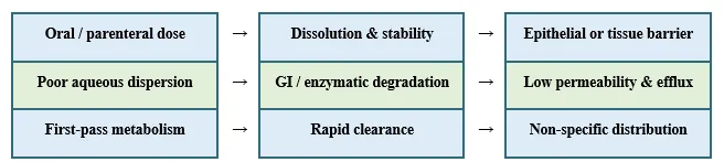


Figure 1. Biopharmaceutical barriers that separate administration of a medicinal-plant bioactive from effective systemic or tissue exposure. The editable boxes represent sequential and interacting failure points.


4. Classification of Next-Generation Nano-Delivery Systems

4.1 Lipid-based platforms

This study focuses on several existing lipid-based formulations and how these formulations can potentially overcome the challenges of delivering phytopharmaceuticals. Liposomes, phytosomes, solid lipid nanoparticles, and emulsions or microemulsions can offer lipid and aqueous phases to facilitate delivery of hydrophilic and lipophilic ingredients. Micelles, when further modified to be self-nanoemulsifying, can carry lipid-based ingredients. They have the potential to facilitate lymphatic transport. Nanostructured lipid carriers (NLCs) and solid lipid nanoparticles (SLNs) can provide controlled release. All of these lipid-based formulations have the potential to be much safer due to their inherently biocompatible components and the various routes of administration that they can offer. However, they are still have numerous risks associated with their formulations due to leakage, oxidation of lipids, transition to a different lipid polymorph, induction of micelle dissociation due to dilution, surfactant toxicity, and similar issues.For the selection of a lipid-based formulation, the rational choice should follow the dominant failure mechanism. When the mechanism of action of a polyphenol is poor membrane partitioning, a phytosome is the rational choice. When a compartment is to be loaded for intravenous delivery, a liposome is required. When a cargo requires protection and controlled release, an NLC should be used. When there is the potential for oral absorption to be enhanced due to intestinal dispersion and digestion of the system, a SNEDDS is the rational choice. Evaluation of the formulation is required for other factors as well, such as the food effect, oxidative stability, the free-drug fraction, and bile-salt interactions.

4.2 Polymeric, Inorganic and Carrier-Free Platforms

Nanoparticles, such as those made from PLGA, chitosan, alginate, gelatin and PEG, can be used to create nanospheres or nanocapsules and can be adapted to include surface ligands and controlled degradation. Chitosan is cationic and increases mucoadhesion but may also increase membrane damage. Dendrimers can have multivalent loading and targeting, but surface charge can be an issue and may require modification or use of biodegradable scaffolds. Mesoporous silica nanocarriers have a large surface area and can include gated pores; metallic systems can provide imaging and can have other multifunctional components. A majority of PLGA will yield nanocrystals that are the active ingredient and that minimize excipients. Nanocrystals can control Ostwald’s ripening (the process whereby crystals grow at the expense of other smaller crystals) and can control precipitation but are also highly aggregation-prone [21, 26–29].Polymeric systems are most useful in controlled release applications; however, the degradation of the polymer can reduce the pH in the local environment, adversely affecting the stability of the therapeutic agent. Mesoporous silica and dendrimers have high loading capabilities but also have complex surface chemistries, which can make impurity characterization and biodegradability assessment challenging. In the case of metallic and metal-oxide carriers, the use of organic carriers can be justified by an additional function such as magnetic targeting, imaging and photothermal stimulation. For highly purified and poorly soluble compounds, nanocrystals may be the most straightforward and most practical option since the carrier is mainly the active compound; however, stabilizer selection, redispersibility and control of a supersaturated state will determine if a loss in solubility will enhance the absorptive capacity [21, 27–29, 35].

4.3 Biomimetic, intelligent and hybrid systems

Exosomes, plant-derived extracellular vesicle-like particles and cell membrane-coated carriers are capable of displaying natural lipids and proteins. As a result, they impose distinct characteristics for vesicle uptake and immune system recognition. Despite their phenotypic sophistication, these particles possess poor purification, compositional heterogeneity, poor reproducibility with cargo-loading, and ambiguity with regulatory classification. Intelligent systems utilize a variety of stimuli, including; pH, redox, enzymatic activity, temperature, light, magnetic fields and reactive oxygen species, to control the release of their cargo. Examples include lipid-polymer systems, theranostic systems, and systems designed to co-deliver drugs and genes or drugs and multiple therapeutic agents. The complexity of these systems is expected to increase the potential immunogenicity and the failure of each production batch [30–39, 90–92].


Responsiveness to a given stimulus should be demonstrated in the presence of realistic gradients. The pH of tumors is not significantly different from the pH of blood, the concentration of intracellular glutathione is not uniform in all cell types, and the penetration of light into tissue is highly variable. A carrier that is stable at a pH of 5 in a buffer and releases cargo upon exposure to endocytosis does not provide a legitimate claim to targeted delivery. The same is true of coatings that provide a biomimetic surface [28–30, 90–92].
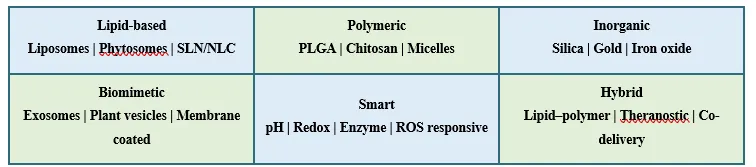


Figure 2. Functional classification of next-generation nano-delivery systems for medicinal-plant bioactives. All categories and labels are editable Word table objects.


5. Mechanisms of Bioavailability Enhancement

Nano-delivery systems utilize a range of sophisticated mechanisms to improve the pharmacokinetic profiles of active pharmaceutical ingredients (APIs). For instance, lipid digestion products can encourage the association of APIs with chylomicrons to be transported through the lymph, where the API first partially avoids the hepatic first-pass metabolism. Additionally, PEGylation of the liposomes’ surfaces, or the application of biomimetic coatings, can improve the liposomes’ circulation time as they promote lipid digestion and lessen the effects of opsonization. Further, therapeutic ligands improve the capacity of the system to promote active drug delivery to the target site. All these integral systems of the nano-delivery system require the appropriate APIs to demonstrate improved pharmacokinetics, such as increased absolute bioavailability and an improved area under the curve (AUC), as well as decreased clearance and an improved half-life. However, the formation of protein coronas can reverse the system’s ability to impart improved pharmacokinetics, and increased concentrations of the API at the target site can indicate the increased retention of the API in tissues, rather than successful intracellular delivery [41-49, 83-87].

When interpreting pharmacokinetics there are some limitations that one must consider based on the metrics of the study. For instance, the total plasma concentration of the API can indicate the presence of the encapsulated API, as well as the protein-bound and free pharmacologically active forms of the API. Additionally, if the API is contained in the lipid delivery system and the system fails to clear through the mononuclear phagocyte system, it cannot be determined if the API was released from the delivery system and entered the cytosol, or if it is contained in the endosomes and is retained in the macrophages. Relative bioavailability may be elevated if the comparator is a poorly-suspended API, but absolute bioavailability may be low if a satisfactory intravenous control is used. To truly discern the system’s ability to transport the active drug, it is critical to determine the concentration of free drug, the mass balance and the pharmacological response. When a local route of administration is used, it may be just as informative to

6. Targeted Therapy and Disease-Specific Applications

6.1 Cancer

Research is abundant in cancer applications since many hydrophobic phytochemicals can be co-delivered with cytotoxic drugs and the nanoparticles take advantage of the leaky vasculature. The enhanced-permeability-and-retention effect is variable in people, hence the use of folate, transferrin, peptides, aptamers, antibodies or hyaluronic acid to improve cellular binding. Formulations of curcumin, quercetin, resveratrol, berberine, artemisinin and ursolic acid target pathways for the modulation of apoptosis, autophagy and angiogenesis, as well as oxidative stress and multidrug-resistance. Most of the evidence is derived from cell-culture and small animal studies. There is no evidence to support that a higher degree of exposure for the other studies demonstrates tumor shrinkage. Effects of binding, receptor occupancy, endosomal escape, immune system interactions, and other effects need to be evaluated in detail [50–61, 72–78].

6.2 Neurological, Cardiometabolic, and Inflammatory Diseases

Intranasal drug delivery systems and receptor-targeted drug delivery systems are designed for use in the treatment of neurological disorders to target the blood–brain barrier (BBB). Curcumin lactoferrin nanoparticles demonstrated increased epithelial permeability and brain-targeting indices in a preclinical study indicating the nanoparticles have value for that specific route of administration [41]. Sustained exposure to the digestive tract or the modulation of oxidative stress is of interest for the treatment of cardiometabolic disorders. Berberine NLCs have demonstrated hepatoprotection and cardioprotection in animal studies [58–60]. There are several delivery systems designed for treatment of rheumatoid arthritis, inflammatory bowel disease, and psoriasis that permit macrophage delivery, permeate inflamed tissue, and allow for local delivery of the therapeutic. The systems may be immunBrain-delivery assertions must be treated cautiously due to the brain-to-plasma ratio being elevated by decreased exposure systemically rather than by increased delivery to the brain. Studies utilizing intranasal delivery should differentiate between the transport via the olfactory and trigeminal systems and the dose that is swallowed. These studies should also assess the clearance of mucus, repeated dosing histology of the nose, and the performance of the nasal delivery system. In the case of metabolic diseases, use of an intranasal system may be effective due to the presence of an effect in the intestines or microbiome, and may not require absorption systemically. Therefore, the site of action should be defined in order to set the desired pharmacokinetic objectives, or to state that the higher level of exposure in plasma is an asset, or is required [13, 41].

6.3 Infectious, dermatological, ocular and wound applications

Nanocarriers may also serve to protect, stabilize and transport antimicrobial agents, or improve their targeting to biofilms and combine with phytochemicals to enable or enhance their synergistic effects toward resistant microorganisms. The use of Carvacrol, Baicalein and Curcumin with metal nanoparticles has improved local activity. These three combinations may create additional mechanisms via membranes and oxidative stress to the host. These nanoparticles may also improve persistence in the environment and local activity with an increase in the local stress and toxicity toward the host [12, 16, 20, 79-89]. Topical lipid carriers improve skin retention and follicular access; ocular systems prolong precorneal residence; and hydrogel-nanoparticle composites may combine antimicrobial and antioxidant functions with control of the rate of release. biodistribution and irritancy are equally important to the endpoints of

7. Comparative Evidence for Major Plant Bioactives

Evidence comparability isn’t uniform. Curcumin leads in published literature on nanoformulations and in human studies. However, due to product and dosage variability, conclusions cannot be applied to the entire class. Strong formulation and anticancer evidence exists for quercetin and resveratrol, although neither has significant clinical research. Most of the other agents still are in the preclinical phase or contain minimal research (thymoquinone, berberine, silymarin, EGCG, andrographolide, artemisinin and ursolic acid). Co-delivery of artemisinin and Paclitaxel serves as an example of an embedded and extended research phase. The albumin-bound formulation of Paclitaxel serves as a translational benchmark. As a regulatory approved nanomedicine, it shows that comparative evidence from clinical studies and the presence of a defined, controllable, and scalable carrier are essential for successful research. It is not sufficient to just label the formulation as a nanocarrier [14–20, 50–73].


Table 1. Major medicinal-plant bioactives, delivery barriers and nanoformulation evidence

Bioactive

Botanical source

Principal activity

Key limitation

Common nano-strategy

Evidence / maturity

Curcumin

Curcuma longa

Anti-inflammatory; anticancer

Very low solubility; rapid conjugation

Liposomes, SLN/NLC, polymeric NPs, nanocrystals

Extensive preclinical; heterogeneous human studies [14–20, 40–49]

Resveratrol

Vitis vinifera; Polygonum cuspidatum

Cardiometabolic; anticancer

Low solubility; rapid metabolism

Liposomes, polymeric and protein NPs

Preclinical; limited formulation-specific clinical evidence [51–52]

Quercetin

Allium cepa; many fruits/plants

Antioxidant; anticancer

Low solubility and permeability

Dendrimers, magnetic and polymeric NPs

Predominantly preclinical [50, 52–57]

Berberine

Berberis spp.; Coptis chinensis

Metabolic; antimicrobial

Low permeability; efflux; first pass

NLC, gold and hybrid lipid carriers

Animal pharmacology; early translation [58–61]

EGCG

Camellia sinensis

Antioxidant; chemopreventive

Oxidation; low stability

SLN, liposomes, protein NPs

Food/pharma preclinical evidence [63–64]

Silibinin / silymarin

Silybum marianum

Hepatoprotective; anticancer

Poor dissolution; variable extract

Polymeric NPs, phytosomes

Formulation and preclinical evidence [65]

Thymoquinone

Nigella sativa

Anti-inflammatory; anticancer

Hydrophobicity; oxidation

Lipid and polymeric carriers

Mostly preclinical [66]

Andrographolide

Andrographis paniculata

Anti-inflammatory; antiviral

Low solubility; rapid metabolism

PLGA and pH-sensitive NPs

Preclinical PK improvement [67–68]

Ursolic acid

Many medicinal plants

Anticancer; anti-inflammatory

Extreme hydrophobicity

Polymeric and lipid NPs

Preclinical; no established nanomedicine [69–70]

Artemisinin

Artemisia annua

Antimalarial; anticancer research

Short exposure; formulation sensitivity

Niosomes, lipid/polymeric co-delivery

Investigational [71]

Paclitaxel

Taxus spp.

Cytotoxic anticancer agent

Poor aqueous solubility

Albumin nanoparticles, liposomes

Albumin-bound formulation clinically approved [22, 75]

Camptothecin

Camptotheca acuminata

Topoisomerase-I inhibition

Lactone instability; poor solubility

Polymeric, lipid and silica carriers

Derivatives translated; parent nanoforms experimental [10, 21]


8. Safety, Toxicity and Biocompatibility

C Safety includes the entirety of the formulation. A natural cargo can be cytotoxic, genotoxic, hepatotoxic, or have other pharmacological interactions. A carrier can activate the complement system, cause membrane damage, be retained in the mononuclear phagocyte system, or leach toxic degradation products. Factors that influence safety include formulations of different sizes and aspect ratios, surface charge, ligand density, residual solvents, surfactants, and metal ions. Consequently, a reasonable safety assessment incorporates testing for the presence of and/or for the following: impurity, cell viability, hemolysis, oxidative stress, gene toxins, immunotoxicity, biocompatibility, biodistribution, acute and chronic toxicity, and if appropriate, developmental and reproductive toxicity and histopathology. Determining the pharmacokinetic–toxicodynamic relationship of the formulation requires an assessment of the safety concerns of prolonged tissue retention versus sequestration versus a fleeting exposure to the formulation. Many of the in-vitro tests provide a useful relative safety assessment of formulation components. However, these tests will not address safety concerns regarding the protein corona, immune system interactions, and the body's compensatory systems at the organ level.Safety assessments must use exposure-matched dosing and the active drug, inactive drug, carrier, and any relevant excipient controls. A reduction in toxicity at an equal nominal dose may be due to a slower release and, therefore, a reduction in the active exposure. It is also possible that the carrier may facilitate a broader distribution and expose target organs that the active compound did not. Infusion reactions, complement activation, thrombosis, and hemolysis are significant safety concerns for parenteral products. However, surfactant-induced changes in permeability and irritation differ for oral or mucosal products and can affect the intestinal microbiome. Long-term safety assessments are especially important for products that contain inorganic or poorly biodegradable materials.

9. Manufacturing, Characterisation and Quality-Control Challenges

Translation halts when an amenable lab scale preparation is not sustainable. Key quality attributes (KQAs) encompass mean and distribution of size, polydispersity index, zeta potential, morphology, drug loading and encapsulation, the crystallinity of the drug, surface chemistry, the free fraction and release rate of the drug, potency, sterility, endotoxins, residual solvents and storage stability. DLS is supplemented with microscopy or NPT; DSC and XRD deal with the state of the solid; FTIR deals with interactions; and stability-indicating HPLC or LC–MS deal with the active and degradation products. The release methods must be bio relevant and discriminatory. The structure of the particles may change with the storage improving techniques of lyophilisation or spry drying. Microfluidic or continuous manufacture gives more precise control, however, the techniques still rely on QBD, PAT, and validated comparability due to material variability and scale dependent mixing [23, 28, 30, 39, 96].The first layer of variability is due to the botanical. The drug must be associated with a species and part of the plant of verified origin, as well as the conditions of cultivation or collection and the phytochemical marker and extraction method. If an extract is used rather than a purified drug, other constituents can compete with the carrier for incorporation and can modify the interfacial properties, thus leading to a misinterpretation of encapsulation efficiency. Stability studies should consider the carrier and relevant phytochemicals, along with oxidation products and microbial quality, and should represent the expected conditions of storage and transport. Thus, a change in extract supplier can have the same effect as a change in polymer or mixing method [1–4, 99–100].

10. Clinical Translation and Regulatory Considerations

Barriers to clinical translation include the challenges posed by the multiple formulations that are produced, poor dose equivalence, translation gaps, short safety evaluation studies, irregular endpoints and uncertain cost-benefit outcome assessments. Nanophytopharmaceuticals are assessed by the regulatory authorities based on the function of the product and its legal classification rather than the promotional claims. A drug product must have complete CMC, validated assessment techniques, justification of particle characteristics, stability, and nonclinical and clinical evaluation of the route and indication. Differences in the manufacturing site, scale and/or excipients may alter biodistribution and therefore require assessment of comparability. FDA regulations consider products that contain nanomaterials as case by case, while guidance on liposomes refers to the importance of the composition, whether the drug is free or encapsulated, drug leakage, and the pharmacokinetics of the system. Guidance on the quality of herbal products refers to the identity of the plant, the control of the active markers and the assessment of contaminants [27, 29, 37 to 39, 93, 98 to 100].The early stages of clinical development should determine if a nanotechnology formulation alters assessment of the pharmacokinetics, and if so, further testing of the wide range of potential therapeutic effects may be warranted. In this case, justification should be provided for the burden of the nanoparticle and the active dose in each case. Where possible, bioanalytical techniques should be able to differentiate free drug from that which is encapsulated. The standard formulation of the drug provided should be a commercially available formulation and not simply a placebo. Surveillance post-marketing authorization is especially important to assess the potential for rare adverse drug reactions, including infusion or immune reactions, interactions with concomitantly administered drugs, as well as effects that may be dependent on the specific drug formulation. While the value of the intellectual property and the acceptability of the patient may be relevant, neither relates to the assessment of comparative benefit [22, 27, 30, 37 to 39].

11. Available for Purchase and Studied in Clinical Trials Formulations

The status of curcumin formulations must be precise. Most formulations of curcumin phytosomes and “nanocurcumin” are marketed as dietary supplements, and such marketing does not confer drug approval or claim clinical advantage. Formulations of curcumin developed for research do have different carriers and therefore different exposures and cannot be considered interchangeable. Unlike most nanoformulations of other phytochemicals and curcumin, which remain investigational, albumin-bound paclitaxel is a regulated anticancer nanomedicine. There is insufficient evidence for the approval of any of the nanoformulations of medicinal plants [2, 7, 15, 22, 30].


Table 2. Comparative characteristics of next-generation nano-delivery platforms

Platform

Typical composition

Principal advantage

Suitable cargo

Targeting potential

Main limitation

Translational maturity

Liposome / phytosome

Phospholipid bilayer or phospholipid complex

Dual-domain loading; membrane interaction

Hydrophilic and lipophilic actives

Ligand or passive

Leakage, oxidation, cost

High for some drugs; variable for phytochemicals

SLN / NLC

Solid lipid; or solid + liquid lipid

Protection and sustained release

Lipophilic actives

Surface ligand feasible

Polymorphism; drug expulsion

Moderate

Polymeric NP

PLGA, chitosan, alginate, gelatin, PEG

Controlled degradation and surface engineering

Broad cargo range

High design flexibility

Residual solvents; scale-up

Moderate–high

Polymeric micelle

Amphiphilic block copolymer

Solubilises hydrophobes

Hydrophobic actives

Ligand feasible

Dilution instability

Moderate

Nanoemulsion / SNEDDS

Oil, surfactant, co-surfactant

Oral dispersion and lymphatic uptake

Lipophilic actives

Mostly non-specific

Surfactant burden; precipitation

Moderate–high

Dendrimer

Branched polymer

Multivalent loading

Small molecules / nucleic acids

High

Cationic toxicity; complex manufacture

Low–moderate

Mesoporous silica

Porous silica

High loading; gated release

Hydrophobic and charged cargo

Surface functionalisation

Biopersistence

Low–moderate

Metal / metal oxide

Au, Ag, Fe3O4, ZnO

Imaging, heat, magnetism or antimicrobial effect

Conjugated bioactives

Magnetic or ligand

ROS, ion release, persistence

Indication-specific

Nanocrystal

Stabilised drug crystal

Very high drug loading; faster dissolution

Poorly soluble actives

Limited intrinsic targeting

Aggregation; precipitation

Moderate–high

Exosome / biomimetic

Natural vesicle or cell membrane

Biological interfaces; immune evasion

Diverse cargo

Native or engineered

Purity, yield, heterogeneity

Early

Stimuli-responsive hybrid

Multi-component smart carrier

Triggered and multifunctional delivery

Drug combinations

High in principle

Analytical and regulatory complexity

Early

Table 3. Representative preclinical and translational evidence

Bioactive

Nanoformulation

Model / indication

Study type

Major reported outcome

Safety interpretation

DOI / identifier

Curcumin

Solid lipid nanoparticles

Oral bioavailability

Formulation / preclinical

Improved stability and exposure versus free curcumin

Requires carrier- and dose-specific toxicity

10.3389/fbioe.2020.00879 [17]

Curcumin

Lactoferrin nanoparticles, intranasal

Neuroprotection / brain delivery

In vitro + animal PK

Higher permeability and brain targeting

Early preclinical evidence only

10.3389/fbioe.2023.1168408 [41]

Curcumin

RGD-liposomal curcumin

Cancer targeting

In vitro / preclinical

Increased target-cell association

Ligand and lipid immunology unresolved

10.1177/0885328220949367 [46]

Quercetin

Magnetite nanoparticles + radiotherapy

Breast cancer

Preclinical

Enhanced anticancer response

Metal-related oxidative toxicity requires controls

10.1177/11782234221086728 [56]

Resveratrol

Cationic liposomes

Hepatocellular carcinoma

In vitro / animal

Improved delivery and antitumour signal

Cationic surface safety is formulation-specific

10.1021/acsbiomaterials.0c00429 [51]

Berberine

NLC

Hepatic ischaemia–reperfusion

Animal

Protective pharmacodynamic effects

Not evidence of human benefit

10.1016/j.biopha.2021.112122 [58]

EGCG

SLN

Stability / oral delivery

Formulation study

Improved encapsulation and stability

Food-grade components still need dose testing

10.1016/j.jfoodeng.2018.09.008 [63]

Andrographolide

PLGA nanoparticles

Controlled delivery

Preclinical

Improved formulation performance

Polymer residuals and chronic safety required

10.3389/fbioe.2021.639409 [67]

Artemisinin + curcumin

Niosomes

Colon cancer cells

In vitro

Enhanced cell-killing versus free combination

No clinical inference from cytotoxicity alone

10.1007/s12032-023-02032-7 [71]

Carvacrol

Chitosan nano-delivery with albumin corona

GI antimicrobial delivery

In vitro biorelevant model

Corona altered mucoadhesion and activity

Demonstrates biological-identity drift

10.1016/j.ijbiomac.2020.12.085 [86]

Paclitaxel

Albumin-bound nanoparticles

Solid tumours

Regulated medicine

Clinical translation of a plant-derived active

Established product-specific safety profile

Regulatory product; context [22, 98]


12. Critical Research Gaps

Priority research gaps include head-to-head comparisons using dose- and exposure-matched controls; validated quantification of phytochemical identity and degradation products; sex, age and disease-specific studies of pharmacokinetics; large-animal and subchronic and chronic toxicity studies; and publication of negative results. Evolution of the protein corona, microbiome interactions, immune memory, reproductive effects, and environmental release are infrequently studied together. Challenges of conducting clinical trials of a given formulation include the need for formulation-specific biomarkers and meaningful comparators as well as the need for pharmacokinetic sampling in the clinical trial framework. The lack of economically and environmentally sustainable carrier systems and the burden of implementation remain challenges, despite the fact that a carrier system may be technically effective [11, 26–39].

FUTURE RESEARCH

A reasonable research agenda can be conducted in stages. The first stage is the application of quality-by-design and machine-learning frameworks to map the composition-process-attribute continuum, incorporating a degree of mechanistic interpretability. The second stage is the integration of physiologically based pharmacokinetic modeling and Organ-on-a-Chip systems and 3D disease models to assist in formulation selection prior to the escalation to large animal models. The third stage is the integration of multi-omics and biomarkers to target specific patients and tissues. The fourth stage is the development of continuous, solvent-free, and energy-efficient manufacturing processes. In the fifth stage, the focus can shift to platform-centered comparability, long-term pharmacovigilance, and patient-centered outcomes. AI can help design experiments, but cannot substitute for validated assays, causal pharmacology, or clinical research [5–8, 21, 28–30, 96].
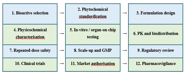


Figure 3. Translational pathway for nano-delivered medicinal-plant bioactives, from molecular selection to lifecycle surveillance. Each stage is an editable Word table cell.


Table 4. Translational barriers and prioritised solutions

Barrier

Why it matters

Current limitation

Proposed solution

Priority

Variable botanical input

Changes dose and impurity profile

Inconsistent markers and extraction

Authenticated source, fingerprinting, potency assays

Immediate

Weak exposure evidence

Dissolution is confused with bioavailability

Sparse absolute PK and tissue data

Exposure-matched PK/PD and mass-balance studies

Immediate

Scale-dependent particle attributes

May change efficacy and toxicity

Bench processes do not translate directly

QbD, PAT, continuous or controlled mixing

Immediate

Inadequate chronic safety

Nanoparticles may accumulate or alter immunity

Short rodent studies dominate

Repeated-dose, immunotoxicity and recovery studies

High

Protein-corona uncertainty

Changes targeting and uptake

Static in-vitro media

Disease-relevant biofluid and longitudinal corona studies

High

Regulatory ambiguity

Delays development and comparability

Herbal, drug and nanomaterial rules intersect

Early scientific advice and product-specific CMC strategy

High

Limited clinical differentiation

No proof of benefit over simpler formulations

Small heterogeneous trials

Dose-equivalent randomised trials with PK endpoints

High

Cost and sustainability

Complex platforms may be inaccessible

Few life-cycle analyses

Technoeconomic and environmental assessment

Medium–high


CONCLUSION

Next-generation controlled and targetable formulations of medicinal plant bioactives can be made using next-generation nano-delivery systems. These formulations allow for precise control of delivery and even make previously poorly dispersible plant molecules bioactive. Next-generation controlled and targetable formulations are made using biocompatible and biodegradable lipid polymer nanoparticles, nanocrystals, and inspired biomimetic systems. Nano-size is a necessary, but not a sufficient, condition for the success of these delivery systems. For positive demonstration of value, delivery systems must protect the therapeutically relevant moieties, allow for preferential local delivery, and provide clinical benefit compared to other delivery systems of greater simplicity. The strongest evidence currently available supports the use of next-generation delivery systems at the formulation and pre-clinical phases, with the case of paclitaxel providing the most evidence of the demands of full translation. Continued advancements will rely on phytochemical input standardization, integration of exposure-focused pharmacology, the use of safe-by-design closed systems, implementation of quality systems, and the completion of chronic safety and formulation-centered clinical trials.

REFERENCES

  1. Dewi, M. K., Chaerunisaa, A. Y., Muhaimin, M., & Joni, I. M. (2022). Improved activity of herbal medicines through nanotechnology. Nanomaterials, 12(22), 4073. https://doi.org/10.3390/nano12224073
  2. Teja, P. K., Mithiya, J., Kate, A. S., Bairwa, K., & Chauthe, S. K. (2022). Herbal nanomedicines: Recent advancements, challenges, opportunities and regulatory overview. Phytomedicine, 96, 153890. https://doi.org/10.1016/j.phymed.2021.153890
  3. Ng, P. Q., Ling, L. S., Chellian, J., Madheswaran, T., Panneerselvam, J., Kunnath, A. P., & Gupta, G. (2020). Applications of nanocarriers as drug delivery vehicles for active phytoconstituents. Current Pharmaceutical Design, 26(36), 4580–4590. https://doi.org/10.2174/1381612826666200610111013
  4. Jalili, A., Bagherifar, R., Nokhodchi, A., Conway, B. R., & Javadzadeh, Y. (2023). Current advances in nanotechnology-mediated delivery of herbal and plant-derived medicines. Advanced Pharmaceutical Bulletin, 13(4), 712–722. https://doi.org/10.34172/apb.2023.087
  5. Lv, Y., et al. (2024). Nano-drug delivery systems based on natural products. International Journal of Nanomedicine, 19, 2591–2622. https://doi.org/10.2147/IJN.S443692
  6. Parvin, N., Aslam, M., Joo, S. W., & Mandal, T. K. (2025). Nano-phytomedicine: Harnessing plant-derived phytochemicals in nanocarriers for targeted human health applications. Molecules, 30(15), 3177. https://doi.org/10.3390/molecules30153177
  7. Najafi, F., et al. (2025). Advancement in nanocarrier-mediated delivery of herbal bioactives: From bench to beside. Natural Products and Bioprospecting, 15, 69. https://doi.org/10.1007/s13659-025-00550-7
  8. Civelek, M., et al. (2024). Nanocarriers for therapeutic phytochemicals. Nanomedicine, 19(21–22), 1711–1716. https://doi.org/10.1080/17435889.2024.2380242
  9. Chavda, V. P., et al. (2023). Advanced phytochemical-based nanocarrier systems for the treatment of breast cancer. Cancers, 15(4), 1023. https://doi.org/10.3390/cancers15041023
  10. Kumari, S., et al. (2022). Bioactive-loaded novel nano-formulations for targeted drug delivery and therapeutic potential. Pharmaceutics, 14(5), 1091. https://doi.org/10.3390/pharmaceutics14051091
  11. Ai, S., et al. (2024). Collision of herbal medicine and nanotechnology: A bibliometric analysis of herbal nanoparticles from 2004 to 2023. Journal of Nanobiotechnology, 22, 140. https://doi.org/10.1186/s12951-024-02426-3
  12. Ghosh, S., Nandi, S., & Basu, T. (2022). Nano-antibacterials using medicinal plant components: An overview. Frontiers in Microbiology, 12, 768739. https://doi.org/10.3389/fmicb.2021.768739
  13. Mishra, K., et al. (2023). Recent advancements in nanocarrier-assisted brain delivery of phytochemicals against neurological diseases. Neurochemical Research, 48(10), 2936–2968. https://doi.org/10.1007/s11064-023-03955-3
  14. Dizaj, S. M., et al. (2022). Curcumin nanoformulations: Beneficial nanomedicine against cancer. Phytotherapy Research, 36(3), 1156–1181. https://doi.org/10.1002/ptr.7389
  15. Silvestre, F., et al. (2023). Pharmacokinetics of curcumin delivered by nanoparticles and relationship with antitumor efficacy: A systematic review. Pharmaceuticals, 16(7), 943. https://doi.org/10.3390/ph16070943
  16. Trigo-Gutierrez, J. K., Vega-Chacón, Y., Soares, A. B., & Mima, E. G. O. (2021). Antimicrobial activity of curcumin in nanoformulations: A comprehensive review. International Journal of Molecular Sciences, 22(13), 7130. https://doi.org/10.3390/ijms22137130
  17. Gupta, T., et al. (2020). Enhancing bioavailability and stability of curcumin using solid lipid nanoparticles. Frontiers in Bioengineering and Biotechnology, 8, 879. https://doi.org/10.3389/fbioe.2020.00879
  18. Beyene, A. M., Moniruzzaman, M., Karthikeyan, A., & Min, T. (2021). Curcumin nanoformulations with metal oxide nanomaterials for biomedical applications. Nanomaterials, 11(2), 460. https://doi.org/10.3390/nano11020460
  19. Kumari, A., et al. (2022). Wound-healing effects of curcumin and its nanoformulations: A comprehensive review. Pharmaceutics, 14(11), 2288. https://doi.org/10.3390/pharmaceutics14112288
  20. Rai, M., et al. (2020). Curcumin and curcumin-loaded nanoparticles: Antipathogenic and antiparasitic activities. Expert Review of Anti-infective Therapy, 18(4), 367–379. https://doi.org/10.1080/14787210.2020.1730815
  21. Mitchell, M. J., Billingsley, M. M., Haley, R. M., Wechsler, M. E., Peppas, N. A., & Langer, R. (2021). Engineering precision nanoparticles for drug delivery. Nature Reviews Drug Discovery, 20, 101–124. https://doi.org/10.1038/s41573-020-0090-8
  22. Anselmo, A. C., & Mitragotri, S. (2019). Nanoparticles in the clinic: An update. Bioengineering & Translational Medicine, 4(3), e10143. https://doi.org/10.1002/btm2.10143
  23. Danaei, M., et al. (2018). Impact of particle size and polydispersity index on the clinical applications of lipidic nanocarrier systems. Pharmaceutics, 10(2), 57. https://doi.org/10.3390/pharmaceutics10020057
  24. Kiaie, S. H., Mojarad-Jabali, S., Khaleseh, F., Allahyari, S., Taheri, E., Zakeri-Milani, P., & Valizadeh, H. (2020). Axial pharmaceutical properties of liposome in cancer therapy: Recent advances and perspectives. International Journal of Pharmaceutics, 581, 119269. https://doi.org/10.1016/j.ijpharm.2020.119269
  25. Harwansh, R. K., Deshmukh, R., & Rahman, M. A. (2019). Nanoemulsion: Promising nanocarrier system for delivery of herbal bioactives. Journal of Drug Delivery Science and Technology, 51, 224–233. https://doi.org/10.1016/j.jddst.2019.03.006
  26. Zielińska, A., et al. (2020). Nanotoxicology and nanosafety: Safety-by-design and testing at a glance. International Journal of Environmental Research and Public Health, 17(13), 4657. https://doi.org/10.3390/ijerph17134657
  27. Foulkes, R., Man, E., Thind, J., Yeung, S., Joy, A., & Hoskins, C. (2020). The regulation of nanomaterials and nanomedicines for clinical application: Current and future perspectives. Biomaterials Science, 8(17), 4653–4664. https://doi.org/10.1039/D0BM00558D
  28. Jiménez, A. S., et al. (2022). Safe(r)-by-design guidelines for the nanotechnology industry. NanoImpact, 25, 100385. https://doi.org/10.1016/j.impact.2022.100385
  29. Havelikar, U., et al. (2024). Comprehensive insights into mechanisms of nanotoxicity, assessment methods and regulatory challenges of nanomedicines. Discover Nano, 19, 165. https://doi.org/10.1186/s11671-024-04118-1
  30. Musazzi, U. M., et al. (2023). Feeding next-generation nanomedicines to Europe. Advanced Healthcare Materials, 13, 2301956. https://doi.org/10.1002/adhm.202301956
  31. Forest, V. (2022). Experimental and computational nanotoxicology: Complementary approaches for nanomaterial hazard assessment. Nanomaterials, 12(8), 1346. https://doi.org/10.3390/nano12081346
  32. Shinde, R. B., et al. (2020). State-of-the-art bioassay systems and electrochemical approaches for nanotoxicity assessment. Frontiers in Bioengineering and Biotechnology, 8, 325. https://doi.org/10.3389/fbioe.2020.00325
  33. Nithiyanandam, H., et al. (2021). Analysis of nanotoxicity with integrated omics and mechanobiology. Nanomaterials, 11(9), 2385. https://doi.org/10.3390/nano11092385
  34. Furxhi, I., et al. (2023). Data-driven quantitative intrinsic hazard criteria for nanoproduct development. ACS Applied Nano Materials, 6(5), 3948–3962. https://doi.org/10.1021/acsanm.3c00173
  35. Damasco, J. A., et al. (2020). Understanding nanoparticle toxicity to direct a safe-by-design approach. Nanomaterials, 10(11), 2186. https://doi.org/10.3390/nano10112186
  36. Schmutz, M., Borges, O., Jesus, S., Borchard, G., Perale, G., Zinn, M., & Di Francesco, T. (2020). A methodological safe-by-design approach for the development of nanomedicines. Frontiers in Bioengineering and Biotechnology, 8, 258. https://doi.org/10.3389/fbioe.2020.00258
  37. Mühlebach, S. (2018). Regulatory challenges of nanomedicines and their follow-on versions. Advanced Drug Delivery Reviews, 131, 122–131. https://doi.org/10.1016/j.addr.2018.06.024
  38. Soares, S., Sousa, J., Pais, A., & Vitorino, C. (2018). Nanomedicine: Principles, properties, and regulatory issues. Frontiers in Chemistry, 6, 360. https://doi.org/10.3389/fchem.2018.00360
  39. Leong, H. S., et al. (2019). On the issue of transparency and reproducibility in nanomedicine. Nature Nanotechnology, 14, 629–635. https://doi.org/10.1038/s41565-019-0496-9
  40. Zielińska, A., et al. (2020). Properties, extraction methods, delivery systems, and bioactivity of curcumin. Medicina, 56(7), 336. https://doi.org/10.3390/medicina56070336
  41. Li, L., et al. (2023). Nose-to-brain delivery of self-assembled curcumin-lactoferrin nanoparticles: Characterization, neuroprotective effect and in vivo pharmacokinetic study. Frontiers in Bioengineering and Biotechnology, 11, 1168408. https://doi.org/10.3389/fbioe.2023.1168408
  42. Rajasekar, A., et al. (2022). Curcumin nanospheres and nanorods: Synthesis, characterization and anticancer activity. Process Biochemistry, 112, 248–253. https://doi.org/10.1016/j.procbio.2021.12.007
  43. dos Santos, R. B., et al. (2021). Curcumin-loaded nanocapsules: Antimalarial activity and toxicity assessment. Materials Science and Engineering C, 118, 111356. https://doi.org/10.1016/j.msec.2020.111356
  44. de Oliveira Pacheco, C., et al. (2022). Surface-functionalized curcumin-loaded polymeric nanocapsules modulate apomorphine-related behavioral changes. Pharmacological Reports, 74(1), 135–147. https://doi.org/10.1007/s43440-021-00331-2
  45. Perera, W. P. T. D., et al. (2020). Curcumin-loaded zinc oxide nanoparticles for biomedical applications. RSC Advances, 10, 30785–30795. https://doi.org/10.1039/D0RA05755J
  46. Mahmoudi, R., et al. (2021). RGD peptide-mediated liposomal curcumin targeted delivery. Journal of Biomaterials Applications, 35(7), 743–753. https://doi.org/10.1177/0885328220949367
  47. Hong, S. C., et al. (2020). Microfluidic assembly of liposomes dual-loaded with catechin and curcumin. Colloids and Surfaces A, 594, 124670. https://doi.org/10.1016/j.colsurfa.2020.124670
  48. Guo, X., et al. (2023). Redox-responsive lipidic prodrug nano-delivery improves the performance of a curcumin derivative. Pharmaceutics, 15(5), 1546. https://doi.org/10.3390/pharmaceutics15051546
  49. Ávila-Gálvez, M. A., et al. (2021). Disposition of dietary polyphenols in breast cancer patients’ tumors. Molecular Nutrition & Food Research, 65(12), e2100163. https://doi.org/10.1002/mnfr.202100163
  50. Ghanbari-Movahed, M., et al. (2022). Quercetin- and rutin-based nanoformulations for cancer treatment: A systematic review. Phytomedicine, 97, 153909. https://doi.org/10.1016/j.phymed.2021.153909
  51. Jagwani, S., Jalalpure, S., Dhamecha, D., Jadhav, K., & Bohara, R. (2020). Resveratrol-loaded cationic liposomes for targeting hepatocellular carcinoma. ACS Biomaterials Science & Engineering, 6(9), 4969–4984. https://doi.org/10.1021/acsbiomaterials.0c00429
  52. Yang, Z., McClements, D. J., Peng, X., et al. (2023). Fabrication of zein–carboxymethyl cellulose nanoparticles for co-delivery of quercetin and resveratrol. Journal of Food Engineering, 341, 111322. https://doi.org/10.1016/j.jfoodeng.2022.111322
  53. Ahmadi, M., et al. (2021). Ultra-pH-sensitive Fe2O3/chitosan/montmorillonite nanocomposite for quercetin delivery. International Journal of Biological Macromolecules, 191, 738–745. https://doi.org/10.1016/j.ijbiomac.2021.09.023
  54. Rehman, K., et al. (2021). Resorcinarene dendrimer-vesicles for improving the antibacterial potential of quercetin. Journal of Molecular Liquids, 341, 116921. https://doi.org/10.1016/j.molliq.2021.116921
  55. Najafabadi, A. P., et al. (2023). pH-sensitive ameliorated quercetin delivery using graphene oxide nanocarriers coated with a gelatin-polyvinylpyrrolidone nanoemulsion. Journal of Drug Delivery Science and Technology, 82, 104339. https://doi.org/10.1016/j.jddst.2023.104339
  56. Askar, M. A., et al. (2022). Quercetin-loaded magnetite nanoparticles combined with radiotherapy in breast cancer. Breast Cancer: Basic and Clinical Research, 16, 11782234221086728. https://doi.org/10.1177/11782234221086728
  57. Vafadar, A., et al. (2020). Quercetin and cancer: New insights into its therapeutic effects on ovarian cancer cells. Cell & Bioscience, 10, 32. https://doi.org/10.1186/s13578-020-00397-0
  58. Gendy, A. M., et al. (2022). Berberine-loaded nanostructured lipid carriers mitigate hepatic ischemia–reperfusion injury. Biomedicine & Pharmacotherapy, 145, 112122. https://doi.org/10.1016/j.biopha.2021.112122
  59. Chiu, C. F., et al. (2021). Berberine-loaded gold nanoparticles induce apoptosis in breast cancer cells. Cancers, 13(21), 5317. https://doi.org/10.3390/cancers13215317
  60. Hosseini, S. H., et al. (2024). Berberine nanoparticles protect against arsenic trioxide-induced cardiotoxicity. Cardiovascular Toxicology, 24, 1311–1316. https://doi.org/10.1007/s12012-024-09927-5
  61. Kabary, D. M., Helmy, M. W., Elkhodairy, K. A., Fang, J. Y., Elzoghby, A. O., & Samaha, M. W. (2018). Hyaluronate/lactoferrin-coated lipid nanocarriers for co-delivery of rapamycin and berberine. Colloids and Surfaces B: Biointerfaces, 169, 183–194. https://doi.org/10.1016/j.colsurfb.2018.05.008
  62. Jia, W., et al. (2023). Nano-based delivery of polyphenolic compounds for cancer treatment. Pharmaceuticals, 16(1), 101. https://doi.org/10.3390/ph16010101
  63. Shtay, R., Keppler, J. K., Schrader, K., & Schwarz, K. (2019). Encapsulation of epigallocatechin gallate in solid lipid nanoparticles. Journal of Food Engineering, 244, 91–100. https://doi.org/10.1016/j.jfoodeng.2018.09.008
  64. Zwolak, I. (2021). Epigallocatechin gallate for protection against heavy-metal-induced oxidative stress. International Journal of Molecular Sciences, 22(8), 4027. https://doi.org/10.3390/ijms22084027
  65. Pourgholi, A., Dadashpour, M., Mousapour, A., Firouzi Amandi, A., & Zarghami, N. (2021). Anticancer potential of silibinin-loaded polymeric nanoparticles against breast cancer cells. Asian Pacific Journal of Cancer Prevention, 22(8), 2587–2596. https://doi.org/10.31557/APJCP.2021.22.8.2587
  66. El-Far, A. H., Al Jaouni, S. K., Li, W., & Mousa, S. A. (2018). Protective roles of thymoquinone nanoformulations: Potential nanonutraceuticals in human diseases. Nutrients, 10(10), 1369. https://doi.org/10.3390/nu10101369
  67. Oseni, B. A., et al. (2021). Encapsulation of andrographolide in PLGA nanoparticles: Formulation, characterization and biological evaluation. Frontiers in Bioengineering and Biotechnology, 9, 639409. https://doi.org/10.3389/fbioe.2021.639409
  68. Chellampillai, B., & Pawar, A. P. (2011). Improved oral bioavailability of andrographolide from pH-sensitive nanoparticles. European Journal of Drug Metabolism and Pharmacokinetics, 35, 123–129. https://doi.org/10.1007/s13318-010-0016-7
  69. Miatmoko, A., et al. (2021). Nanoparticles for delivering ursolic acid in cancer therapy: A scoping review. Frontiers in Pharmacology, 12, 787226. https://doi.org/10.3389/fphar.2021.787226
  70. Khwaza, V., Oyedeji, O. O., & Aderibigbe, B. A. (2020). Ursolic acid-based derivatives as potential anti-cancer agents: An update. International Journal of Molecular Sciences, 21(16), 5920. https://doi.org/10.3390/ijms21165920
  71. Firouzi Amandi, A., et al. (2023). Enhanced anti-cancer effect of artemisinin- and curcumin-loaded niosomal nanoparticles against human colon cancer cells. Medical Oncology, 40(6), 170. https://doi.org/10.1007/s12032-023-02032-7
  72. Chavda, V. P., et al. (2022). Nano-drug delivery systems entrapping natural bioactive compounds for cancer treatment. Frontiers in Oncology, 12, 867655. https://doi.org/10.3389/fonc.2022.867655
  73. Liu, M., et al. (2022). Nanotechnology-based drug delivery for anticancer active ingredients from traditional Chinese medicine. American Journal of Chinese Medicine, 50(8), 2011–2032. https://doi.org/10.1142/S0192415X22500860
  74. Bozzuto, G., et al. (2024). Phytocompounds and nanoformulations for anticancer therapy. Molecules, 29(16), 3784. https://doi.org/10.3390/molecules29163784
  75. Andreani, T., et al. (2024). Natural-compound-based nanomedicines for cancer treatment: Future directions and challenges. Drug Delivery and Translational Research, 14(10), 2845–2916. https://doi.org/10.1007/s13346-024-01649-z
  76. Zou, B., et al. (2024). Nanodelivery systems for bioactive compounds from traditional Chinese medicine in prostate cancer. Phytomedicine, 128, 155554. https://doi.org/10.1016/j.phymed.2024.155554
  77. Xu, Z., et al. (2023). Why traditional herbal medicine promotes wound healing: Immune response, microbiome and controlled delivery. Advanced Drug Delivery Reviews, 195, 114764. https://doi.org/10.1016/j.addr.2023.114764
  78. Ben-Shabat, S., Yarmolinsky, L., Porat, D., & Dahan, A. (2020). Antiviral effect of phytochemicals from medicinal plants: Applications and drug delivery strategies. Drug Delivery and Translational Research, 10, 354–367. https://doi.org/10.1007/s13346-019-00691-6
  79. Stan, D., et al. (2021). Natural compounds with antimicrobial and antiviral effects and nanocarriers used for their transportation. Frontiers in Pharmacology, 12, 723233. https://doi.org/10.3389/fphar.2021.723233
  80. Gafur, A., et al. (2020). From bulk to nano-delivery of essential phytochemicals: Recent progress and strategies for antibacterial resistance. Journal of Materials Chemistry B, 8, 9825–9835. https://doi.org/10.1039/D0TB01671C
  81. Guo, Y., et al. (2023). Nanotechnology-based drug delivery systems to control bacterial-biofilm-associated lung infections. Pharmaceutics, 15(11), 2582. https://doi.org/10.3390/pharmaceutics15112582
  82. Aflakian, F., et al. (2023). Nanoparticle therapeutics for bacterial infections: From preclinical evidence to FDA-approved products and clinical trials. European Journal of Pharmaceutical Sciences, 188, 106515. https://doi.org/10.1016/j.ejps.2023.106515
  83. Gao, Y., et al. (2023). Hyaluronic-acid-modified curcumin–copper complexes accelerate healing in bacterial prostatitis. Carbohydrate Polymers, 310, 120668. https://doi.org/10.1016/j.carbpol.2023.120668
  84. Liu, Q., et al. (2022). Optimization, antimicrobial activity and safety evaluation of a carvacrol nanoemulsion. Industrial Crops and Products, 181, 114816. https://doi.org/10.1016/j.indcrop.2022.114816
  85. Niaz, T., Sarkar, A., Mackie, A., & Imran, M. (2021). Impact of albumin corona on mucoadhesion and antimicrobial activity of carvacrol-loaded chitosan nano-delivery systems under simulated gastrointestinal conditions. International Journal of Biological Macromolecules, 169, 171–182. https://doi.org/10.1016/j.ijbiomac.2020.12.085
  86. Guo, Q., et al. (2021). Co-delivery of an antibiotic and baicalein using polymeric nanoparticles for synergistic antibacterial therapy. International Journal of Pharmaceutics, 599, 120419. https://doi.org/10.1016/j.ijpharm.2021.120419
  87. Liu, Y., et al. (2025). Glabrol and colistin micelle co-delivery against multidrug-resistant bacterial pathogens. Phytomedicine, 137, 156371. https://doi.org/10.1016/j.phymed.2025.156371
  88. Mollazadeh, S., et al. (2021). Redox-responsive drug delivery nanoplatforms: A chemical perspective. Materials Science and Engineering C, 118, 111536. https://doi.org/10.1016/j.msec.2020.111536
  89. Fu, C. P., et al. (2023). Hyaluronic-acid-based nanocarriers for anticancer drug delivery. Polymers, 15(10), 2317. https://doi.org/10.3390/polym15102317
  90. Mazumdar, S., Chitkara, D., & Mittal, A. (2021). Exploration and insights into the cellular internalization and intracellular fate of amphiphilic polymeric nanocarriers. Acta Pharmaceutica Sinica B, 11(4), 903–924. https://doi.org/10.1016/j.apsb.2021.02.019
  91. Sainz, V., Conniot, J., Matos, A. I., Peres, C., Zupančič, E., Moura, L., Silva, L. C., Florindo, H. F., & Gaspar, R. S. (2015). Regulatory aspects on nanomedicines. Biochemical and Biophysical Research Communications, 468(3), 504–510. https://doi.org/10.1016/j.bbrc.2015.08.023
  92. Tirumala, M. G., et al. (2021). Novel methods for the safety evaluation of nanoparticle formulations using in vitro and adverse-outcome-pathway approaches. Frontiers in Pharmacology, 12, 612659. https://doi.org/10.3389/fphar.2021.612659
  93. Cai, X., et al. (2020). Nanoparticle biotransformation and implications for nanosafety. Small, 16(36), e1907663. https://doi.org/10.1002/smll.201907663
  94. Matthew, S. A. L., et al. (2022). Scalability of microfluidic and semi-batch silk nanoprecipitation. Molecules, 27(7), 2368. https://doi.org/10.3390/molecules27072368
  95. Page, M. J., et al. (2021). The PRISMA 2020 statement: An updated guideline for reporting systematic reviews. BMJ, 372, n71. https://doi.org/10.1136/bmj.n71
  96. Croitoru, A.-M., Moroșan, A., Tihăuan, B., Oprea, O., Motelică, L., Trușcă, R., Nicoară, A. I., Popescu, R.-C., Savu, D., Mihăiescu, D. E., & Ficai, A. (2022). Novel graphene oxide/quercetin and graphene oxide/juglone nanostructured platforms as effective drug delivery systems with biomedical applications. Nanomaterials, 12(11), 1943. https://doi.org/10.3390/nano12111943
  97. Firouzi Amandi, A., Bahmanyar, Z., Dadashpour, M., et al. (2024). Fabrication of magnetic niosomal platform for delivery of resveratrol: Potential anticancer activity against human pancreatic cancer Capan-1 cells. Cancer Cell International, 24, 46. https://doi.org/10.1186/s12935-024-03219-2
  98. U.S. Food and Drug Administration. (2022). Drug products, including biological products, that contain nanomaterials: Guidance for industry. U.S. Department of Health and Human Services. https://www.fda.gov/regulatory-information/search-fda-guidance-documents/drug-products-including-biological-products-contain-nanomaterials
  99. U.S. Food and Drug Administration. (2018). Liposome drug products: Chemistry, manufacturing, and controls; human pharmacokinetics and bioavailability; and labeling documentation. U.S. Department of Health and Human Services. https://www.fda.gov/regulatory-information/search-fda-guidance-documents/liposome-drug-products
  100. European Medicines Agency. (2022). Guideline on quality of herbal medicinal products/traditional herbal medicinal products (EMA/HMPC/CHMP/CVMP/201116/2005 Rev. 3). European Medicines Agency. https://www.ema.europa.eu/en/documents/scientific-guideline/guideline-quality-herbal-medicinal-products-traditional-herbal-medicinal-products-revision-3_en.pdf

Reference

  1. Dewi, M. K., Chaerunisaa, A. Y., Muhaimin, M., & Joni, I. M. (2022). Improved activity of herbal medicines through nanotechnology. Nanomaterials, 12(22), 4073. https://doi.org/10.3390/nano12224073
  2. Teja, P. K., Mithiya, J., Kate, A. S., Bairwa, K., & Chauthe, S. K. (2022). Herbal nanomedicines: Recent advancements, challenges, opportunities and regulatory overview. Phytomedicine, 96, 153890. https://doi.org/10.1016/j.phymed.2021.153890
  3. Ng, P. Q., Ling, L. S., Chellian, J., Madheswaran, T., Panneerselvam, J., Kunnath, A. P., & Gupta, G. (2020). Applications of nanocarriers as drug delivery vehicles for active phytoconstituents. Current Pharmaceutical Design, 26(36), 4580–4590. https://doi.org/10.2174/1381612826666200610111013
  4. Jalili, A., Bagherifar, R., Nokhodchi, A., Conway, B. R., & Javadzadeh, Y. (2023). Current advances in nanotechnology-mediated delivery of herbal and plant-derived medicines. Advanced Pharmaceutical Bulletin, 13(4), 712–722. https://doi.org/10.34172/apb.2023.087
  5. Lv, Y., et al. (2024). Nano-drug delivery systems based on natural products. International Journal of Nanomedicine, 19, 2591–2622. https://doi.org/10.2147/IJN.S443692
  6. Parvin, N., Aslam, M., Joo, S. W., & Mandal, T. K. (2025). Nano-phytomedicine: Harnessing plant-derived phytochemicals in nanocarriers for targeted human health applications. Molecules, 30(15), 3177. https://doi.org/10.3390/molecules30153177
  7. Najafi, F., et al. (2025). Advancement in nanocarrier-mediated delivery of herbal bioactives: From bench to beside. Natural Products and Bioprospecting, 15, 69. https://doi.org/10.1007/s13659-025-00550-7
  8. Civelek, M., et al. (2024). Nanocarriers for therapeutic phytochemicals. Nanomedicine, 19(21–22), 1711–1716. https://doi.org/10.1080/17435889.2024.2380242
  9. Chavda, V. P., et al. (2023). Advanced phytochemical-based nanocarrier systems for the treatment of breast cancer. Cancers, 15(4), 1023. https://doi.org/10.3390/cancers15041023
  10. Kumari, S., et al. (2022). Bioactive-loaded novel nano-formulations for targeted drug delivery and therapeutic potential. Pharmaceutics, 14(5), 1091. https://doi.org/10.3390/pharmaceutics14051091
  11. Ai, S., et al. (2024). Collision of herbal medicine and nanotechnology: A bibliometric analysis of herbal nanoparticles from 2004 to 2023. Journal of Nanobiotechnology, 22, 140. https://doi.org/10.1186/s12951-024-02426-3
  12. Ghosh, S., Nandi, S., & Basu, T. (2022). Nano-antibacterials using medicinal plant components: An overview. Frontiers in Microbiology, 12, 768739. https://doi.org/10.3389/fmicb.2021.768739
  13. Mishra, K., et al. (2023). Recent advancements in nanocarrier-assisted brain delivery of phytochemicals against neurological diseases. Neurochemical Research, 48(10), 2936–2968. https://doi.org/10.1007/s11064-023-03955-3
  14. Dizaj, S. M., et al. (2022). Curcumin nanoformulations: Beneficial nanomedicine against cancer. Phytotherapy Research, 36(3), 1156–1181. https://doi.org/10.1002/ptr.7389
  15. Silvestre, F., et al. (2023). Pharmacokinetics of curcumin delivered by nanoparticles and relationship with antitumor efficacy: A systematic review. Pharmaceuticals, 16(7), 943. https://doi.org/10.3390/ph16070943
  16. Trigo-Gutierrez, J. K., Vega-Chacón, Y., Soares, A. B., & Mima, E. G. O. (2021). Antimicrobial activity of curcumin in nanoformulations: A comprehensive review. International Journal of Molecular Sciences, 22(13), 7130. https://doi.org/10.3390/ijms22137130
  17. Gupta, T., et al. (2020). Enhancing bioavailability and stability of curcumin using solid lipid nanoparticles. Frontiers in Bioengineering and Biotechnology, 8, 879. https://doi.org/10.3389/fbioe.2020.00879
  18. Beyene, A. M., Moniruzzaman, M., Karthikeyan, A., & Min, T. (2021). Curcumin nanoformulations with metal oxide nanomaterials for biomedical applications. Nanomaterials, 11(2), 460. https://doi.org/10.3390/nano11020460
  19. Kumari, A., et al. (2022). Wound-healing effects of curcumin and its nanoformulations: A comprehensive review. Pharmaceutics, 14(11), 2288. https://doi.org/10.3390/pharmaceutics14112288
  20. Rai, M., et al. (2020). Curcumin and curcumin-loaded nanoparticles: Antipathogenic and antiparasitic activities. Expert Review of Anti-infective Therapy, 18(4), 367–379. https://doi.org/10.1080/14787210.2020.1730815
  21. Mitchell, M. J., Billingsley, M. M., Haley, R. M., Wechsler, M. E., Peppas, N. A., & Langer, R. (2021). Engineering precision nanoparticles for drug delivery. Nature Reviews Drug Discovery, 20, 101–124. https://doi.org/10.1038/s41573-020-0090-8
  22. Anselmo, A. C., & Mitragotri, S. (2019). Nanoparticles in the clinic: An update. Bioengineering & Translational Medicine, 4(3), e10143. https://doi.org/10.1002/btm2.10143
  23. Danaei, M., et al. (2018). Impact of particle size and polydispersity index on the clinical applications of lipidic nanocarrier systems. Pharmaceutics, 10(2), 57. https://doi.org/10.3390/pharmaceutics10020057
  24. Kiaie, S. H., Mojarad-Jabali, S., Khaleseh, F., Allahyari, S., Taheri, E., Zakeri-Milani, P., & Valizadeh, H. (2020). Axial pharmaceutical properties of liposome in cancer therapy: Recent advances and perspectives. International Journal of Pharmaceutics, 581, 119269. https://doi.org/10.1016/j.ijpharm.2020.119269
  25. Harwansh, R. K., Deshmukh, R., & Rahman, M. A. (2019). Nanoemulsion: Promising nanocarrier system for delivery of herbal bioactives. Journal of Drug Delivery Science and Technology, 51, 224–233. https://doi.org/10.1016/j.jddst.2019.03.006
  26. Zieli?ska, A., et al. (2020). Nanotoxicology and nanosafety: Safety-by-design and testing at a glance. International Journal of Environmental Research and Public Health, 17(13), 4657. https://doi.org/10.3390/ijerph17134657
  27. Foulkes, R., Man, E., Thind, J., Yeung, S., Joy, A., & Hoskins, C. (2020). The regulation of nanomaterials and nanomedicines for clinical application: Current and future perspectives. Biomaterials Science, 8(17), 4653–4664. https://doi.org/10.1039/D0BM00558D
  28. Jiménez, A. S., et al. (2022). Safe(r)-by-design guidelines for the nanotechnology industry. NanoImpact, 25, 100385. https://doi.org/10.1016/j.impact.2022.100385
  29. Havelikar, U., et al. (2024). Comprehensive insights into mechanisms of nanotoxicity, assessment methods and regulatory challenges of nanomedicines. Discover Nano, 19, 165. https://doi.org/10.1186/s11671-024-04118-1
  30. Musazzi, U. M., et al. (2023). Feeding next-generation nanomedicines to Europe. Advanced Healthcare Materials, 13, 2301956. https://doi.org/10.1002/adhm.202301956
  31. Forest, V. (2022). Experimental and computational nanotoxicology: Complementary approaches for nanomaterial hazard assessment. Nanomaterials, 12(8), 1346. https://doi.org/10.3390/nano12081346
  32. Shinde, R. B., et al. (2020). State-of-the-art bioassay systems and electrochemical approaches for nanotoxicity assessment. Frontiers in Bioengineering and Biotechnology, 8, 325. https://doi.org/10.3389/fbioe.2020.00325
  33. Nithiyanandam, H., et al. (2021). Analysis of nanotoxicity with integrated omics and mechanobiology. Nanomaterials, 11(9), 2385. https://doi.org/10.3390/nano11092385
  34. Furxhi, I., et al. (2023). Data-driven quantitative intrinsic hazard criteria for nanoproduct development. ACS Applied Nano Materials, 6(5), 3948–3962. https://doi.org/10.1021/acsanm.3c00173
  35. Damasco, J. A., et al. (2020). Understanding nanoparticle toxicity to direct a safe-by-design approach. Nanomaterials, 10(11), 2186. https://doi.org/10.3390/nano10112186
  36. Schmutz, M., Borges, O., Jesus, S., Borchard, G., Perale, G., Zinn, M., & Di Francesco, T. (2020). A methodological safe-by-design approach for the development of nanomedicines. Frontiers in Bioengineering and Biotechnology, 8, 258. https://doi.org/10.3389/fbioe.2020.00258
  37. Mühlebach, S. (2018). Regulatory challenges of nanomedicines and their follow-on versions. Advanced Drug Delivery Reviews, 131, 122–131. https://doi.org/10.1016/j.addr.2018.06.024
  38. Soares, S., Sousa, J., Pais, A., & Vitorino, C. (2018). Nanomedicine: Principles, properties, and regulatory issues. Frontiers in Chemistry, 6, 360. https://doi.org/10.3389/fchem.2018.00360
  39. Leong, H. S., et al. (2019). On the issue of transparency and reproducibility in nanomedicine. Nature Nanotechnology, 14, 629–635. https://doi.org/10.1038/s41565-019-0496-9
  40. Zieli?ska, A., et al. (2020). Properties, extraction methods, delivery systems, and bioactivity of curcumin. Medicina, 56(7), 336. https://doi.org/10.3390/medicina56070336
  41. Li, L., et al. (2023). Nose-to-brain delivery of self-assembled curcumin-lactoferrin nanoparticles: Characterization, neuroprotective effect and in vivo pharmacokinetic study. Frontiers in Bioengineering and Biotechnology, 11, 1168408. https://doi.org/10.3389/fbioe.2023.1168408
  42. Rajasekar, A., et al. (2022). Curcumin nanospheres and nanorods: Synthesis, characterization and anticancer activity. Process Biochemistry, 112, 248–253. https://doi.org/10.1016/j.procbio.2021.12.007
  43. dos Santos, R. B., et al. (2021). Curcumin-loaded nanocapsules: Antimalarial activity and toxicity assessment. Materials Science and Engineering C, 118, 111356. https://doi.org/10.1016/j.msec.2020.111356
  44. de Oliveira Pacheco, C., et al. (2022). Surface-functionalized curcumin-loaded polymeric nanocapsules modulate apomorphine-related behavioral changes. Pharmacological Reports, 74(1), 135–147. https://doi.org/10.1007/s43440-021-00331-2
  45. Perera, W. P. T. D., et al. (2020). Curcumin-loaded zinc oxide nanoparticles for biomedical applications. RSC Advances, 10, 30785–30795. https://doi.org/10.1039/D0RA05755J
  46. Mahmoudi, R., et al. (2021). RGD peptide-mediated liposomal curcumin targeted delivery. Journal of Biomaterials Applications, 35(7), 743–753. https://doi.org/10.1177/0885328220949367
  47. Hong, S. C., et al. (2020). Microfluidic assembly of liposomes dual-loaded with catechin and curcumin. Colloids and Surfaces A, 594, 124670. https://doi.org/10.1016/j.colsurfa.2020.124670
  48. Guo, X., et al. (2023). Redox-responsive lipidic prodrug nano-delivery improves the performance of a curcumin derivative. Pharmaceutics, 15(5), 1546. https://doi.org/10.3390/pharmaceutics15051546
  49. Ávila-Gálvez, M. A., et al. (2021). Disposition of dietary polyphenols in breast cancer patients’ tumors. Molecular Nutrition & Food Research, 65(12), e2100163. https://doi.org/10.1002/mnfr.202100163
  50. Ghanbari-Movahed, M., et al. (2022). Quercetin- and rutin-based nanoformulations for cancer treatment: A systematic review. Phytomedicine, 97, 153909. https://doi.org/10.1016/j.phymed.2021.153909
  51. Jagwani, S., Jalalpure, S., Dhamecha, D., Jadhav, K., & Bohara, R. (2020). Resveratrol-loaded cationic liposomes for targeting hepatocellular carcinoma. ACS Biomaterials Science & Engineering, 6(9), 4969–4984. https://doi.org/10.1021/acsbiomaterials.0c00429
  52. Yang, Z., McClements, D. J., Peng, X., et al. (2023). Fabrication of zein–carboxymethyl cellulose nanoparticles for co-delivery of quercetin and resveratrol. Journal of Food Engineering, 341, 111322. https://doi.org/10.1016/j.jfoodeng.2022.111322
  53. Ahmadi, M., et al. (2021). Ultra-pH-sensitive Fe2O3/chitosan/montmorillonite nanocomposite for quercetin delivery. International Journal of Biological Macromolecules, 191, 738–745. https://doi.org/10.1016/j.ijbiomac.2021.09.023
  54. Rehman, K., et al. (2021). Resorcinarene dendrimer-vesicles for improving the antibacterial potential of quercetin. Journal of Molecular Liquids, 341, 116921. https://doi.org/10.1016/j.molliq.2021.116921
  55. Najafabadi, A. P., et al. (2023). pH-sensitive ameliorated quercetin delivery using graphene oxide nanocarriers coated with a gelatin-polyvinylpyrrolidone nanoemulsion. Journal of Drug Delivery Science and Technology, 82, 104339. https://doi.org/10.1016/j.jddst.2023.104339
  56. Askar, M. A., et al. (2022). Quercetin-loaded magnetite nanoparticles combined with radiotherapy in breast cancer. Breast Cancer: Basic and Clinical Research, 16, 11782234221086728. https://doi.org/10.1177/11782234221086728
  57. Vafadar, A., et al. (2020). Quercetin and cancer: New insights into its therapeutic effects on ovarian cancer cells. Cell & Bioscience, 10, 32. https://doi.org/10.1186/s13578-020-00397-0
  58. Gendy, A. M., et al. (2022). Berberine-loaded nanostructured lipid carriers mitigate hepatic ischemia–reperfusion injury. Biomedicine & Pharmacotherapy, 145, 112122. https://doi.org/10.1016/j.biopha.2021.112122
  59. Chiu, C. F., et al. (2021). Berberine-loaded gold nanoparticles induce apoptosis in breast cancer cells. Cancers, 13(21), 5317. https://doi.org/10.3390/cancers13215317
  60. Hosseini, S. H., et al. (2024). Berberine nanoparticles protect against arsenic trioxide-induced cardiotoxicity. Cardiovascular Toxicology, 24, 1311–1316. https://doi.org/10.1007/s12012-024-09927-5
  61. Kabary, D. M., Helmy, M. W., Elkhodairy, K. A., Fang, J. Y., Elzoghby, A. O., & Samaha, M. W. (2018). Hyaluronate/lactoferrin-coated lipid nanocarriers for co-delivery of rapamycin and berberine. Colloids and Surfaces B: Biointerfaces, 169, 183–194. https://doi.org/10.1016/j.colsurfb.2018.05.008
  62. Jia, W., et al. (2023). Nano-based delivery of polyphenolic compounds for cancer treatment. Pharmaceuticals, 16(1), 101. https://doi.org/10.3390/ph16010101
  63. Shtay, R., Keppler, J. K., Schrader, K., & Schwarz, K. (2019). Encapsulation of epigallocatechin gallate in solid lipid nanoparticles. Journal of Food Engineering, 244, 91–100. https://doi.org/10.1016/j.jfoodeng.2018.09.008
  64. Zwolak, I. (2021). Epigallocatechin gallate for protection against heavy-metal-induced oxidative stress. International Journal of Molecular Sciences, 22(8), 4027. https://doi.org/10.3390/ijms22084027
  65. Pourgholi, A., Dadashpour, M., Mousapour, A., Firouzi Amandi, A., & Zarghami, N. (2021). Anticancer potential of silibinin-loaded polymeric nanoparticles against breast cancer cells. Asian Pacific Journal of Cancer Prevention, 22(8), 2587–2596. https://doi.org/10.31557/APJCP.2021.22.8.2587
  66. El-Far, A. H., Al Jaouni, S. K., Li, W., & Mousa, S. A. (2018). Protective roles of thymoquinone nanoformulations: Potential nanonutraceuticals in human diseases. Nutrients, 10(10), 1369. https://doi.org/10.3390/nu10101369
  67. Oseni, B. A., et al. (2021). Encapsulation of andrographolide in PLGA nanoparticles: Formulation, characterization and biological evaluation. Frontiers in Bioengineering and Biotechnology, 9, 639409. https://doi.org/10.3389/fbioe.2021.639409
  68. Chellampillai, B., & Pawar, A. P. (2011). Improved oral bioavailability of andrographolide from pH-sensitive nanoparticles. European Journal of Drug Metabolism and Pharmacokinetics, 35, 123–129. https://doi.org/10.1007/s13318-010-0016-7
  69. Miatmoko, A., et al. (2021). Nanoparticles for delivering ursolic acid in cancer therapy: A scoping review. Frontiers in Pharmacology, 12, 787226. https://doi.org/10.3389/fphar.2021.787226
  70. Khwaza, V., Oyedeji, O. O., & Aderibigbe, B. A. (2020). Ursolic acid-based derivatives as potential anti-cancer agents: An update. International Journal of Molecular Sciences, 21(16), 5920. https://doi.org/10.3390/ijms21165920
  71. Firouzi Amandi, A., et al. (2023). Enhanced anti-cancer effect of artemisinin- and curcumin-loaded niosomal nanoparticles against human colon cancer cells. Medical Oncology, 40(6), 170. https://doi.org/10.1007/s12032-023-02032-7
  72. Chavda, V. P., et al. (2022). Nano-drug delivery systems entrapping natural bioactive compounds for cancer treatment. Frontiers in Oncology, 12, 867655. https://doi.org/10.3389/fonc.2022.867655
  73. Liu, M., et al. (2022). Nanotechnology-based drug delivery for anticancer active ingredients from traditional Chinese medicine. American Journal of Chinese Medicine, 50(8), 2011–2032. https://doi.org/10.1142/S0192415X22500860
  74. Bozzuto, G., et al. (2024). Phytocompounds and nanoformulations for anticancer therapy. Molecules, 29(16), 3784. https://doi.org/10.3390/molecules29163784
  75. Andreani, T., et al. (2024). Natural-compound-based nanomedicines for cancer treatment: Future directions and challenges. Drug Delivery and Translational Research, 14(10), 2845–2916. https://doi.org/10.1007/s13346-024-01649-z
  76. Zou, B., et al. (2024). Nanodelivery systems for bioactive compounds from traditional Chinese medicine in prostate cancer. Phytomedicine, 128, 155554. https://doi.org/10.1016/j.phymed.2024.155554
  77. Xu, Z., et al. (2023). Why traditional herbal medicine promotes wound healing: Immune response, microbiome and controlled delivery. Advanced Drug Delivery Reviews, 195, 114764. https://doi.org/10.1016/j.addr.2023.114764
  78. Ben-Shabat, S., Yarmolinsky, L., Porat, D., & Dahan, A. (2020). Antiviral effect of phytochemicals from medicinal plants: Applications and drug delivery strategies. Drug Delivery and Translational Research, 10, 354–367. https://doi.org/10.1007/s13346-019-00691-6
  79. Stan, D., et al. (2021). Natural compounds with antimicrobial and antiviral effects and nanocarriers used for their transportation. Frontiers in Pharmacology, 12, 723233. https://doi.org/10.3389/fphar.2021.723233
  80. Gafur, A., et al. (2020). From bulk to nano-delivery of essential phytochemicals: Recent progress and strategies for antibacterial resistance. Journal of Materials Chemistry B, 8, 9825–9835. https://doi.org/10.1039/D0TB01671C
  81. Guo, Y., et al. (2023). Nanotechnology-based drug delivery systems to control bacterial-biofilm-associated lung infections. Pharmaceutics, 15(11), 2582. https://doi.org/10.3390/pharmaceutics15112582
  82. Aflakian, F., et al. (2023). Nanoparticle therapeutics for bacterial infections: From preclinical evidence to FDA-approved products and clinical trials. European Journal of Pharmaceutical Sciences, 188, 106515. https://doi.org/10.1016/j.ejps.2023.106515
  83. Gao, Y., et al. (2023). Hyaluronic-acid-modified curcumin–copper complexes accelerate healing in bacterial prostatitis. Carbohydrate Polymers, 310, 120668. https://doi.org/10.1016/j.carbpol.2023.120668
  84. Liu, Q., et al. (2022). Optimization, antimicrobial activity and safety evaluation of a carvacrol nanoemulsion. Industrial Crops and Products, 181, 114816. https://doi.org/10.1016/j.indcrop.2022.114816
  85. Niaz, T., Sarkar, A., Mackie, A., & Imran, M. (2021). Impact of albumin corona on mucoadhesion and antimicrobial activity of carvacrol-loaded chitosan nano-delivery systems under simulated gastrointestinal conditions. International Journal of Biological Macromolecules, 169, 171–182. https://doi.org/10.1016/j.ijbiomac.2020.12.085
  86. Guo, Q., et al. (2021). Co-delivery of an antibiotic and baicalein using polymeric nanoparticles for synergistic antibacterial therapy. International Journal of Pharmaceutics, 599, 120419. https://doi.org/10.1016/j.ijpharm.2021.120419
  87. Liu, Y., et al. (2025). Glabrol and colistin micelle co-delivery against multidrug-resistant bacterial pathogens. Phytomedicine, 137, 156371. https://doi.org/10.1016/j.phymed.2025.156371
  88. Mollazadeh, S., et al. (2021). Redox-responsive drug delivery nanoplatforms: A chemical perspective. Materials Science and Engineering C, 118, 111536. https://doi.org/10.1016/j.msec.2020.111536
  89. Fu, C. P., et al. (2023). Hyaluronic-acid-based nanocarriers for anticancer drug delivery. Polymers, 15(10), 2317. https://doi.org/10.3390/polym15102317
  90. Mazumdar, S., Chitkara, D., & Mittal, A. (2021). Exploration and insights into the cellular internalization and intracellular fate of amphiphilic polymeric nanocarriers. Acta Pharmaceutica Sinica B, 11(4), 903–924. https://doi.org/10.1016/j.apsb.2021.02.019
  91. Sainz, V., Conniot, J., Matos, A. I., Peres, C., Zupan?i?, E., Moura, L., Silva, L. C., Florindo, H. F., & Gaspar, R. S. (2015). Regulatory aspects on nanomedicines. Biochemical and Biophysical Research Communications, 468(3), 504–510. https://doi.org/10.1016/j.bbrc.2015.08.023
  92. Tirumala, M. G., et al. (2021). Novel methods for the safety evaluation of nanoparticle formulations using in vitro and adverse-outcome-pathway approaches. Frontiers in Pharmacology, 12, 612659. https://doi.org/10.3389/fphar.2021.612659
  93. Cai, X., et al. (2020). Nanoparticle biotransformation and implications for nanosafety. Small, 16(36), e1907663. https://doi.org/10.1002/smll.201907663
  94. Matthew, S. A. L., et al. (2022). Scalability of microfluidic and semi-batch silk nanoprecipitation. Molecules, 27(7), 2368. https://doi.org/10.3390/molecules27072368
  95. Page, M. J., et al. (2021). The PRISMA 2020 statement: An updated guideline for reporting systematic reviews. BMJ, 372, n71. https://doi.org/10.1136/bmj.n71
  96. Croitoru, A.-M., Moro?an, A., Tih?uan, B., Oprea, O., Motelic?, L., Tru?c?, R., Nicoar?, A. I., Popescu, R.-C., Savu, D., Mih?iescu, D. E., & Ficai, A. (2022). Novel graphene oxide/quercetin and graphene oxide/juglone nanostructured platforms as effective drug delivery systems with biomedical applications. Nanomaterials, 12(11), 1943. https://doi.org/10.3390/nano12111943
  97. Firouzi Amandi, A., Bahmanyar, Z., Dadashpour, M., et al. (2024). Fabrication of magnetic niosomal platform for delivery of resveratrol: Potential anticancer activity against human pancreatic cancer Capan-1 cells. Cancer Cell International, 24, 46. https://doi.org/10.1186/s12935-024-03219-2
  98. U.S. Food and Drug Administration. (2022). Drug products, including biological products, that contain nanomaterials: Guidance for industry. U.S. Department of Health and Human Services. https://www.fda.gov/regulatory-information/search-fda-guidance-documents/drug-products-including-biological-products-contain-nanomaterials
  99. U.S. Food and Drug Administration. (2018). Liposome drug products: Chemistry, manufacturing, and controls; human pharmacokinetics and bioavailability; and labeling documentation. U.S. Department of Health and Human Services. https://www.fda.gov/regulatory-information/search-fda-guidance-documents/liposome-drug-products
  100. European Medicines Agency. (2022). Guideline on quality of herbal medicinal products/traditional herbal medicinal products (EMA/HMPC/CHMP/CVMP/201116/2005 Rev. 3). European Medicines Agency. https://www.ema.europa.eu/en/documents/scientific-guideline/guideline-quality-herbal-medicinal-products-traditional-herbal-medicinal-products-revision-3_en.pdf

Photo
Anjali Kandekar
Corresponding author

SNJB'S shriman Sureshdada jain College of Pharmacy, chandwad, nashik

Photo
Shrinath Chandak
Co-author

SNJB'S shriman Sureshdada jain College of Pharmacy, chandwad, nashik

Anjali Kandekar, Shrinath Chandak, Next-Generation Nano-Delivery Systems for Medicinal Plant Bioactives: An Analytical Review of Bioavailability Enhancement, Targeted Therapy, Safety, and Clinical Translation, Int. J. of Pharm. Sci., 2026, Vol 4, Issue 9, 3338-3358, https://doi.org/10.5281/zenodo.22960089

More related articles
Pharmaceutical Formulation Using 3D Printing: Adva...
Anjali Rai, Satyam Sharma, Dr. Sudha Rathod...
Formulation and Evaluation of a Lip Balm Prepared ...
Abhijit Jankar, Vaibhavi Pandit, Mohini Patil, Tanuja Ghadge, Aa...
Hidden in the blood: NGS revealed Aspergillus Waynelawii in a critically ill neu...
Gouri B.G, Shaiju Dharan, Grace Raju, Shebin C. E., Arathy Gopal...
Artificial Intelligence in Clinical and Community Pharmacy Practice: Transformin...
Yogesh Wankhede, Srushti Tribhuvan, Prashant Chaturvedi, Lavanya Sawarkar, Nisha Fangal...
Related Articles
Formulation And Evaluation of Poly Herbal Hair Gel...
Mahankali Rajitha, Gurrapu niharika, Donthireddy anjali, Nagunuri mangadevi, Erugulu shivanandini, G...
The Miracle Tree: Unveiling the Nutritional Medicinal Wonders of Moringa oleifer...
Rameshwar Pawar, A. Shinde , G. Patharkar, R. Pandhare, V. Deshmukh...
Comparative Assessment Of Hormonal Contraceptive Methods: Subdermal Implants, Ho...
Bhavya S., Jiji K., Srushti K. V., Hrushitha Minchu K .S., Deeksha R., Asmitha B....
Assessment of the Effect of Squash Juice on Hematological parameters (Hemoglobin...
Wala Eldin Osman Alradi, Safa Wdidi, Amira Hassan AbdAlrahman Arman, Weam Abdo M. Abdalgani, Mortada...