
We use cookies to ensure our website works properly and to personalise your experience. Cookies policy
Department of Pharmaceutical Science, Faculty of Pharmacy, Madhyanchal Professional University, Bhopal (M.P.).
Background: Hepatocellular carcinoma (HCC) is one of the most prevalent and lethal malignancies worldwide. Chronic inflammation, oxidative stress, fibrosis, and dysregulated angiogenesis contribute significantly to its pathogenesis. Emerging evidence suggests that modulation of the renin–angiotensin–aldosterone system may offer a novel therapeutic approach in cancer management.Objective: The present study aimed to investigate the protective and therapeutic effects of Perindopril, an angiotensin-converting enzyme inhibitor, against diethylnitrosamine (DEN)-induced hepatocellular carcinoma in Wistar rats.Methods: HCC was induced in albino Wistar rats using DEN (100 mg/kg, i.p.). Animals were divided into normal control, carcinogen control, positive control (5-fluorouracil), and Perindopril treatment groups (1 and 10 mg/kg, p.o.). Physiological, biochemical, oxidative stress, inflammatory, and histopathological parameters were evaluated. Serum liver function markers, antioxidant enzyme status, lipid peroxidation, cytokine levels, and liver morphology were assessed.Results: DEN administration produced significant hepatic injury characterized by loss of body weight, increased liver weight, elevated tumor incidence, increased serum ALT, AST, and LDH levels, severe oxidative stress, dyslipidemia, altered pigment markers, and increased inflammatory cytokines. Perindopril treatment significantly attenuated these pathological alterations in a dose-dependent manner. Restoration of antioxidant defenses, suppression of inflammatory mediators, normalization of liver function markers, and improvement in hepatic architecture were observed in treated groups. Histopathological findings further confirmed the hepatoprotective and anticarcinogenic effects of Perindopril.Conclusion: Perindopril demonstrated significant chemopreventive and therapeutic activity against DEN-induced hepatocellular carcinoma. The beneficial effects appear to be mediated through attenuation of oxidative stress, inflammation, and hepatic injury. These findings support the potential repurposing of Perindopril as an adjunct therapeutic strategy for hepatocellular carcinoma.
Hepatocellular carcinoma (HCC) is the most common primary malignancy of the liver and accounts for approximately 75–85% of all liver cancers. It represents a major global health challenge and remains among the leading causes of cancer-related mortality worldwide.¹˒² Despite advances in diagnosis and treatment, the incidence and mortality of HCC continue to rise due to the increasing prevalence of chronic liver diseases, including viral hepatitis B and C infections, alcohol-induced liver injury, and metabolic dysfunction-associated fatty liver disease (MAFLD).²˒³
The pathogenesis of HCC is a multifactorial process involving chronic inflammation, oxidative stress, fibrosis, angiogenesis, and genomic instability.⁴ Persistent hepatic injury promotes excessive generation of reactive oxygen species (ROS), leading to lipid peroxidation, DNA damage, mitochondrial dysfunction, and activation of various inflammatory signaling pathways.⁵˒⁶ These molecular events contribute to hepatocyte transformation, uncontrolled cellular proliferation, and tumor progression. Furthermore, chronic inflammatory responses within the liver microenvironment facilitate fibrosis and cirrhosis, which are recognized as major predisposing factors for HCC development.⁴˒⁵
Recent evidence suggests that the renin–angiotensin–aldosterone system (RAAS), traditionally known for its role in cardiovascular regulation, also plays a significant role in cancer biology.⁷ Angiotensin II, the principal effector peptide of RAAS, has been shown to stimulate angiogenesis, inflammation, fibrosis, and cellular proliferation through activation of multiple intracellular signaling pathways, including nuclear factor-kappa B (NF-κB), mitogen-activated protein kinase (MAPK), and transforming growth factor-beta (TGF-β)-mediated pathways.⁸˒⁹ These effects contribute to tumor growth, invasion, and metastatic potential. Consequently, pharmacological inhibition of RAAS has emerged as a promising strategy for suppressing tumor progression and improving cancer outcomes.⁷˒⁸
Perindopril is a long-acting angiotensin-converting enzyme (ACE) inhibitor widely prescribed for the management of hypertension, heart failure, and other cardiovascular disorders.¹⁰ In addition to its established cardiovascular benefits, Perindopril exhibits several pleiotropic pharmacological effects, including antioxidant, anti-inflammatory, anti-fibrotic, and anti-angiogenic activities.¹¹˒¹² By reducing the formation of angiotensin II and enhancing the bioavailability of protective mediators such as bradykinin, Perindopril may interfere with critical molecular mechanisms involved in hepatocarcinogenesis. These properties suggest that Perindopril could provide therapeutic benefits beyond cardiovascular disease and may represent a potential candidate for drug repurposing in cancer management.¹¹˒¹²
Therefore, the present study was undertaken to evaluate the protective and therapeutic potential of Perindopril against diethylnitrosamine (DEN)-induced hepatocellular carcinoma in Wistar rats through assessment of physiological, biochemical, oxidative stress, inflammatory, and histopathological parameters.
2. MATERIALS AND METHODS
2.1 Chemicals and Reagents
Diethylnitrosamine (DEN) was procured from Sigma-Aldrich (India) and used for induction of hepatocellular carcinoma. Perindopril was obtained from a certified pharmaceutical source and used as the test drug. 5-Fluorouracil (5-FU) was employed as the standard anticancer agent. All chemicals and reagents used in the study were of analytical grade.
2.2 Experimental Animals
Healthy adult male albino Wistar rats weighing 150–200 g and aged 3–4 months were used in the study. Animals were obtained from a CPCSEA-approved breeding facility and housed in polypropylene cages under standard laboratory conditions (25 ± 1°C, 12 h light/dark cycle, relative humidity 50–60%). Animals had free access to standard pellet diet and water ad libitum throughout the experimental period.
All experimental procedures were conducted in accordance with CPCSEA guidelines and were approved by the Institutional Animal Ethics Committee (IAEC).¹³
2.3 Induction of Hepatocellular Carcinoma
Hepatocellular carcinoma was induced by intraperitoneal administration of diethylnitrosamine (DEN) at a dose of 100 mg/kg body weight once weekly for six weeks. Animals were monitored regularly for changes in body weight, behavioral abnormalities, and signs of disease progression.¹⁴˒¹⁵
2.4 Experimental Design
Animals were randomly divided into five groups (n = 6 per group):
Group I (Normal Control): Received 0.25% CMC orally.
Group II (Carcinogen Control): Received DEN (100 mg/kg, i.p.).
Group III (Positive Control): Received DEN along with 5-Fluorouracil (10 mg/kg, i.p.).
Group IV (Perindopril Low Dose): Received DEN along with Perindopril (1 mg/kg/day, p.o.).
Group V (Perindopril High Dose): Received DEN along with Perindopril (10 mg/kg/day, p.o.).
Treatment was continued for 15 days following successful induction of hepatocellular carcinoma.
2.5 Assessment of Physiological Parameters
Body weight of animals was recorded weekly throughout the study. At the end of the experimental period, animals were sacrificed and livers were excised, washed with ice-cold saline, and weighed. Tumor incidence and visible hepatic nodules were recorded.
2.6 Biochemical Analysis
Blood samples were collected and centrifuged to obtain serum. Liver function biomarkers including alanine aminotransferase (ALT), aspartate aminotransferase (AST), and lactate dehydrogenase (LDH) were estimated using commercially available diagnostic kits according to the manufacturer’s instructions.²⁴
2.7 Assessment of Oxidative Stress Markers
Liver tissues were homogenized in phosphate buffer (0.1 M, pH 7.4) and centrifuged. The resulting supernatant was used for biochemical estimations. Superoxide dismutase (SOD) activity was measured based on inhibition of epinephrine auto-oxidation.¹⁶ Catalase (CAT) activity was determined by monitoring decomposition of hydrogen peroxide.¹⁷ Reduced glutathione (GSH) levels were estimated using Ellman’s reagent (DTNB).¹⁸ Malondialdehyde (MDA), a marker of lipid peroxidation, was determined using the thiobarbituric acid reactive substances (TBARS) assay.¹⁹ Protein carbonyl content was quantified using the 2,4-dinitrophenylhydrazine (DNPH) method.²⁰
2.8 Estimation of Pigment Markers and Lipid Profile
Conjugated bilirubin and biliverdin levels were determined spectrophotometrically. Serum lipid profile including total cholesterol (TC), triglycerides (TG), high-density lipoprotein (HDL), low-density lipoprotein (LDL), and very-low-density lipoprotein (VLDL) was analyzed using standard enzymatic methods.²⁴
2.9 Estimation of Inflammatory Cytokines
Levels of inflammatory cytokines including IL-2, IL-6, IL-10, and IL-1β were quantified using commercially available ELISA kits according to the manufacturer’s protocol. Cytokine concentrations were calculated using standard calibration curves.²¹
2.10 Histopathological Examination
Liver tissues were fixed in 10% neutral buffered formalin, processed through graded alcohols, cleared in xylene, and embedded in paraffin wax. Sections of 4–5 μm thickness were prepared using a rotary microtome and stained with hematoxylin and eosin (H&E). Histological examination was performed under a light microscope to assess hepatocellular degeneration, necrosis, inflammatory infiltration, fibrosis, and neoplastic changes.²²
2.11 Statistical Analysis
Data were expressed as mean ± SD. Statistical analysis was performed using one-way analysis of variance (ANOVA) followed by Bonferroni’s multiple comparison test. A value of p < 0.05 was considered statistically significant. Statistical analyses were performed using GraphPad Prism software.
3.1 Effect of Perindopril on Body Weight, Liver Weight, and Tumor Incidence
DEN administration produced a marked reduction in body weight and significantly increased liver weight and tumor incidence compared with the normal control group. Perindopril treatment attenuated DEN-induced body weight loss and reduced tumor burden in a dose-dependent manner. The higher dose (10 mg/kg) demonstrated greater efficacy, improving body weight gain (14 ± 1 g) and reducing tumor incidence (10.00 ± 1.97) compared with the low-dose group (1 mg/kg). These findings indicate a protective effect of Perindopril against DEN-induced hepatocarcinogenesis.
Table 1. Effect of Perindopril on Physiological Parameters (Body Weight Variation, Liver Weight, and Tumor Incidence) in DEN-Induced HCC in Albino Wistar Rats.
|
Sr. No. |
Groups |
Weight variation (Mean ± SD) (g) |
Liver weight (Mean ± SD) (g) |
Tumor Incidence (No.) |
|
1 |
NC |
23 ± 1 g |
2.81 ± 0.73 |
0.00 ± 0.00 |
|
2 |
CC |
-19 ± 1 g |
3.88 ± 0.67 |
26.00 ± 2.71 |
|
3 |
PC |
17 ± 1 g |
4.68 ± 0.58 |
8.00 ± 1.37* |
|
4 |
T1 |
9 ± 1 g |
6.09 ± 0.56 |
12.00 ± 2.83* |
|
5 |
T2 |
14 ± 1 g |
4.45 ± 0.48 |
10.00 ± 1.97* |
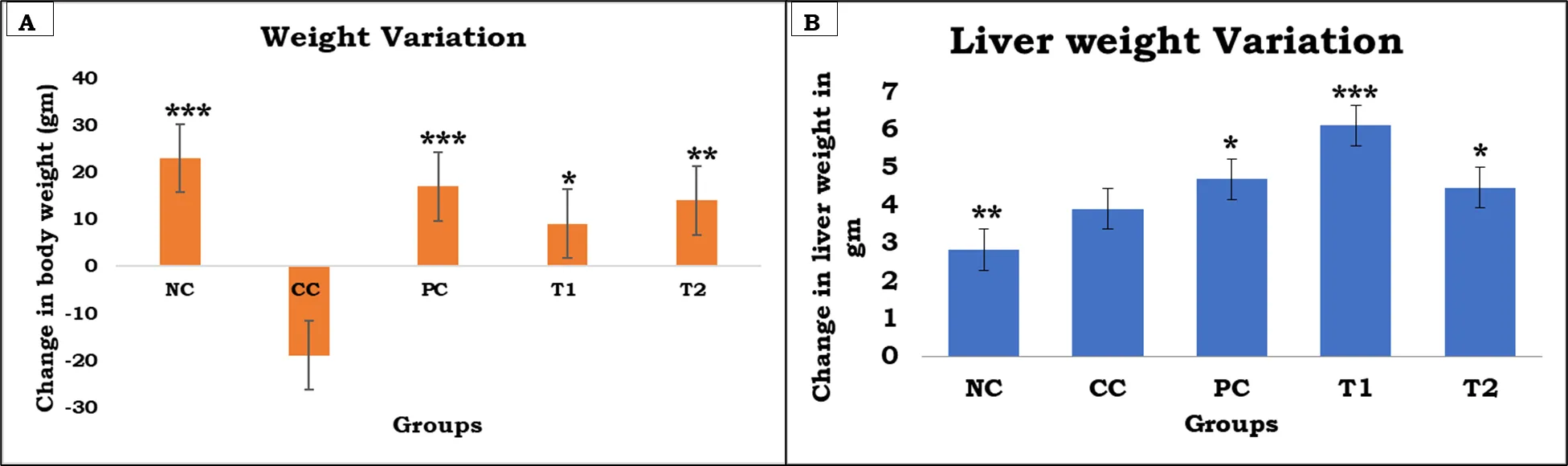
Figure 1. Effect of Perindopril on physiological parameters in DEN-induced HCC in Wistar rats
(A) Body weight variation and (B) Liver weight variation. Data are expressed as Mean ± SD (n = 6). DEN administration resulted in a significant reduction in body weight and alteration in liver weight in the carcinogen control (CC) group compared with the normal control (NC) group, indicating disease progression and hepatic dysfunction. Treatment with Perindopril (1 and 10 mg/kg, p.o.) and 5-fluorouracil (5-FU) improved body weight and modulated liver weight compared with the CC group. Statistical analysis was performed using one-way ANOVA followed by Tukey's multiple comparison test. *P < 0.05, **P < 0.01 and ***P < 0.001 compared with the CC group. NC: Normal Control; CC: Carcinogen Control; PC: Positive Control (5-FU); T1: Perindopril (1 mg/kg); T2: Perindopril (10 mg/kg).
3.2 Effect of Perindopril on Serum Liver Function Biomarkers
DEN administration significantly increased serum ALT, AST, and LDH levels compared with normal controls, indicating severe hepatic injury. Perindopril treatment attenuated these elevations in a dose-dependent manner. The 10 mg/kg dose produced greater reductions in ALT (38.96 ± 4.79 U/L), AST (45.39 ± 6.70 U/L), and LDH (20.34 ± 1.15 U/L) than the 1 mg/kg dose and showed efficacy comparable to 5-FU. These findings demonstrate the hepatoprotective effect of Perindopril against DEN-induced HCC.
Table 3. Effect of Perindopril on Serum Enzymatic Biomarkers (ALT, AST, and LDH) in DEN-Induced HCC in Wistar Rats.
|
Sr. No. |
Group |
ALT (Mean ± SD) (U/L) |
AST (Mean ± SD) (U/L) |
LDH (Mean ± SD) (U/L) |
|
1 |
NC |
20.57 ± 1.81 |
31.13 ± 2.03 |
15.17 ± 4.54 |
|
2 |
CC |
70.14 ± 2.03 |
83.90 ± 3.10 |
59.23 ± 5.72 |
|
3 |
PC |
45.27 ± 2.85 |
51.43 ± 6.56 |
22.34 ± 1.51 |
|
4 |
T1 |
51.06 ± 4.97 |
55.35 ± 6.70 |
47.22 ± 2.52 |
|
5 |
T2 |
38.96 ± 4.79 |
45.39 ± 6.70 |
20.34 ± 1.15 |
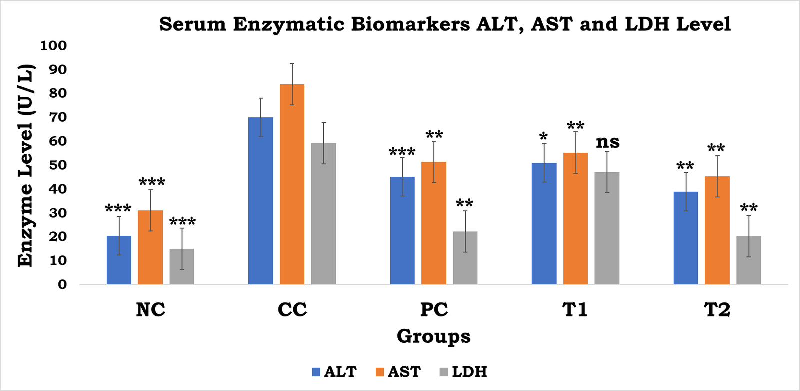
Figure 2. Effect of Perindopril on serum enzymatic biomarkers (ALT, AST, and LDH) in DEN-induced HCC in Wistar rats.
Data are expressed as Mean ± SD (n = 6). DEN administration significantly elevated serum ALT, AST, and LDH levels in the CC group compared with the NC group, indicating hepatocellular injury and impaired liver function. Treatment with Perindopril (3 and 6 mg/kg, p.o.) and 5-FU significantly attenuated DEN-induced elevations in serum enzyme levels. Statistical analysis was performed using one-way ANOVA followed by Bonferroni multiple comparison test. ns = non-significant, *P < 0.05, **P < 0.01 and *P < 0.001 compared with the CC group. NC: Normal Control; CC: Carcinogen Control; PC: Positive Control (5-FU); T1: Perindopril (3 mg/kg); T2: Perindopril (6 mg/kg).
3.3 Effect of Perindopril on Antioxidant Defense System
DEN administration produced marked oxidative stress, as evidenced by significant reductions in hepatic SOD, CAT, and GSH levels compared with the normal control group. SOD activity decreased from 4.12 ± 0.06 U/µg protein in the NC group to 1.79 ± 0.28 U/µg protein in the CC group, while CAT and GSH levels were reduced from 7.25 ± 2.42 to 2.19 ± 1.45 nM H₂O₂/min/µg protein and from 10.97 ± 1.76 to 4.03 ± 0.13 µM/µg protein, respectively. Treatment with Perindopril significantly restored antioxidant enzyme activities and endogenous glutathione levels. The higher dose of Perindopril (10 mg/kg) demonstrated greater efficacy, increasing SOD, CAT, and GSH levels to 3.93 ± 0.07 U/µg protein, 6.89 ± 1.75 nM H₂O₂/min/µg protein, and 9.56 ± 1.84 µM/µg protein, respectively, approaching normal values.
Table 4. Effect of Perindopril on Hepatic Antioxidant Enzyme Levels in DEN-Induced HCC in Wistar Rats.
|
Sr. No. |
Group |
SOD (U/µg protein) |
CAT (nM H₂O₂/min/µg protein) |
GSH (µM/µg protein) |
|
1 |
NC |
4.12 ± 0.06 |
7.25 ± 2.42 |
10.97 ± 1.76 |
|
2 |
CC |
1.79 ± 0.28 |
2.19 ± 1.45 |
4.03 ± 0.13 |
|
3 |
PC |
3.29 ± 0.06*** |
6.02 ± 1.94* |
7.98 ± 1.62*** |
|
4 |
T1 |
2.97 ± 0.08*** |
4.91 ± 2.36ns |
6.66 ± 0.89** |
|
5 |
T2 |
3.93 ± 0.07*** |
6.89 ± 1.75** |
9.56 ± 1.84*** |
Values are expressed as Mean ± SD (n = 6). Statistical analysis was performed using one-way ANOVA followed by Bonferroni multiple comparison test. ns = non-significant, *P < 0.05, **P < 0.01 and *P < 0.001 compared with the CC group. NC: Normal Control; CC: Carcinogen Control; PC: Positive Control (5-FU); T1: Perindopril (1 mg/kg); T2: Perindopril (10 mg/kg).
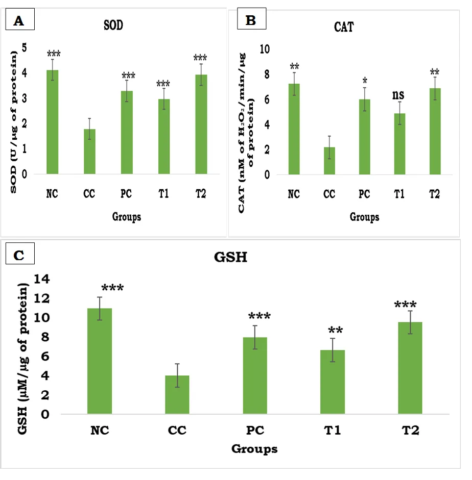
Figure 3. Effect of Perindopril on hepatic antioxidant enzyme levels in DEN-induced HCC in Wistar rats.
(A) Superoxide dismutase (SOD), (B) Catalase (CAT), and (C) Reduced glutathione (GSH). Data are expressed as Mean ± SD (n = 6). DEN administration significantly decreased hepatic SOD, CAT, and GSH levels in the carcinogen control (CC) group compared with the normal control (NC) group, indicating enhanced oxidative stress and impaired antioxidant defense mechanisms. Treatment with Perindopril (1 and 10 mg/kg, p.o.) and 5-FU restored antioxidant enzyme activities and GSH levels toward normal values. Statistical analysis was performed using one-way ANOVA followed by Bonferroni multiple comparison test. ns = non-significant, *P < 0.05, **P < 0.01 and ***P < 0.001 compared with the CC group. NC: Normal Control; CC: Carcinogen Control; PC: Positive Control (5-FU); T1: Perindopril (1 mg/kg); T2: Perindopril (10 mg/kg).
3.4 Effect of Perindopril on Oxidative Damage Markers
Oxidative damage markers were markedly elevated in DEN-treated animals, as evidenced by significant increases in TBARS/MDA and protein carbonyl levels compared with normal controls. Perindopril treatment attenuated oxidative damage in a dose-dependent manner. The higher dose (10 mg/kg) significantly reduced TBARS/MDA levels from 48.78 ± 2.69 to 30.97 ± 3.90 nM/µg protein and decreased protein carbonyl content from 0.38 ± 0.01 to 0.19 ± 0.02 µM/µg protein. These findings indicate that Perindopril effectively protects hepatic tissue against DEN-induced lipid peroxidation and oxidative protein modification.
Table 5. Effect of Perindopril on Oxidative Damage Markers in DEN-Induced HCC in Wistar Rats. **Values are expressed as Mean ± SD (n = 6). Statistical analysis was performed using one-way ANOVA followed by Bonferroni multiple comparison test. ns = non-significant, *P < 0.05, and *P < 0.001 compared with the CC group. NC: Normal Control; CC: Carcinogen Control; PC: Positive Control (5-FU); T1: Perindopril (1 mg/kg); T2: Perindopril (10 mg/kg).
|
Sr. No. |
Group |
TBARS/MDA (nM/µg protein) |
Protein Carbonyl (µM/µg protein) |
|
1 |
NC |
27.73 ± 0.50 |
0.11 ± 0.01 |
|
2 |
CC |
48.78 ± 2.69 |
0.38 ± 0.01 |
|
3 |
PC |
32.72 ± 1.29*** |
0.16 ± 0.04*** |
|
4 |
T1 |
45.13 ± 4.01ns |
0.29 ± 0.08* |
|
5 |
T2 |
30.97 ± 3.90*** |
0.19 ± 0.02*** |
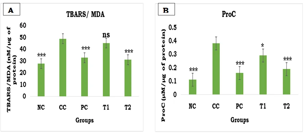
Figure 7.5. Effect of Perindopril on hepatic oxidative stress markers in DEN-induced HCC in Wistar rats
. (A) TBARS/MDA and (B) ProC. Data are expressed as Mean ± SD (n = 6). DEN administration significantly increased hepatic TBARS/MDA and ProC levels in the carcinogen control (CC) group compared with the normal control (NC) group, indicating enhanced lipid peroxidation and oxidative protein damage. Treatment with Perindopril (1 and 10 mg/kg, p.o.) and 5-FU reduced oxidative stress markers and attenuated DEN-induced hepatic oxidative damage. Statistical analysis was performed using one-way ANOVA followed by Bonferroni multiple comparison test. ns = non-significant, *P < 0.05 and ***P < 0.001 compared with the CC group. NC: Normal Control; CC: Carcinogen Control; PC: Positive Control (5-FU); T1: Perindopril (1 mg/kg); T2: Perindopril (10 mg/kg).
3.5 Effect of Perindopril on Pigment Markers
|
Sr. No. |
Group |
Bilirubin (ng/µg protein) |
Biliverdin (ng/µg protein) |
|
1 |
NC |
12.93 ± 2.17 |
10.78 ± 2.21 |
|
2 |
CC |
31.06 ± 1.06 |
26.35 ± 3.96 |
|
3 |
PC |
15.75 ± 0.71*** |
16.13 ± 1.18*** |
|
4 |
T1 |
18.15 ± 2.49** |
20.07 ± 2.52* |
|
5 |
T2 |
13.73 ± 9.73ns |
13.47 ± 3.52** |
DEN administration significantly increased hepatic conjugated bilirubin and biliverdin levels compared with normal controls, indicating impaired bilirubin metabolism, enhanced heme degradation, and hepatic dysfunction. Perindopril treatment reduced both pigment markers in a dose-dependent manner. The higher dose (10 mg/kg) decreased bilirubin and biliverdin levels to 13.73 ± 9.73 and 13.47 ± 3.52 ng/µg protein, respectively, approaching normal values. These findings suggest that Perindopril improves hepatic metabolic function and attenuates oxidative stress associated with DEN-induced HCC.
Table 6. Effect of Perindopril on Hepatic Pigment Markers (Conjugated Bilirubin and Biliverdin) in DEN-Induced HCC in Wistar Rats. **Values are expressed as Mean ± SD (n = 6). Statistical analysis was performed using one-way ANOVA followed by Bonferroni multiple comparison test. ns = non-significant, *P < 0.05, **P < 0.01 and *P < 0.001 compared with the CC group. NC: Normal Control; CC: Carcinogen Control; PC: Positive Control (5-FU); T1: Perindopril (1 mg/kg); T2: Perindopril (10 mg/kg).
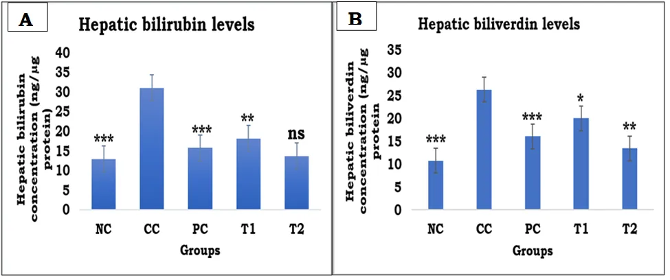
Figure 7.6. Effect of Perindopril on hepatic pigment markers in DEN-induced HCC in Wistar rats.
(A) Hepatic conjugated bilirubin levels and (B) Hepatic biliverdin levels. Data are expressed as Mean ± SD (n = 6). DEN administration significantly increased hepatic bilirubin and biliverdin concentrations in the carcinogen control (CC) group compared with the normal control (NC) group, indicating impaired bilirubin metabolism, disturbed hepatic excretory function, and enhanced oxidative stress. Treatment with Perindopril (1 and 10 mg/kg, p.o.) and 5-FU reduced pigment marker levels and improved hepatic metabolic function. Statistical analysis was performed using one-way ANOVA followed by Bonferroni multiple comparison test. ns = non-significant, *P < 0.05, **P < 0.01 and ***P < 0.001 compared with the CC group. NC: Normal Control; CC: Carcinogen Control; PC: Positive Control (5-FU); T1: Perindopril (1 mg/kg); T2: Perindopril (10 mg/kg).
3.6 Effect of Perindopril on Serum Lipid Profile
DEN administration induced significant dyslipidemia, characterized by elevated TC, TG, LDL, and VLDL levels along with reduced HDL levels compared with normal controls. Perindopril treatment improved all lipid profile parameters in a dose-dependent manner. The higher dose (10 mg/kg) markedly reduced TC, TG, LDL, and VLDL levels while significantly increasing HDL levels, indicating restoration of lipid homeostasis and improvement of hepatic metabolic function in DEN-induced HCC.
Table 7. Effect of Perindopril on Serum Lipid Profile Parameters in DEN-Induced HCC in Wistar Rats. Values are expressed as Mean ± SD (n = 6). Statistical analysis was performed using one-way ANOVA followed by Bonferroni multiple comparison test. NC: Normal Control; CC: Carcinogen Control; PC: Positive Control (5-FU); T1: Perindopril (1 mg/kg); T2: Perindopril (10 mg/kg).
|
Sr. No. |
Group |
TC (mg/dL) |
TG (mg/dL) |
HDL (mg/dL) |
LDL (mg/dL) |
VLDL (mg/dL) |
|
1 |
NC |
184.20 ± 5.58 |
148.61 ± 22.85 |
56.61 ± 6.89 |
98.34 ± 5.88 |
29.72 ± 4.57 |
|
2 |
CC |
306.07 ± 15.37 |
220.13 ± 17.71 |
40.31 ± 10.89 |
195.73 ± 0.93 |
44.02 ± 3.54 |
|
3 |
PC (5-FU) |
163.73 ± 12.58*** |
147.91 ± 34.42* |
54.82 ± 3.45* |
79.32 ± 2.24*** |
29.58 ± 6.88*** |
|
4 |
T1 |
240.02 ± 23.84* |
173.61 ± 44.50** |
75.83 ± 21.59** |
129.46 ± 6.65*** |
34.72 ± 8.90* |
|
5 |
T2 |
187.92 ± 16.11*** |
156.94 ± 10.27*** |
79.11 ± 10.84*** |
77.41 ± 3.20*** |
31.38 ± 2.05*** |
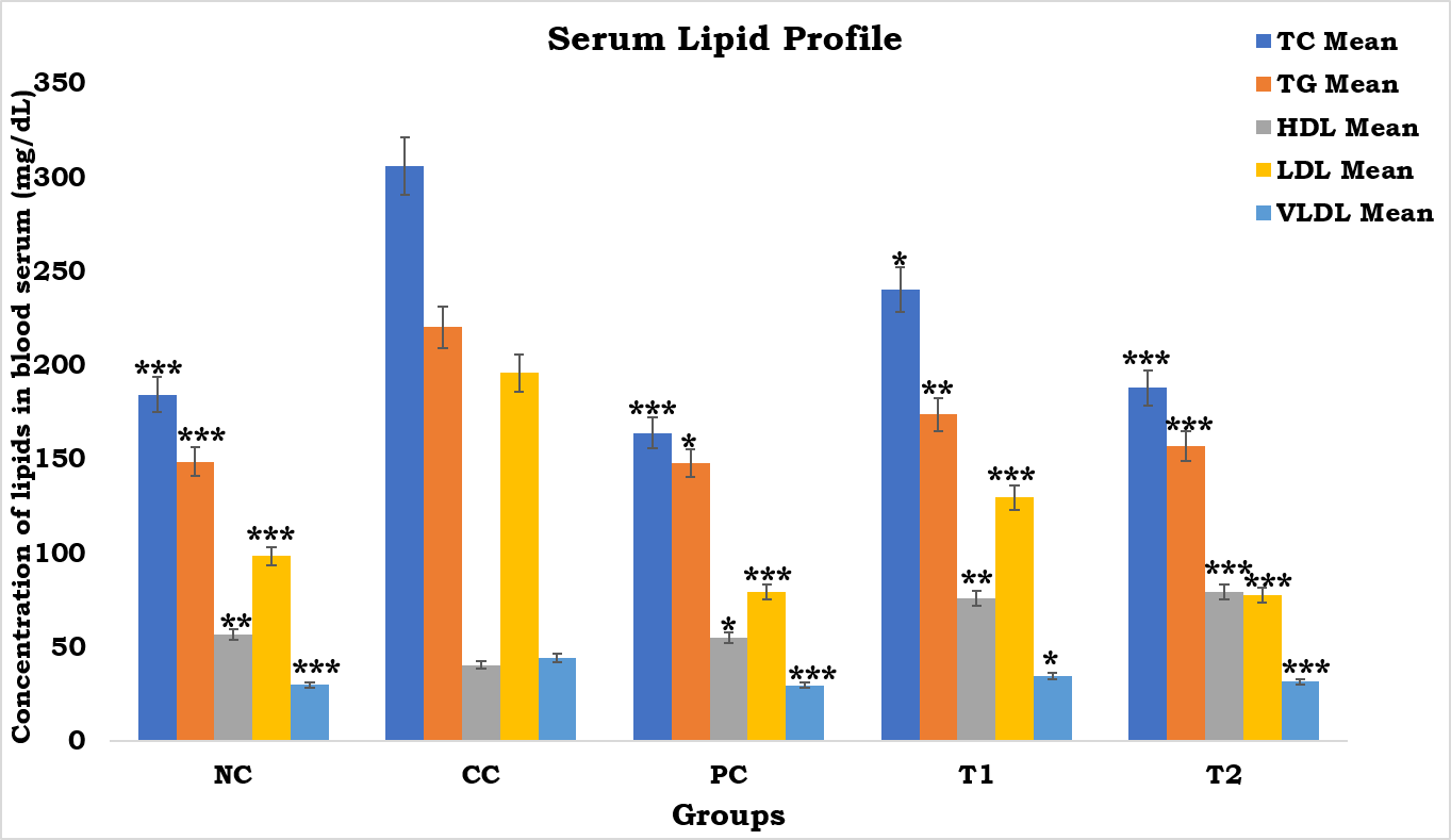
Figure …. Effect of Perindopril on serum lipid profile parameters in DEN-induced HCC in Wistar rats.
Data are expressed as Mean ± SD (n = 6). DEN administration significantly altered serum lipid metabolism, as evidenced by increased total cholesterol (TC), triglycerides (TG), low-density lipoprotein (LDL), and very low-density lipoprotein (VLDL) levels, along with a reduction in high-density lipoprotein (HDL) levels in the CC group compared with the NC group. Treatment with Perindopril (1 and 10 mg/kg, p.o.) and 5-FU significantly restored lipid profile parameters toward normal values. Statistical analysis was performed using one-way ANOVA followed by Bonferroni multiple comparison test. ns = non-significant, *P < 0.05, **P < 0.01 and *P < 0.001 compared with the CC group. NC: Normal Control; CC: Carcinogen Control; PC: Positive Control (5-FU); T1: Perindopril (1 mg/kg); T2: Perindopril (10 mg/kg).
3.7 Effect of Perindopril on Inflammatory Cytokines
DEN administration significantly increased hepatic IL-2, IL-6, IL-10, and IL-1β levels compared with normal controls, indicating severe inflammatory responses associated with HCC progression. Perindopril treatment attenuated the production of all measured cytokines in a dose-dependent manner. The higher dose (10 mg/kg) markedly reduced IL-2, IL-6, IL-10, and IL-1β levels to 420.18 ± 6.23, 488.23 ± 10.15, 364.16 ± 4.23, and 245.02 ± 6.72 pg/mL, respectively. These findings demonstrate the potent anti-inflammatory and immunomodulatory effects of Perindopril in DEN-induced HCC.
Table 6. Effect of Perindopril on Hepatic Inflammatory Cytokine Levels in DEN-Induced HCC in Wistar Rats. **Values are expressed as Mean ± SD (n = 6). Statistical analysis was performed using one-way ANOVA followed by Bonferroni multiple comparison test. *P < 0.05 and *P < 0.001 compared with the CC group. NC: Normal Control; CC: Carcinogen Control; PC: Positive Control (5-FU); T1: Perindopril (1 mg/kg); T2: Perindopril (10 mg/kg).
|
Sr. No. |
Group |
IL-2 (pg/mL) |
IL-6 (pg/mL) |
IL-10 (pg/mL) |
IL-1β (pg/mL) |
|
1 |
NC |
316.12 ± 9.76 |
398.12 ± 9.83 |
231.00 ± 2.69 |
193.17 ± 8.26 |
|
2 |
CC |
791.91 ± 15.19 |
689.51 ± 14.36 |
887.75 ± 15.38 |
573.42 ± 11.28 |
|
3 |
PC |
427.14 ± 10.16*** |
501.02 ± 10.67*** |
392.25 ± 6.38*** |
276.75 ± 8.15*** |
|
4 |
T1 |
489.31 ± 9.42*** |
573.07 ± 9.92* |
402.83 ± 7.51*** |
387.59 ± 8.84*** |
|
5 |
T2 |
420.18 ± 6.23*** |
488.23 ± 10.15*** |
364.16 ± 4.23*** |
245.02 ± 6.72*** |
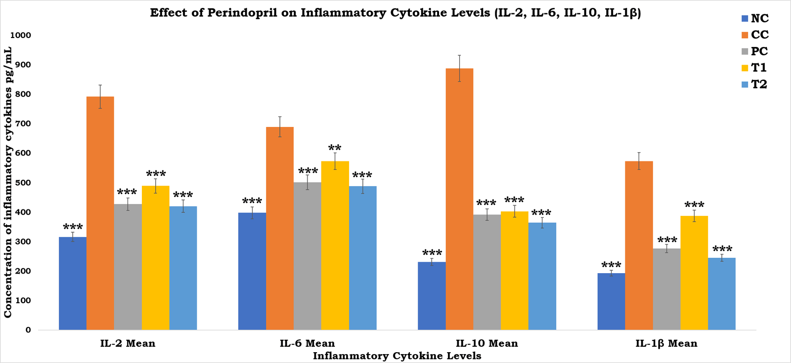
Figure 7.12. Effect of Perindopril on hepatic inflammatory cytokine levels (IL-2, IL-6, IL-10, and IL-1β) in DEN-induced HCC in Wistar rats.
Data are expressed as Mean ± SD (n = 6). DEN administration significantly elevated hepatic cytokine levels in the CC group compared with the NC group, indicating severe inflammatory responses associated with hepatocarcinogenesis. Treatment with Perindopril (1 and 10 mg/kg, p.o.) and 5-FU significantly attenuated cytokine production and restored inflammatory markers toward normal values. Statistical analysis was performed using one-way ANOVA followed by Bonferroni multiple comparison test. *P < 0.05, **P < 0.01 and *P < 0.001 compared with the CC group. NC: Normal Control; CC: Carcinogen Control; PC: Positive Control (5-FU); T1: Perindopril (1 mg/kg); T2: Perindopril (10 mg/kg).
3.8 Gross Morphological Evaluation
Gross morphological examination revealed marked differences among the experimental groups. The normal control group exhibited smooth liver surfaces, uniform reddish-brown coloration, and intact hepatic lobes. In contrast, the carcinogen control group displayed severe morphological abnormalities, including enlarged lobes, rough liver surfaces, and multiple visible tumor nodules, confirming successful induction of HCC. Treatment with 5-FU markedly reduced tumor burden and improved liver morphology. Perindopril treatment produced dose-dependent improvements, with the 10 mg/kg group exhibiting near-normal liver appearance and a marked reduction in nodular lesions.
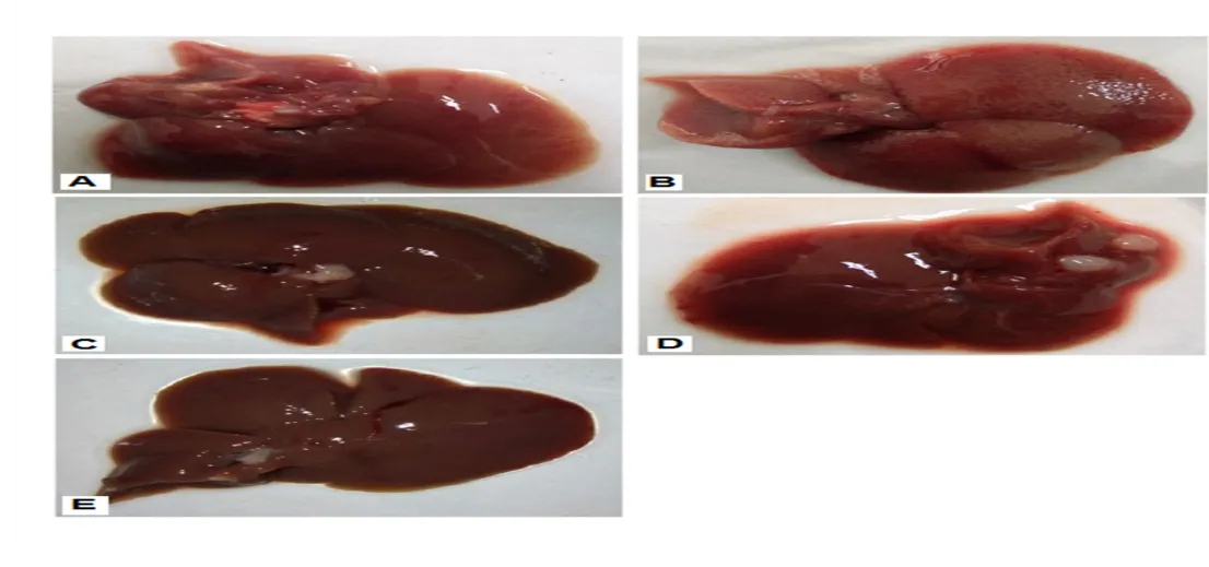
Figure 6. Gross morphological evaluation of liver tissues in experimental groups.
(A) Normal control (NC) group showing smooth surface, normal coloration, and intact hepatic lobes; (B) carcinogen control (CC) group exhibiting rough liver surface, enlarged lobes, multiple visible nodules, and severe tumor burden; (C) positive control (5-FU) group showing marked reduction in nodularity and partial restoration of normal liver morphology; (D) Perindopril-treated group (1 mg/kg) displaying moderate improvement in liver architecture with reduced nodular lesions; (E) Perindopril-treated group (10 mg/kg) showing near-normal liver morphology with smooth surface, uniform coloration, and marked reduction in tumor nodules.
3.9 Histopathological Evaluation
Histopathological examination further confirmed the gross morphological findings. Liver sections from the normal control group showed preserved hepatic architecture with intact hepatocytes and normal nuclei. In contrast, the carcinogen control group exhibited extensive architectural disruption characterized by inflammatory cell infiltration, tumor aggregate cells, regenerative clusters, nuclear degeneration, and necrotic changes. Treatment with 5-FU partially restored hepatic architecture and reduced tumor-associated lesions. Perindopril administration improved hepatic histology in a dose-dependent manner, with the 10 mg/kg group showing near-complete restoration of hepatic architecture, minimal inflammatory changes, and marked reduction in neoplastic alterations. These findings demonstrate the potent hepatoprotective and anticarcinogenic effects of Perindopril against DEN-induced HCC.
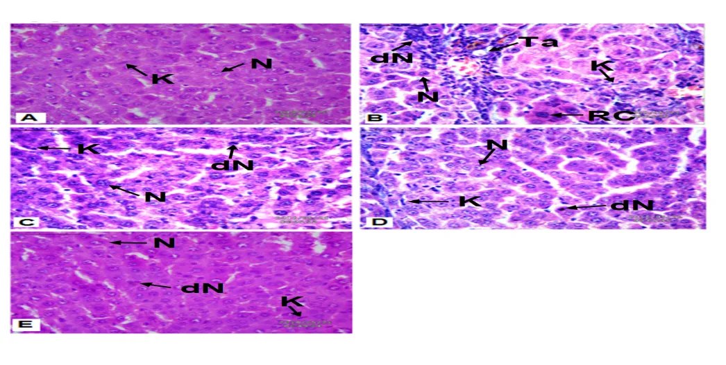
Figure 7. Histopathological evaluation of liver tissues (H&E staining, 400×; scale bar = 50 µm).
(A) Normal control liver showing preserved hepatic architecture, intact hepatocytes, normal nuclei (N), and prominent Kupffer cells (K); (B) carcinogen control liver displaying severe architectural distortion, inflammatory infiltration, tumor aggregate cells (Ta), regenerative clusters (RC), degenerated nuclei (dN), and necrotic areas; (C) 5-FU-treated group showing partial restoration of hepatic architecture with reduced tumor burden and preserved hepatocyte morphology; (D) Perindopril-treated group (1 mg/kg) demonstrating improved hepatocyte organization and reduced inflammatory changes; (E) Perindopril-treated group (10 mg/kg) exhibiting near-normal hepatic architecture with intact hepatocytes, well-defined nuclei, prominent Kupffer cells, and minimal degenerative alterations.
DISCUSSION
Hepatocellular carcinoma (HCC) is a multifactorial disease characterized by oxidative stress, chronic inflammation, metabolic disturbances, and progressive deterioration of liver function.⁴˒⁵ In the present study, DEN administration successfully induced HCC, as evidenced by alterations in physiological, biochemical, oxidative stress, inflammatory, and histopathological parameters. Treatment with Perindopril significantly attenuated these changes, suggesting its potential hepatoprotective and chemopreventive activity.
Body weight loss is a common feature of experimental carcinogenesis and is often associated with metabolic dysfunction, reduced nutrient utilization, and tumor-associated cachexia.²³ In the present study, DEN-treated animals showed a marked reduction in body weight along with increased liver weight and tumor burden. These findings indicate successful induction of hepatic tumors and progressive liver injury. Perindopril treatment improved body weight gain and reduced tumor incidence, suggesting its ability to counteract the systemic effects of carcinogenesis and suppress tumor progression.
Serum enzymatic biomarkers such as ALT, AST, and LDH are widely recognized indicators of hepatocellular damage.²⁴ The significant elevation of these enzymes in the carcinogen control group reflects loss of membrane integrity and leakage of intracellular enzymes into the circulation. Perindopril treatment significantly reduced ALT, AST, and LDH levels, indicating stabilization of hepatocyte membranes and preservation of liver function. The higher dose produced greater improvement and showed effects comparable to those observed with the standard drug treatment.
Oxidative stress is considered a major contributor to HCC development.⁵˒⁶ Excessive generation of reactive oxygen species can damage cellular lipids, proteins, and nucleic acids, thereby promoting carcinogenesis. In the present study, DEN administration significantly reduced the activities of antioxidant enzymes such as SOD and CAT and depleted GSH levels, indicating impairment of the endogenous antioxidant defense system. Similar reductions in antioxidant defenses have been reported in DEN-induced hepatocarcinogenesis models.¹⁴˒²⁵ Perindopril treatment restored these antioxidant markers, suggesting enhanced scavenging of reactive oxygen species and improved cellular defense against oxidative injury. Restoration of antioxidant status may have contributed substantially to the protective effects observed in liver tissue.
The increased levels of TBARS/MDA and protein carbonyl content observed in DEN-treated animals further confirmed the presence of severe oxidative damage. Elevated TBARS/MDA levels indicate enhanced lipid peroxidation, whereas increased protein carbonyl content reflects oxidative modification of cellular proteins.¹⁹˒²⁰ Perindopril significantly reduced both markers, indicating protection against oxidative degradation of lipids and proteins. These findings suggest that the antioxidant properties of Perindopril play an important role in preventing cellular injury associated with hepatocarcinogenesis.¹¹˒¹²
Alterations in bilirubin and biliverdin metabolism are commonly associated with hepatic dysfunction and oxidative stress.²⁴ The significant elevation of these pigment markers in the carcinogen control group indicated impaired hepatic metabolic activity and disturbed heme degradation pathways. Perindopril treatment effectively normalized bilirubin and biliverdin levels, suggesting improvement in hepatic functional integrity and restoration of normal metabolic processes.
Liver cancer is frequently associated with abnormalities in lipid metabolism.²⁶˒²⁷ The dyslipidemia observed in DEN-treated animals, characterized by elevated TC, TG, LDL, and VLDL levels along with reduced HDL concentrations, is consistent with impaired hepatic lipid handling during carcinogenesis. Perindopril significantly corrected these abnormalities and restored lipid homeostasis. Improvement in lipid profile may not only reflect better liver function but may also contribute to limiting tumor progression by reducing metabolic disturbances associated with cancer development.
Chronic inflammation is a key driving force in HCC initiation and progression.²⁸˒²⁹ Elevated levels of IL-2, IL-6, IL-10, and IL-1β in the carcinogen control group indicated a pronounced inflammatory response within hepatic tissue. Among these cytokines, IL-6 and IL-1β are known to promote tumor growth, survival, angiogenesis, and cellular proliferation.²⁸˒²⁹ Perindopril treatment significantly reduced the levels of all measured cytokines, demonstrating potent anti-inflammatory activity. Suppression of inflammatory mediators may represent an important mechanism through which Perindopril exerts its protective effects against DEN-induced hepatocarcinogenesis.¹¹˒¹²
The biochemical findings were further supported by gross morphological and histopathological observations. DEN-treated animals exhibited multiple tumor nodules, distorted liver architecture, inflammatory infiltration, degenerative changes, and necrotic lesions. In contrast, Perindopril-treated groups showed marked improvement in liver morphology and histological organization. The high-dose group demonstrated near-normal hepatic architecture with reduced tumor burden and minimal degenerative changes. These observations strongly support the biochemical evidence of hepatoprotection and indicate effective attenuation of carcinogen-induced hepatic injury.
Overall, the findings of the present study suggest that Perindopril exerts significant protective effects against DEN-induced HCC through multiple mechanisms, including enhancement of antioxidant defenses, reduction of oxidative damage, suppression of inflammatory responses, normalization of lipid metabolism, and preservation of hepatic architecture.⁷˒¹¹˒¹² The greater efficacy observed at the higher dose further indicates a dose-dependent protective effect. These results highlight the potential of Perindopril as a promising candidate for further investigation in the prevention and management of hepatocellular carcinoma.
CONCLUSION
The present study demonstrates that Perindopril exerts significant protective effects against DEN-induced hepatocellular carcinoma in Wistar rats. Treatment with Perindopril effectively reduced tumor burden, improved body weight, normalized liver function biomarkers, and restored hepatic architecture. In addition, Perindopril enhanced endogenous antioxidant defenses by increasing SOD, CAT, and GSH levels while reducing lipid peroxidation and protein oxidation, indicating a substantial attenuation of oxidative stress.
Perindopril also improved hepatic metabolic function by normalizing bilirubin, biliverdin, and lipid profile parameters. Furthermore, the marked reduction in pro-inflammatory cytokines, including IL-2, IL-6, IL-10, and IL-1β, highlights its anti-inflammatory and immunomodulatory properties. These biochemical findings were strongly supported by gross morphological and histopathological observations, which revealed significant protection against DEN-induced hepatic damage and neoplastic alterations.
Overall, the findings suggest that the hepatoprotective and chemopreventive effects of Perindopril are mediated through the combined suppression of oxidative stress, inflammation, and metabolic disturbances associated with hepatocarcinogenesis. The higher dose of Perindopril demonstrated superior efficacy throughout the study. These results indicate that Perindopril may represent a promising therapeutic candidate for the prevention and management of hepatocellular carcinoma. However, further mechanistic studies and clinical investigations are required to fully establish its potential role in cancer therapy.
DECLARATIONS
Ethics Approval and Consent to Participate
All animal experimental procedures were performed in accordance with the guidelines of the CPCSEA, Government of India, and were approved by the IAEC Approval No. 1698/PO/Re/S/13/CPCSEA.
Consent for Publication
Not applicable.
Availability of Data and Materials
The datasets generated and/or analyzed during the current study are available from the corresponding author upon reasonable request.
Funding
The authors declare that no external funding was received for this study.
Conflict of Interest
The authors declare that they have no competing interests.
Authors' Contributions
Virendra Kumar Baheliya conceived and designed the study, performed the experiments, analyzed the data, and drafted the manuscript. Dharmendra Singh Rajput, Naveen Gupta, Ganesh Prasad Patel, and Brajmohan Kaushal contributed to the experimental work, data analysis, and manuscript review. Ritesh Suresh Bhate supervised the study, validated the methodology, and critically revised the manuscript. All authors read and approved the final manuscript.
Acknowledgements
The authors express sincere gratitude to the Department of Pharmaceutical Sciences, Faculty of Pharmacy, Madhyanchal Professional University, Bhopal, for providing the necessary facilities and support to carry out this research work. The authors also acknowledge the valuable guidance and encouragement provided by faculty members and research colleagues throughout the study.
REFERENCES


Virendra Kumar Baheliya, Dharmendra Singh Rajput, Naveen Gupta, Ganesh Prasad Patel, Brajmohan Kaushal, Ritesh Bhate, Perindopril Attenuates Diethylnitrosamine-Induced Hepatocellular Carcinoma in Wistar Rats Through Modulation of Oxidative Stress, Inflammatory Cytokines, And Hepatic Injury Biomarkers, Int. J. of Pharm. Sci., 2026, Vol 4, Issue 6, 6795-6810, https://doi.org/10.5281/zenodo.20928596
 10.5281/zenodo.20928596