

We use cookies to ensure our website works properly and to personalise your experience. Cookies policy
1Department of Pharmaceutical Quality Assurance, D.S.T.S. Mandal’s College of Pharmacy, Solapur, 413004 Maharashtra, India.
2Department of Pharmaceutical Chemistry, D.S.T.S. Mandal’s College of Pharmacy, Solapur, 413004 Maharashtra, India
Ritonavir is an antiretroviral medication that has limited water solubility, and its low solubility along with its tendency to form different crystal structures significantly hinders its ability to dissolve and be absorbed through the mouth. This study aimed to develop and test a ternary co-amorphous system to improve the solubility and dissolution properties of ritonavir. The ternary co-amorphous formulations were made by mixing ritonavir with two selected low-molecular-weight co-formers, L-arginine and nicotinamide, using a solvent evaporation method. The goal was to create a stable amorphous form by using strong interactions between the drug and co-formers. Before starting the formulation, some preliminary studies were carried out to understand the properties of the pure drug, such as its melting point and solubility. The prepared formulations were analyzed using various solid-state techniques, including Differential Scanning Calorimetry (DSC), Powder X-Ray Diffraction (PXRD), Fourier Transform Infrared Spectroscopy (FTIR), and Scanning Electron Microscopy (SEM), to confirm the amorphous state and the interactions between the drug and co-formers. The solubility and dissolution behavior of the formulations were tested to evaluate how much better the drug release was compared to the original crystalline form of ritonavir. The best formulation showed a big increase in solubility and a much better dissolution rate. This was due to lower crystallinity, better surface wettability, and improved molecular dispersion.DSC and PXRD results confirmed that the crystalline ritonavir had been transformed into an amorphous form. FTIR analysis showed that hydrogen bonds formed between ritonavir and the co-formers, which helped in maintaining the stability of the system. These findings show that ternary co-amorphous systems are a promising strategy for improving the solubility, dissolution, and possibly the bioavailability. of drugs with low solubility like ritonavir. This method could be a valuable approach in developing more effective oral drug delivery systems for drugs classified as BCS class II
Poor aqueous solubility is a major challenge in pharmaceutical formulation development because it directly affects dissolution, absorption, and oral bioavailability of active pharmaceutical ingredients (APIs). Approximately 40–70% of newly discovered drug molecules show poor water solubility, making solubility enhancement an important area of research in drug delivery science [1, 2].
Is a protease inhibitor used in antiretroviral therapy for the treatment of Human Immunodeficiency Virus (HIV) infection. It also acts as a potent inhibitor of cytochrome P450 3A4 (CYP3A4), enhancing the plasma concentration of co-administered drugs [3]. However, ritonavir exhibits poor aqueous solubility, high lipophilicity, polymorphism, and variable oral bioavailability, creating major formulation challenges [4].
The poor dissolution behavior of ritonavir is mainly due to its hydrophobic crystalline structure, which reduces therapeutic efficiency. Ritonavir is also known for polymorphism, where conversion to a more stable crystalline form can significantly reduce solubility and bioavailability [5, 6].
Several techniques such as micronization, solid dispersions, liquisolid systems, and lipid-based formulations have been investigated to improve ritonavir solubility. Although these approaches improve dissolution, they often face limitations such as recrystallization, poor stability, and complex manufacturing [7, 8].
Amorphous drug delivery systems are widely used to enhance solubility and dissolution by converting crystalline drugs into high-energy amorphous forms. However, amorphous systems are thermodynamically unstable and may recrystallize during storage [9].
To overcome this limitation, co-amorphous systems have emerged as a promising strategy. These systems combine the drug with low-molecular-weight co-formers to form a stable amorphous phase through strong intermolecular interactions such as hydrogen bonding and ionic interactions [10]. Binary co-amorphous systems improve solubility but may still have limited long-term stability [11].
Recently, ternary co-amorphous systems containing one drug and two co-formers have shown improved stability and dissolution due to stronger molecular interactions and higher glass transition temperature [12]. Therefore, the present study aims to formulate and evaluate a ternary co-amorphous system of ritonavir to enhance its solubility, dissolution, and physical stability.
MATERIALS AND METHODS
Drug and coformers
Table 1: Drug and Coformers
|
Sr. No |
Drug and Coformers |
Manufacturer |
|
1 |
Ritonavir |
Hetero labs Hyderabad |
|
2 |
Nicotinamide |
SRL Chemicals |
|
3 |
L-Arginine |
Research lab |
Equipments
Table 2: Equipments and Suppliers
|
Sr. No |
Name of Equipments |
Supplier |
|
1 |
Weighing Balance |
Contech CA233 |
|
2 |
Sonicator |
Oscar 103 |
|
3 |
Water Bath Shaker |
REMIRSB-12 |
|
4 |
UV Spectrophotometer |
Systronic UV 2102 |
|
5 |
Tablet Punching Machine |
Karnavati 12 Station Tablet Compression Machine |
|
6 |
Friabilator |
Electrolab |
|
7 |
Dissolution Apparatus |
Electrolab |
|
8 |
FTIR |
Bruker Alpha ? |
|
9 |
DSC |
Mettler Star SW 12.10 |
|
10 |
XRD |
Ultima ?, Riga Corporation, Japan |
Calibration curve of Ritonavir
A series of standard solutions of at concentrations of 10, 20, 30, 40, 50, and 60 µg/mL were prepared in 0.1 N HCl, and the absorbance of each solution was measured at 240 nm using a UV-visible spectrophotometer.
Solvent evaporation method for preparation of co amorphous
The required molecular ratio of and selected co-formers was dissolved in ml methanol under continuous stirring for 20 min to obtain a homogeneous solution. The solution was then allowed to evaporate at room temperature, resulting in the formation of a free-flowing co-amorphous powder.
Table 3: Composition of Ternary Co-Amorphous Formulations
|
Formulation Code |
Drug (Ritonavir) mg |
Co-former 1 (L-Arginine) mg |
Co-former 2 (Nicotinamide) mg |
Ratio (D:C1:C2) |
|
F1 |
200 |
48.3 |
33.8 |
1:1:1 |
|
F2 |
200 |
48.3 |
67.6 |
1:1:2 |
|
F3 |
200 |
48.3 |
101.4 |
1:1:3 |
|
F4 |
200 |
96.6 |
33.8 |
1:2:1 |
|
F5 |
200 |
96.6 |
67.6 |
1:2:2 |
|
F6 |
200 |
96.6 |
101.4 |
1:2:3 |
|
F7 |
200 |
144.9 |
33.8 |
1:3:1 |
|
F8 |
200 |
144.9 |
67.6 |
1:3:2 |
|
F9 |
200 |
144.9 |
101.4 |
1:3:3 |
Saturation solubility study
An excess quantity of sample was introduced into 10 mL of 0.1 N hydrochloric acid to evaluate saturation solubility. The dispersion was continuously mixed on a Magnetic Stirring system at 300–500 rpm while maintaining the temperature at 37 ± 0.5°C for 24 h to establish equilibrium. After equilibration, the undissolved particles were allowed to sediment, and the supernatant was separated by filtration through a 0.45 μm membrane filter. The filtrate was appropriately diluted and quantified using UV-Visible Spectroscopy at 240 nm.
Fourier transform infrared spectroscopy
Fourier Transform Infrared spectroscopy analysis was carried out to characterize the co amorphous using a Bruker Alpha ? FTIR spectrometer.
Differential scanning calorimetry
Differential Scanning Calorimetry analysis of optimized formulation was carried out using a METTLER TOLEDO DSC1 STARe System.
Powder x-ray diffraction
The crystalline nature of the sample was evaluated by powder X-ray Diffraction employing an Ultima ?, Riga Corporation, Japan.
Scanning electron microscopy
Surface morphology of the sample was examined using Scanning Electron Microscopy.
Pre-compression study
Bulk density
Bulk density of a powder may vary considerably depending on factors such as crystallization method, milling process, and formulation characteristics. It was determined by carefully transferring the powder blend into a graduated cylinder through a wide funnel, followed by measurement of the occupied volume and sample weight. The bulk density was then calculated as the ratio of powder mass to bulk volume.
Tapped density
The tapped density was measured by putting the powder mixture into a graduated cylinder using a large funnel, then tapping the cylinder 50 times, and finally recording the volume and weight of the compacted powder.
Angle of repose
The angle of repose was determined by allowing the powder to flow through a funnel onto a flat surface, forming a cone, and then measuring the angle between the powder slope and the horizontal plane.
Hausner’s ratio
This parameter indicates powder flow properties and is calculated from the ratio of tapped density to bulk density.
Carr’s index
The compressibility index was determined using the measured bulk density and tapped density values.
Post compression study
Hardness
Tablet hardness refers to the force needed to break a tablet under diametric compression. It was measured using a Monsanto hardness tester.
Tablet size and thickness
The thickness of the tablets was measured using a Vernier caliper.
Friability
The friability test was carried out to assess the ability of tablets to resist abrasion during packaging, handling, and transportation. An initial weight of 20 tablets was recorded and placed in a friabilator operating at 25 rpm for 4 minutes. The weight loss after the test was calculated and expressed as a percentage. The acceptable friability range is preferably between 0.5% and 1.0%.
Weight variation test
Uniformity of tablet weight is an important quality parameter for a batch. Any deviation in weight must comply with the prescribed limits. A total of 20 tablets were randomly chosen and weighed precisely for analysis.
In-vitro dissolution studies
In vitro dissolution studies of pure Ritonavir and the optimized formulation were carried out using a USP type II dissolution apparatus with paddle assembly operated at 50 rpm. The dissolution test was performed in 900 mL of 0.1 N HCl, which served as the dissolution medium, while maintaining the temperature at 37 ± 0.5°C. At predetermined time intervals, 5 mL aliquots were withdrawn and replaced with an equal volume of fresh 0.1 N HCl to maintain sink conditions. The collected samples were analyzed using UV-Visible Spectroscopy, and the dissolution profile was evaluated accordingly.
RESULTS AND DISCUSSTION
Calibration curve of Ritonavir
Table 4: Concentration and Absorbance of Ritonavir in 0.1 N HCL
|
Concentration (µg/ml) |
Absorbance |
|
10 |
0.1372 |
|
20 |
0.2751 |
|
30 |
0.3918 |
|
40 |
0.5326 |
|
50 |
0.6748 |
|
60 |
0.8106 |
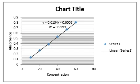
Figure 1: Calibration Curve of Ritonavir
The calibration curve of Ritonavir in 0.1 N HCl showed a linear increase in absorbance with increasing concentration in the range of 10–60 µg/mL. The absorbance values increased from 0.1372 at 10 µg/mL to 0.8106 at 60 µg/mL, indicating good compliance with the Beer–Lambert law. This confirms that the analytical method is suitable for quantitative estimation of the drug.
Saturation solubility study
Table 5: Saturation Solubility Study Co-Amorphous System
|
Batch |
Enhanced solubility Mg/mL |
Fold increases |
|
F1 |
0.017 |
3.6 |
|
F2 |
0.023 |
4.8 |
|
F3 |
0.030 |
6.2 |
|
F4 |
0.038 |
7.8 |
|
F5 |
0.047 |
9.6 |
|
F6 |
0.067 |
12.2 |
|
F7 |
0.073 |
14.8 |
|
F8 |
0.095 |
19.2 |
|
F9 |
0.086 |
17.4 |
Among all formulated batches of Ritonavir, batch F8 (1:3:2) was identified as the optimized formulation due to its highest solubility and better dissolution performance. It showed a solubility of 0.096 mg/mL, representing a 19.2-fold increase over pure drug. Further studies such as Fourier-transform infrared spectroscopy, Differential scanning calorimetry, and X-ray diffraction are required for confirmation.
Infrared spectroscopy
Comparison of the Infrared spectroscopy spectra of the pure drug and co-amorphous system indicated hydrogen bond formation between the drug and coformer, suggesting the formation of a new supramolecular synthon.
IR spectrum of Ritonavir
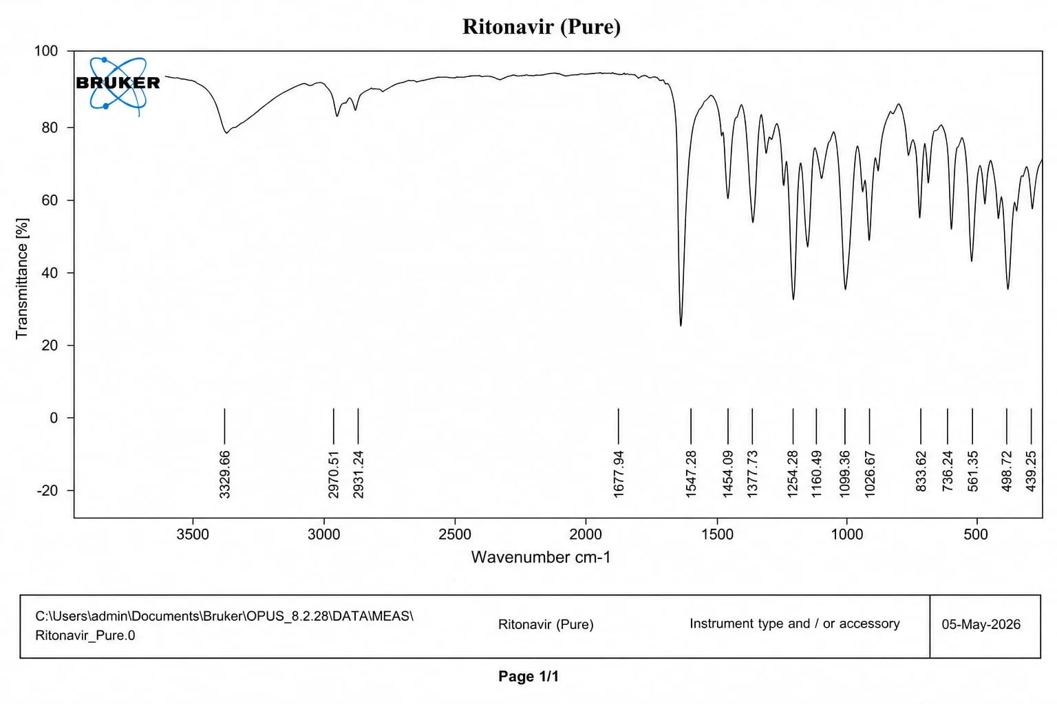
Figure 2: IR Spectra of Ritonavir
Table 6: Characteristic peaks from Ritonavir
|
Wavenumber (cm-1) |
Functional Group |
|
3329.66 |
N-H stretching |
|
2970.51 |
Aliphatic C-H stretching |
|
1677.94 |
C=O stretching |
|
1547.28 |
N-H bending |
|
1454.09 |
Aromatic C=C stretching |
|
1254.28 |
C-N stretching |
|
1160.49 |
C-O stretching |
IR spectra of Ritonavir+L-Arginine+Nicotinamide
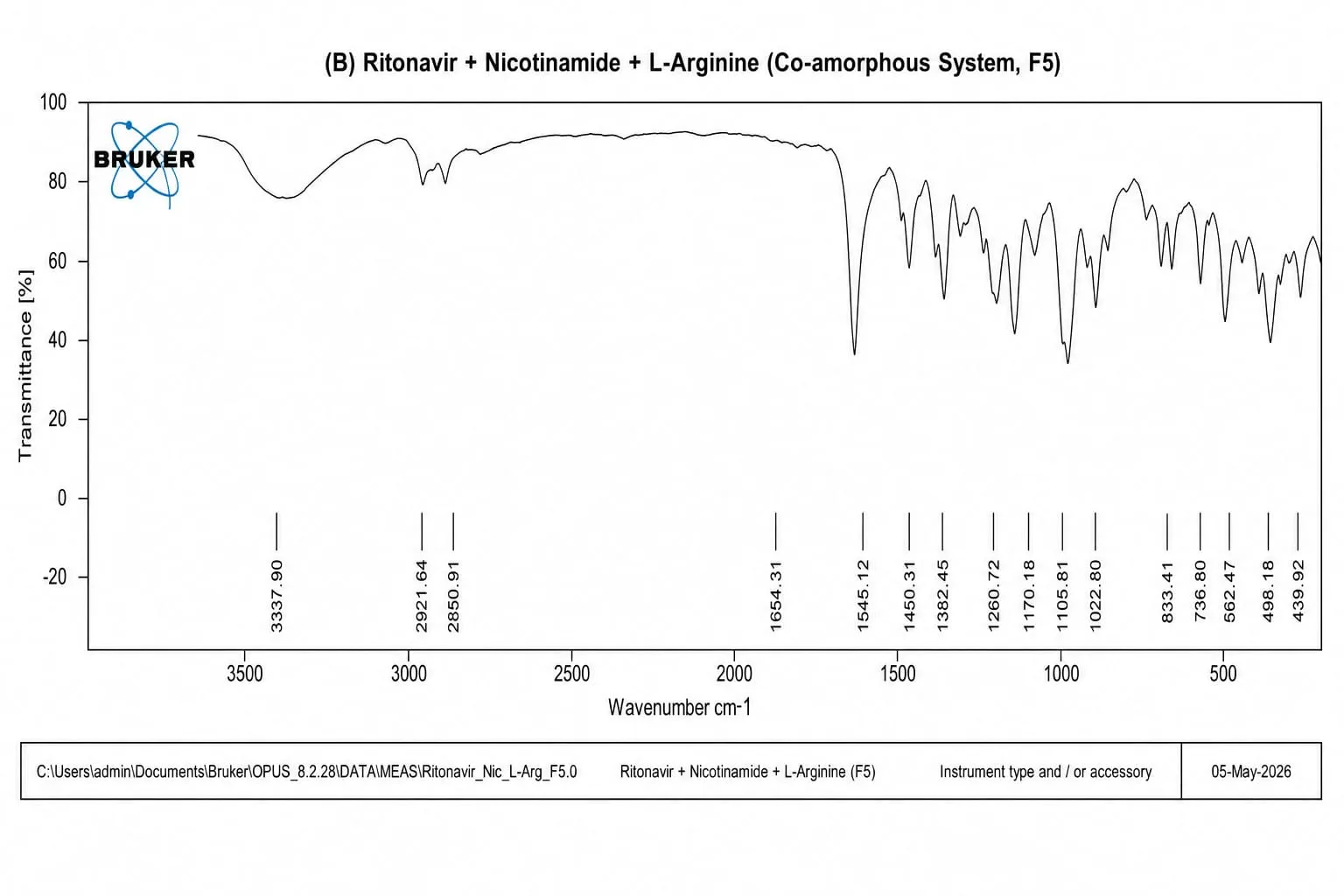
Figure 3: FT-IR spectrum of Ritonavir+L-Arginine+Nicotinamide
Table 7: FT-IR spectrum of Ritonavir+L-Arginine+Nicotinamide
|
Wavenumber (cm-1) |
Interpretation |
|
|
Ritonavir peaks |
Co-Amorphous peaks |
|
|
3329.66 |
3337.90 |
Shift in N–H stretching indicates hydrogen bonding interaction |
|
2970.51 |
2921.64 |
Shift in aliphatic C–H stretching confirms molecular interaction |
|
1677.94 |
1654.31 |
Shift in C=O stretching suggests strong intermolecular interaction and amorphization |
|
1547.28 |
1545.12 |
Slight shift in amide II/N–H bending indicates drug–carrier interaction |
|
1454.09 |
1450.31 |
Aromatic C=C stretching peak shift indicates change in molecular environment |
|
1254.28 |
1260.72 |
Shift in C–O stretching suggests interaction with co-formers |
|
1160.49 |
1170.18 |
C–O stretching peak shift confirms intermolecular interaction in co-amorphous system |
Differential scanning calorimetry
Differential scanning calorimetry of Ritonavir
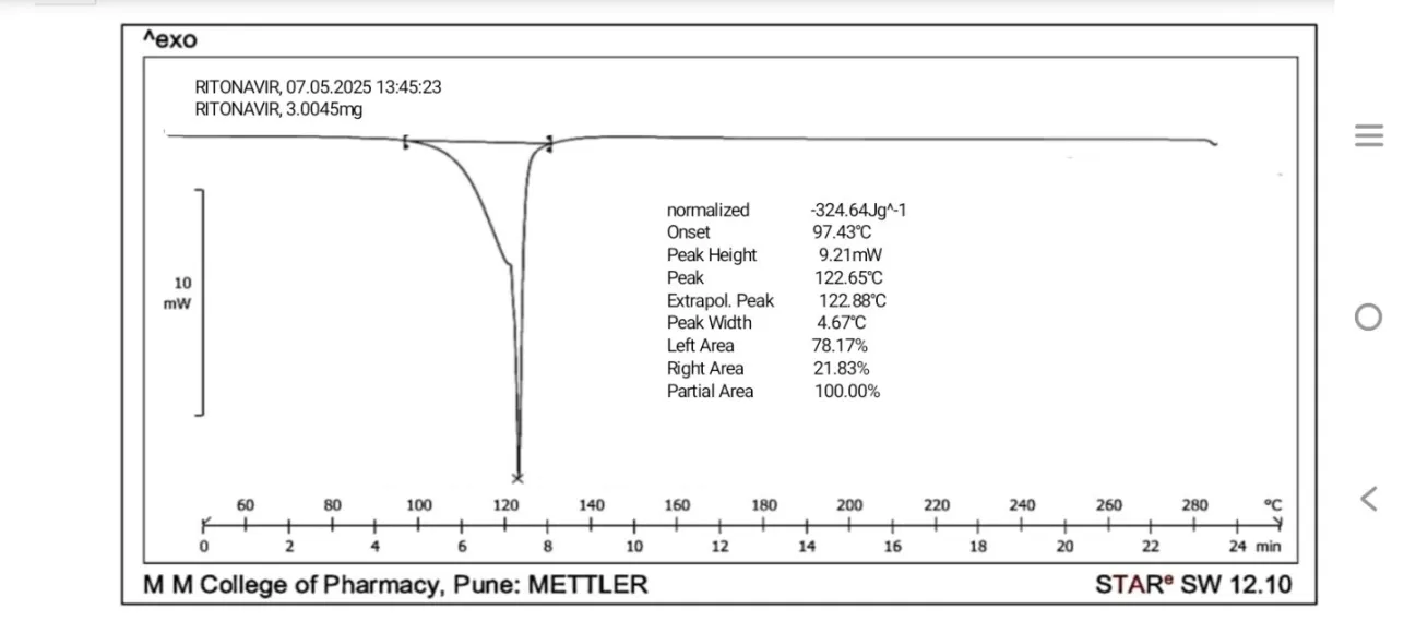
Figure 4: DSC of Ritonavir
Differential scanning calorimetry of Ritonavir+L-Arginine+Nicotinamide
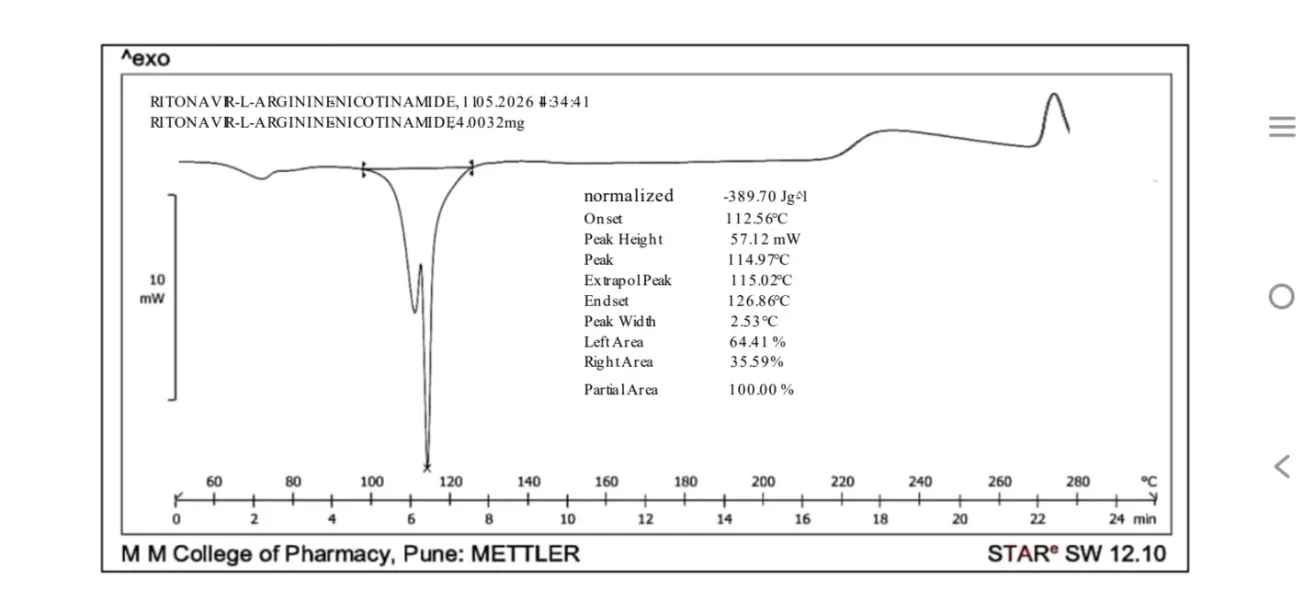
Figure 5: DSC of Ritonavir+L-Arginine+Nicotinamide
The co-amorphous formulation exhibited a noticeable change in thermal behavior compared to the pure drug. The melting temperature shifted from 122.65°C for the pure drug to 114.97°C for the co-amorphous system. Additionally, the onset temperature increased from 97.43°C in the pure drug to 112.56°C in the co-amorphous formulation. These variations in thermal characteristics suggest the successful formation of a co-amorphous phase and indicate molecular interactions between the drug and the co-former.
Powder x-ray diffraction
Powder x-ray diffraction of Ritonavir
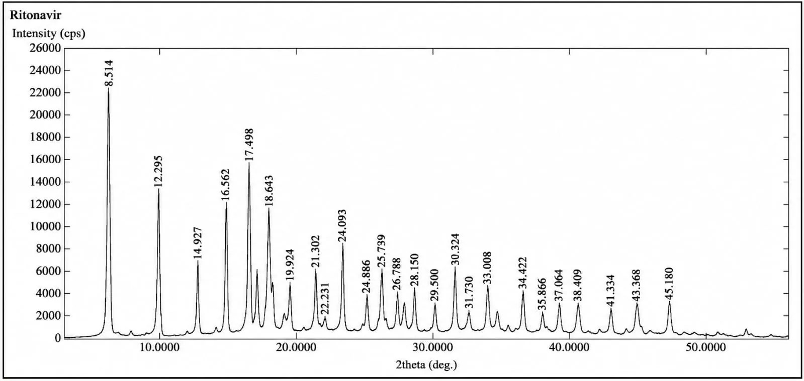
Figure 6: PXRD of Ritonavir
Powder x-ray diffraction of Ritonavir+L-Arginine+Nicotinamide
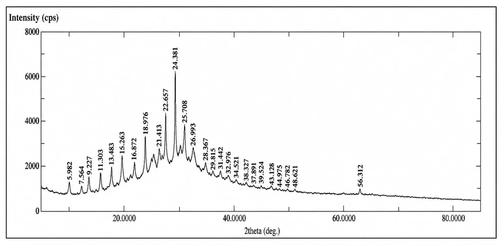
Figure 7: PXRD of Ritonavir+L-Arginine+Nicotinamide
The PXRD pattern of the co-amorphous formulation containing Ritonavir, L-Arginine, and Nicotinamide exhibited significant changes compared with that of pure Ritonavir. The characteristic sharp diffraction peaks of crystalline Ritonavir were markedly reduced in intensity or disappeared, resulting in a broad halo pattern. This reduction in crystallinity indicates the transformation of the drug into an amorphous state. The absence of distinct crystalline peaks and the appearance of diffuse scattering suggest the successful formation of a co-amorphous system through molecular interactions among Ritonavir, L-Arginine, and Nicotinamide. These findings confirm modifications in the solid-state properties of the drug and support the development of a stable co-amorphous phase.
Scanning electron microscopy
Scanning electron microscopy of Ritonavir
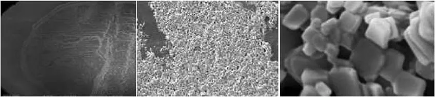
Figure 8: SEM of Ritonavir
Scanning electron microscopy Ritonavir+L-Arginine+Nicotinamide
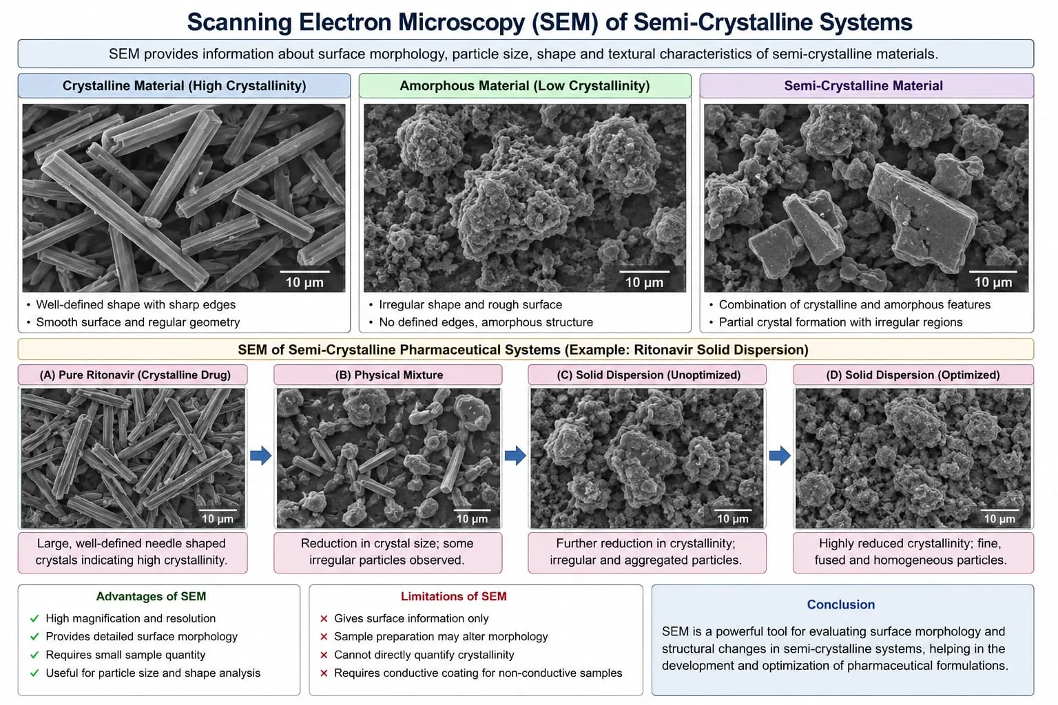
Figure 9: SEM of Ritonavir+L-Arginine+Nicotinamide
The surface morphology of the Ritonavir–L-Arginine–Nicotinamide system was examined by SEM, and the micrographs are presented in Figure X. This formulation was selected for SEM analysis due to its improved solubility. The SEM images revealed distinct morphological changes compared with pure Ritonavir, showing irregular and non-uniform particles. These observations suggest modification of the original solid-state structure and support the formation of a new drug–co-former system.
Pre compression studies of pure drug and co-amorphous formulation
Table 8: Pre compression study of pure drug and co-amorphous system
|
Sample |
Angle of Repose (?) |
Bulk Density |
Tapped Density |
Carr’s Index |
Hausner’s Ratio |
|
Ritonavir |
30.39 (±0.030) |
0.259 (±0.020) |
0.383 (±0.016) |
32.38 (±0.89) |
1.48 (±0.13) |
|
Ritonavir+L-Arginine+Nicotinamide |
29.030 (±0.020) |
0.313 (±0.015) |
0.438 (±0.013) |
28.54 (±1.2) |
1.40 (±0.08) |
(No. of samples, n = 3)
The pre-compression evaluation demonstrated that the Ritonavir–L-Arginine–Nicotinamide co-amorphous system possessed better flow and packing characteristics than pure Ritonavir. The observed improvement in flowability and compressibility may be attributed to the modification of particle properties following co-amorphization, indicating its suitability for further tablet formulation and manufacturing processes.
Post compression parameters of pure drug and co-amorphous tablets
Table 9: Post Compression parameters of tablets
|
Parameter |
Result |
|
Weight Variation (g) |
Complies |
|
Hardness (kg/cm2) |
5.00 (±0.02) |
|
Thickness (mm) |
4.00 (±0.04) |
|
Friability (%) |
0.56 (±0.03) |
|
Disintegration time (min) |
12.00 (±0.02) |
(No. of samples, n = 3)
Dissolution studies
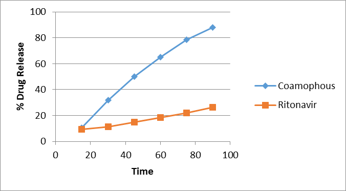
Figure 10: Cumulative % drug release of Ritonavir+L-Arginine+Nicotinamide co amorphous Tablet
The dissolution study revealed a substantial enhancement in drug release from the Ritonavir–L-Arginine–Nicotinamide co-amorphous system compared to pure Ritonavir. The co-amorphous formulation achieved 87.83% drug release at 90 min, while pure Ritonavir exhibited only 26.31% release. The improved dissolution behavior confirms the effectiveness of co-amorphization in enhancing the dissolution characteristics of Ritonavir.
CONCLUSION
The ternary co-amorphous system of Ritonavir with L-Arginine and Nicotinamide was successfully formulated by the solvent evaporation method. The optimized formulation (F8) exhibited a significant increase in solubility and dissolution rate compared with pure Ritonavir. FTIR, DSC, PXRD, and SEM studies confirmed the formation of a stable co-amorphous system with reduced crystallinity and strong intermolecular interactions. Thus, the ternary co-amorphous approach proved to be an effective strategy for enhancing the solubility and dissolution performance of Ritonavir, with potential to improve its oral bioavailability.
ACKNOWLEDGEMENT
The author gratefully acknowledges the support and assistance provided by the Principal, faculty, and staff of D.S.T.S. Mandal’s College of Pharmacy, Solapur, for facilitating this research work. Sincere appreciation is extended to the project guide for valuable guidance, constructive suggestions, and continuous encouragement throughout the study. The author also wishes to thank family members and friends for their unwavering motivation and support during the successful completion of this work.
REFERENCES


Ganesh Narayane*, Dr. V.S. Tegeli, To Formulate And Evaluate Of Ternary Co-Amorphous System For Solubility Enhancement Of Ritonavir, Int. J. of Pharm. Sci., 2026, Vol 4, Issue 8, 1429-1441. https://doi.org/10.5281/zenodo.21842558
 10.5281/zenodo.21842558