The skin is the largest organ of the human body and serves as a vital protective barrier against environmental, microbial, and chemical insults while contributing to sensation, thermoregulation, hydration, and vitamin D synthesis. Its physiological properties and pH significantly influence topical drug delivery and therapeutic effectiveness. Fungal infections of the skin, including superficial and mucosal infections are common and require effective topical antifungal therapy. Antifungal agents primarily act by disrupting fungal cell membranes or inhibiting ergosterol biosynthesis. Pharmaceutical gels are semisolid preparations widely used for topical and transdermal drug delivery because of their ease of application, patient acceptability, and ability to provide controlled drug release. Their three-dimensional polymeric network enables swelling, drug incorporation, controlled release, and enhanced residence time at the application site. Gels may be formed through chemical, physical, or ionic cross-linking mechanisms. They offer advantages such as ease of application, non-greasy feel, cooling effect, and improved bioavailability, but may also exhibit stability and microbial contamination challenges. Thus, gels represent promising carriers for topical antifungal drug delivery. This review summarizes the classification of gels based on colloidal phase, solvent, rheological properties, and physical nature. It also discusses commonly used natural, semisynthetic, synthetic, inorganic, and surfactant-based gelling agents. Various methods of gel preparation, including thermal, chemical, fusion, cold, and dispersion methods, are described. Evaluation parameters such as homogeneity, pH, drug content, viscosity, spreadability, extrudability, grittiness, in vitro drug diffusion, in vivo study and skin irritation are reviewed.
Keywords
Skin, Topical drug delivery, Fungal infections, Antifungal agents, Pharmaceutical Gel.
Introduction
×
SKIN
With an amazing surface area of about 2 m2, the skin is the largest organ in the body and making up around 20% of an adult's total body weight. It serves both passive and active functions as a crucial barrier between the human body and the environment against microbial, chemical, and physical assaults.
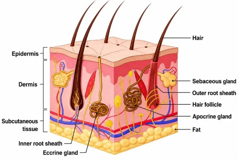
Skin is composed of an upper layer (the epidermis), and a lower layer (the dermis) which is divided by a membrane in the basement.
Epidermis - The epidermis is formed of five layers:
The stratum corneum is the outermost layer of the five layers that make up the epidermis. stratum lucidum, (present only in certain body parts like the fingertips, palms, and soles of the feet), the stratum granulosum, the stratum spinosum, and the stratum basale (the inner most layer that contains epidermal stem cells).
Dermis - Based on the thickness of its collagen content, the dermis is split into two layers:
an upper stratum papillare made of thin fibers, and a lower stratum reticulare containing thick fibers.
Hypodermis or subcutaneous tissue - The hypodermis is a layer of loose connective tissue and elastin that serves as a reservoir for nutrients and energy, shock adsorbent, and insulation against cold temperatures. The thickest hypodermis is found in the hands, buttocks, and soles of the feet. The hypodermis starts to atrophy as we age, which contributes to the thin wrinkles appearance of the skin as it ages.
Additionally, the skin contains certain innate and adaptive immune cells that make up the skin immune system, including mast cells, macrophages, natural killers (NKs), highly specialized antigen presenting cells (APCs), epidermal dendritic cells (EDCs, sometimes called Langerhans cells), dermal dendritic cells (DCs, also called interstitial or migratory DCs), αβ T cells, γδ T cells and B cells.
Figure 1: Skin Anatomy
Upon the disruption or compromise of the cutaneous barrier by trauma, a restorative process is initiated to recover skin integrity and function, a timeline requiring approximately one week for mild injuries. 1
The skin serves as the body's interface with its surroundings. The flexibility and mobility of the skin is necessary for joints to move normally, and the strength of the skin is critical in places such as the hands and feet, which frequently sustain minor injuries. In addition to this, the skin overlying every part of the body has unique adaptations for specific functions; the skin of the fingertip is thick, sensitive, and moist, making it perfect for gripping, while the thin and flexible skin of the eyelid is highly movable and also provides good protection for the globe.
The skin of infants, particularly premature neonates, is characterized by a relatively immature and thin structure, which results in increased transepidermal water loss and reduced protection against microbial invasion and environmental insults. With maturation, the epidermal barrier and associated protective mechanisms progressively develop, and the skin of healthy individuals ultimately achieves an effective level of protection against external challenges.
Ageing produces significant structural and functional changes in the skin. The rate of cellular proliferation within the epidermal germinative layer progressively declines with advancing age, contributing to gradual epidermal thinning, which generally becomes evident after the third decade of life. Concurrently, dermal collagen content decreases by approximately 1% annually. This age-associated reduction is primarily related to diminished collagen synthesis, resulting in decreased dermal elasticity and consequently contributing to the development of wrinkles and skin laxity. Although this decline in collagen is an intrinsic feature of ageing and can occur in areas not exposed to sunlight, chronic exposure to ultraviolet radiation can accelerate the process and further compromise dermal integrity.
Adult female skin generally has lower collagen levels than adult male skin; however, women with hirsutism may exhibit increased dermal collagen. The role of androgens in regulating collagen production is supported by observations in individuals receiving androgen therapy or androgen blockade for various medical conditions. In addition to hormonal influences, several medications can alter dermal collagen content. Corticosteroids are particularly notable because prolonged exposure to these agents is associated with cutaneous thinning.
Roles of Skin
Protection - The skin creates a waterproof layer that shields the body from deeper structures and keeps you from becoming dehydrated.
Thermoregulation - The skin and its appendages, including hair, are essential for thermoregulation.
Sensation - The extensive network of sensory nerve endings within the skin enables the detection of environmental stimuli and provides rapid sensory information to the central nervous system, thereby facilitating appropriate reflex responses.
Water storage - The skin's barrier function is crucial for both protection and also to keep the human body's water and electrolyte levels within normal ranges.
Absorption - The skin can absorb water and some substances that are soluble in water. However, the skin can absorb lipophilic substances more readily than hydrophilic ones.
Expression - The skin, especially over the face, is an essential part of social interaction, attracting attention and is therefore essential to the survival of the species.
Synthesis of vitamin D - It occurs in sun-exposed skin and is the major site of production in good health. 2
Physiologic skin pH
Healthy skin possesses a mildly acidic surface environment, with the pH typically ranging from approximately 4 to 6, while the body’s internal environment maintains a slightly neutral pH values of 7-9. This difference establishes a substantial pH gradient of 2-3 units between the stratum corneum and the deeper epidermal and dermal layers. Historically, the acidic nature of the skin surface, often referred to as the acid mantle, was primarily considered a protective mechanism that restricted the growth or invasion of potentially harmful microorganisms. Increasing evidence, however, indicates that skin pH has broader biological significance. Several enzymes involved in the formation, organization, and maintenance of the epidermal barrier are influenced by pH. Therefore, regulation of the skin's acidic environment is increasingly recognized as an important determinant of barrier function, structural integrity, and overall cutaneous homeostasis. 3
FUNGAL INFECTION
Fungal infections of the skin represent a common dermatological problem caused by microscopic fungal organisms that colonize and invade epithelial tissues. The fungal kingdom comprises diverse organisms, including yeasts, molds, rusts, and mushrooms. Excessive proliferation of these organisms can result in symptomatic infections involving the skin as well as mucosal sites such as the mouth, vagina, and intestine.
A broad range of pharmaceutical dosage forms is available for the management of fungal infections, including solid, semisolid, and liquid preparations. Among topical formulations, clear and transparent gels have gained considerable attention and are widely utilized in both cosmetic and pharmaceutical applications.
Topical drug delivery refers to the application of a drug-containing formulation directly onto the skin, primarily to treat localized cutaneous disorders such as acne or cutaneous manifestations of systemic diseases such as psoriasis. The principal objective of topical administration is to localize the pharmacological action of the drug on the skin surface or within the underlying skin tissues. Semisolid formulations constitute the major dosage forms used for topical drug delivery; however, other systems, including foams, sprays, medicated powders, solutions, and medicated adhesive systems, are also employed.
Topical administration is particularly advantageous for drugs that undergo significant first-pass metabolism, as it can improve local drug availability while avoiding hepatic first-pass degradation. Consequently, topical therapy is considered an effective approach for the treatment of various fungal infections. 4
Types of fungal infection:
Skin infection: Foot, hand (ring worm)
Mucosal infection: Infection of the vagina
Systemic infection: Blood-borne fungal infection
Among fungal pathogens, Candida is one of the most commonly encountered organisms and may cause infections involving various mucosal membranes and the skin. candida albicans is a normal component of the human microbiota but can become pathogenic when favorable conditions promote its excessive growth or invasion. Such infections can produce significant clinical complications and may also involve sensitive sites, including the eyes. 5
ANTIFUNGAL AGENTS
An antifungal agent is a medication that selectively eliminates fungal infections from a host with minimal toxicity to the host.
Echinocandins: Inhibit β-(1,3)-D-glucan synthesis, resulting in weakening and destabilization of the fungal cell wall.
Allylamines: Inhibit squalene epoxidase, an important enzyme in the ergosterol biosynthetic pathway, leading to impaired ergosterol production.
Pyrimidine Analogues: Interfere with fungal nucleic acid synthesis through active metabolites, thereby inhibiting DNA and RNA synthesis.
Antifungal Resistance Mechanisms:
Efflux pumps: Increase the extrusion of antifungal drugs from fungal cells, resulting in reduced intracellular drug concentrations.
Target alteration: Genetic mutations or modifications in antifungal drug targets, such as lanosterol 14α-demethylase and β-(1,3)-D-glucan synthase, reduce drug binding and effectiveness.
Ergosterol pathway alterations: Modification or upregulation of alternative steps in the ergosterol biosynthetic pathway enables fungi to compensate for drug-induced inhibition.
Biofilm formation: Biofilms provide a protective environment that limits antifungal penetration and enhances fungal survival and resistance to treatment. 7
PHARMACEUTICAL GEL
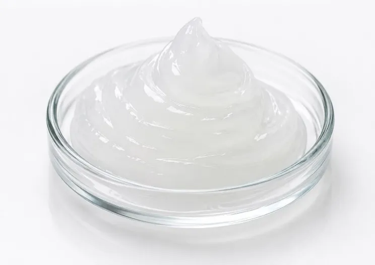
Pharmaceutical gels are semisolid dosage forms designed for application to the skin or accessible mucosal surfaces. They consist of two interpenetrating systems in which colloidal particles, commonly referred to as gelling agents or gelators, are uniformly dispersed within a solvent. These components interact to form a continuous three-dimensional network or matrix, resulting in the characteristic gel structure.
Figure 2: Pharmaceutical Gel
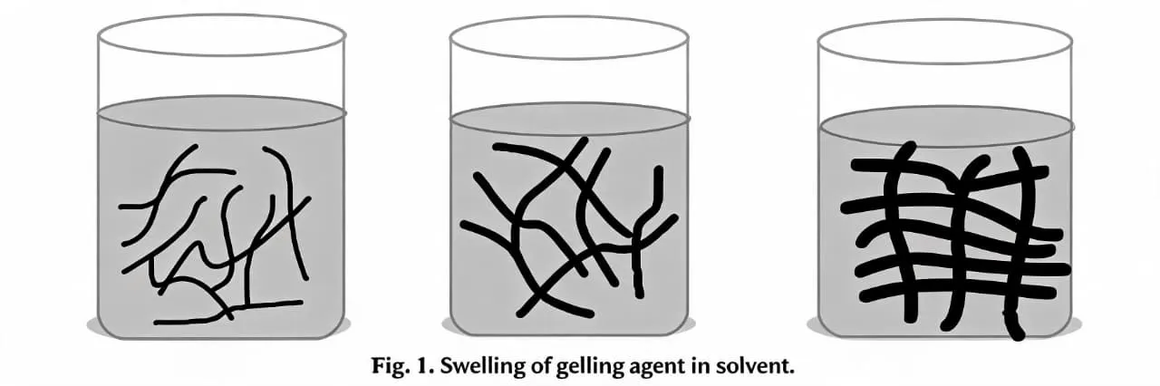
The gels are created by adding a gelling agent which could be natural, synthetic or semi-synthetic polymer or low molecular weight small molecules, into an organic, inorganic or aqueous solvent systems (Figure 3). The polymer in gels acts as the backbone of the gel matrix. The polymeric meshwork gives gel its structural strength, improved adhesion to the surface where applied and decreased absorption of the bigger molecules thereby making the retention possible.
During the gel formation, swelling happens as a result of solvent penetration causing the polymer network to stretch and keep its structure and entwine the drug particles in them. Viscosity plays an integral function in the creation of a gel. A gel in its solution form requires a specific concentration of polymer to increase the viscosity of the gel.
Figure 3: Swelling of gelling agent in solvent
Depending on the kind of bonding, gels can be either irreversible or reversible. Irreversible gels are often covalently bonded, while reversible gels are typically hydrogen bonded systems. A gel might appear as a two-phase system with discrete particle floccules or as a single system with no discernible boundaries.
Advantages of Gels
Compared to other semisolid dosage forms, gels are simpler to make.
A gel is a sophisticated, non-greasy composition.
Gels have good adhesion to the application site.
They are both biocompatible and biodegradable.
Compared to other topical dosage forms, gels have a longer retention period.
They are very tolerant of some stressful situations.
On the application location, they create a protective coating.
They are harmless and washable.
Due to solvent evaporation, they offer superior spreadibility and cooling effect.
They have somewhat less long-term stability problems.
Both polar and non-polar medications can be administered with them.
By repeatedly entwining the polymer, it can be utilized as a controlled release formulation.
Disadvantages of Gels
Gels generally exhibit a relatively slow onset of action.
Certain excipients and gelling agents incorporated into gel formulations may cause irritation at the site of application.
The high water content of gels can provide favorable conditions for microbial and fungal contamination, thereby affecting formulation stability.
Syneresis, characterized by the separation or expulsion of the liquid phase from the gel network, may occur during storage.
Loss of solvent through evaporation can lead to dehydration and drying of the gel formulation.
In certain gel systems, the presence of strong covalent interactions may make the gel network difficult to disrupt, potentially entrapping the drug within the matrix and limiting its release.
Flocculation may occur in some gel formulations, resulting in physical instability and deterioration of the gel structure.
The rheological characteristics of gels can be influenced by environmental conditions such as temperature and humidity, potentially altering their performance and stability.
Precipitation of gelling agents may occur under certain conditions, leading to salting-out phenomena and compromising formulation stability.
The incorporation of polymers may adversely affect the stability of certain active pharmaceutical ingredients, potentially resulting in drug degradation.
Mechanisms of Gel Formulation
Gels can be formed via three types of crosslinking which are described as follows:
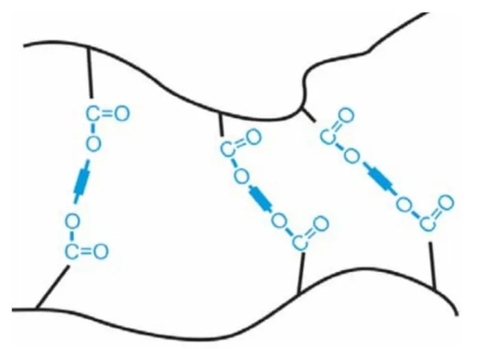
Chemical cross-linking - When a polymer contains dual or multifunctional monomers, an irreversible chemical cross-linking with a large molecular mass can emerge (Figure 4). These polymers are usually insoluble in the solvent but certain solvents, when incorporated, results in only swelling and hence forming a gel, for e.g. polyacrylamide gels. These gels are irreversible due to their covalent bonding. Polymers with unbonded groups in their structure can also undergo chemical cross-linking. When a cross-linking compound is added to these polymers, the free group and the new component undergo an irreversible chemical reaction. This irreversible process raises the viscosity, and once a particular concentration is reached, a gel is created. For example, glycols with hydroxyl groups and polyacrylic acid with multiple carboxylic acids make these kinds of chemical cross-linking gels.
Figure 4: Chemically cross-linked gel
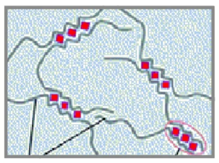
Physical cross-linking - The production of hydrogen bonds, the solubilization of crystalline components, concentration variation, temperature variation transition, or hydrophobic interactions can all cause a solution to gel transition. These gels include cellulose, dextran, and poly (N-isopropyl acrylamide) gels. Figure 5 illustrates physical cross-linking.
Figure 5: Physically cross-linked gel
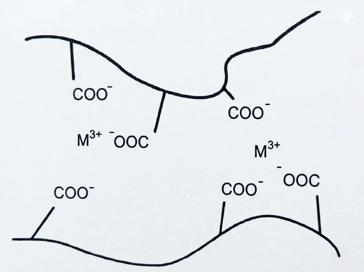
Ionic cross-linking - This type of cross-linking can also be accomplished by creating charges on a polymer or other molecules (solvent) that may draw one another to create a gel (Figure 6). The charges on these molecules lead to the formation of ionic bonds. For instance, when calcium ions are present, polysaccharide alginate forms a gel matrix that can encapsulate specific elements (enzymes, etc.). Another way to achieve ionic gelation is to change the medium's (solvent's) pH. Gelation occurs when the pH of such mixes is changed; for example, pectin gels when exposed to an acidic pH in an appropriate medium.
Figure 6: Ionic cross-linking in gel
Uses of Gels
The following is a list of some typical applications for gel formulations:
Gels are used to produce sustained release dosage form.
They serve as both transporters and lubricants for several medicinal drugs.
One gel can be used to manufacture both polar and non-polar medicines.
They can be applied topically, intraocularly, intranasally, vaginally, rectally, and sometimes parenterally and intramuscularly.
They are extensively utilized in the food and cosmetics industries. 8
Structure of gel
Gel is made of a natural or synthetic polymer that forms a three-dimensional matrix across a dispersion medium or hydrophilic liquid after application, the liquid evaporates, trapping the medication in a thin layer of the gel-forming matrix that physically covers the skin. The stiffness of a gel is caused by a network created by the gelling agent's particles interacting. The structure of the network and the properties of the gel are determined by the type of form that is responsible for the connection and the nature of the particles. Strong primary valencies, as in silic acid gels, as well as weaker hydrogen bonds and stray wall forces, are examples of the forces of attraction that link the particles of the gelling agent. The fact that a small temperature increase frequently results in gel liquefaction indicates the weaker character of these latter forces.
Properties of Gels:
The gel should be chemically inert, biocompatible, and compatible with other components of the formulation.
It should exhibit adequate stability under the specified storage conditions.
It should maintain all rheological properties
The gel should not adversely alter the biological activity, stability, or therapeutic properties of the incorporated drug.
It should exhibit adequate antimicrobial activity to prevent microbial contamination and ensure formulation stability.
Characteristics of Gels
Swelling - Swelling refers to the increase in volume of a gelling agent when it comes into contact with a suitable solvent that can solvate the material. During this process, the solvent penetrates into the polymeric or particulate matrix, resulting in the uptake of a considerable amount of liquid and consequent expansion of the gel. The extent of swelling is influenced by the number and strength of intermolecular linkages within the gelling agent.
Syneresis - Syneresis is the spontaneous contraction of a gel accompanied by the expulsion of part of the liquid phase upon standing. The extent of syneresis generally increases with a decrease in the concentration of the gelling agent. Its occurrence suggests that the initially formed gel is thermodynamically unstable. Gel contraction is attributed to the relaxation of elastic stresses generated during gel formation. As these stresses dissipate, the volume of the interstitial spaces within the network decreases, resulting in the expulsion of the entrapped liquid.
Ageing - Ageing is a gradual and spontaneous process of aggregation commonly observed in colloidal systems. In gels, ageing leads to the progressive development of a more compact and denser network of the gelling agent. According to Theimer, this phenomenon may be considered a continuation of the initial gelation process, during which further structural rearrangement and aggregation occur, accompanied by the gradual loss of the liquid medium from the gel network.
Structure - The mechanical rigidity of a gel is primarily attributed to the formation of a three-dimensional network through the interconnection of particles or molecules of the gelling agent. The characteristics of these constituent particles and the nature of the forces responsible for their interconnection determine the overall network structure and, consequently, the physical properties of the gel.
Rheology - The rheological properties of gels and gelling-agent dispersions are generally characterized by pseudoplastic or shear-thinning behaviour, which is a type of non-Newtonian flow. In such systems, the apparent viscosity decreases with increasing shear rate due to the disruption and rearrangement of the weak three-dimensional network formed by the dispersed particles. The extent of this structural breakdown influences the flow behaviour and consistency of the gel.
Classification of Gels
Gels can be classified based on colloidal phases, nature of solvent used, physical nature and rheological properties.
Based on Colloidal phase
Inorganic (Two phase system) - Inorganic gels are two-phase systems in which the dispersed particles are relatively large and form an interconnected three-dimensional network throughout the dispersion medium. The gel structure is primarily composed of aggregates or floccules of fine particles rather than large molecular chains. These systems may exhibit limited stability and commonly possess thixotropic behavior, in which they form a semisolid structure under quiescent conditions and become more fluid when subjected to agitation or shear.
Organic (single phase system) - Organic gels are single-phase systems composed of large molecules dispersed or dissolved within a continuous phase. These molecules, which may be derived from natural or synthetic polymers, are commonly known as gel-forming agents. They form a three-dimensional network through molecular entanglement and intermolecular interactions, including van der Waals forces, resulting in the development of the characteristic gel structure.
Based on nature of solvent
Hydro gels (water based) - Water is the continuous liquid phase of hydro gels, which are water-based. E.g. – Gelatin, cellulose derivatives.
Organic gels (non-aqueous solvent) - the continuous phase of these gels contains non-aqueous solvent. For instance, the dispersion of metallic stearate in oils and olag gel. E.g. - Olag gel and dispersion of metallic stearate in oils.
Xerogels - Solid gels with little solvent concentration are known as xerogels. E.g. - Tragacanth ribbons.
Based on rheological properties
Usually gels exhibit non- Newtonian properties
Plastic gels - E.g. - Bingham Bodies, flocculated suspensions of Aluminium hydroxide show a plastic flow; the rheogram plot indicates the yield value of the gels, at which the elastic gel deforms and starts to flow.
Pseudo plastic gels - These gels have no yield value and their viscosity falls as the shear rate increases. The disorganized molecules start to align their long axis in the direction of flow as the shearing force increases, releasing the solvent matrix. E.g. - Liquid dispersion of tragacanth, Na CMC.
Thixotropic gels - These gels have extremely weak particle connections that can be broken by shaking. Because of the particles' collision and subsequent re-linking, the resultant solution will return to gel (the reversible isothermal gel-sol-gel transformation). E.g. - Kaolin, bentonite, agar.
Based on physical nature
Elastic gels - In these gels, the fibrous molecules are being linked at the point of junction by comparatively weak bonds like dipole attraction and hydrogen bonds. A salt bridge of the type COO-X-COO forms an extra bond between two neighboring strand networks if the molecule has a free COOH group. E.g. - Alginate and Carbopol.
Rigid gels - These can be made from macromolecules with primary valence bonds connecting the framework. E.g. - Silica gel.
Gel forming agents
Polymers are used to give the structural network, which is essential for the preparation of gels. Gel forming polymers are classified as follows:
Formulation considerations for pharmaceutical gels
The choice of vehicle/ solvent - Purified water is typically utilized as a solvent. Co-solvent may be used to boost medication penetration through the skin or to increase the therapeutic agent's solubility in the dose form. E.g.- alcohol, glycerol, PG, PEG400
Inclusion of buffers - Buffers can be used in hydroalcholic and aqueous-based gels to regulate the formulation's pH. Buffer salts become less soluble in hydroalcholic-based vehicles. E.g.- Phosphate, citrate
Preservatives - By working with the hydrophilic polymers used to make gels, certain preservatives lower the amount of free (anti-microbial active) preservative present in the mixture. Thus, the initial concentration of these preservatives should be increased to make up for this. E.g. - Parabens, phenolic
Antioxidants - It may be involved in the formulation to improve the chemical stability of therapeutic agents that are susceptible to oxidative degradation. The type of vehicle utilized to prepare the gel determines its selection. Since most gels are aqueous-based, water-soluble antioxidants are typically utilized. E.g. - Sodium metabisulphite, sodium formaldehyde sulfoxylate
METHODS
Gels can be prepared by following methods:
Thermal changes - Solvated polymers, particularly lipophilic colloids, may undergo gelation in response to changes in temperature. Several polymers capable of hydrogen bonding exhibit greater solubility in hot water than in cold water. Upon cooling, the extent of polymer hydration decreases, promoting intermolecular interactions and subsequent gel formation. Thus, concentrated polymer solutions may form gels upon cooling. Examples include gelatin, agar, sodium oleate, guar gum, and cellulose derivatives. However, certain cellulose ethers exhibit temperature-dependent solubility due to hydrogen bonding with water. An increase in temperature disrupts these interactions, decreases polymer solubility, and consequently induces gelation. Therefore, temperature-induced gelation is not universally applicable to all polymeric systems.
Flocculation – Gelation can also be achieved through controlled flocculation by adding an appropriate quantity of a precipitating agent. The concentration of the precipitant should be sufficient to induce partial precipitation and formation of a gel-like structure, but insufficient to cause complete precipitation. Rapid and uniform mixing is essential to prevent localized areas of excessive precipitant concentration. For example, solutions of ethyl cellulose or polystyrene in benzene can undergo gelation upon rapid addition of an appropriate non solvent, such as petroleum ether. Although salts generally induce coagulation in hydrophobic colloidal systems, gel formation is less commonly observed. Gels produced by flocculation are typically characterized by thixotropic behavior, in which the gel undergoes reversible sol–gel transformation under shear.
Chemical reaction - In this method gel is formed by chemical interaction between the solute and solvent. E.g.- aluminium hydroxide gel can be prepared through the reaction of an aluminium salt with sodium carbonate in an aqueous medium. Increasing the concentration of the reactants promotes the formation of a more developed three-dimensional gel network. 9
Fusion technique - This method involves combining the drug, gelling agents, formulation components, and suitable vehicles under elevated temperature. The components are blended until a homogeneous formulation with the desired semi-solid or semi-firm consistency is obtained. This approach is particularly useful when the formulation components require heat to facilitate uniform mixing and incorporation.
Cold method - In this method, all of the ingredients, excluding the active pharmaceutical component, are heated and mixed simultaneously. The temperature of the mixture is then lowered, the drug is added, and the blending process is repeated until the gel is formed.
Dispersion approach - This procedure involves the gelling polymer is dispersed gradually in water and allowed to hydrate and swell. The drug, previously dissolved in an appropriate medium, is then incorporated into the hydrated polymer dispersion with continuous mixing to obtain a homogeneous gel. When required, a suitable buffer may be added to adjust the pH and achieve the desired physicochemical properties of the gel. 10
EVALUATION OF GELS
Homogeneity - Once the prepared gels are transferred to the container, they are visually inspected for homogeneity.
pH Measurement - A digital pH meter is used to measure the gel formulation's pH. After dissolving 1 gram of gel in 100 ml of distilled water, the mixture is kept for two hours. The pH of each formulation is measured three times, and the average values are computed.
Drug Content – 1 gram of gel is dissolved in 100 ml of an appropriate solvent. Using a UV visible spectrophotometer, filter the stock solution and measure absorbance at λ max nm after an appropriate dilution.
Viscosity Study - The prepared gel formulation's viscosity can be determined using a Brookfield digital viscometer. The rotational speeds of gels are 0.3, 0.6, and 1.5 revolutions per minute. The dial reading is recorded at each matching speed. The dial measurement is multiplied by a factor found in the viscometer catalogs to determine viscosity.
Spreadability - Spreadability indicates the degree to which gel readily spreads on application to the skin or affected area. Spreadability generally performs in a glass slide. Spreadability is defined as the amount of time, measured in seconds, that two slides take to separate from gel that is positioned between them under a specific stress. Lesser the time is taken for separation of two slides better the spreadability.
It can be expressed as- Spreadability (S) = M×L/T
Where, S = Spreadability
L= Length of glass slides
T = Time taken to separate the slides
Extrudability study - After the gel formulations are set in the container, they are put into collapsible tubes. The weight in grams needed to extrude a 0.5 cm gel ribbon in 10 seconds is used to calculate extrudability.
Grittiness - Every gel formulation is examined under a microscope to determine whether any particle debris is present.
In vitro drug diffusion study - It can be carried out in a Franz diffusion cell. 05 gram of gel is taken in a cellophane membrane. Carried out the dissolution studies with 250 ml of phosphate buffer (pH 7.4) as the dissolution medium at 37±1° C.
In vivo study - A mercury plethysmometer is used to examine the inhibition of carrageenan-induced rat paw edema in male Wistar albino rats. The experimental animals unilateral hind paw volume is assessed both before and after carrageenan is administered. Percentage inhibition is observed.
Skin Irritation test - Skin irritation studies were conducted using guinea pigs weighing approximately 400–500 gram of either sex, maintained under standard laboratory conditions. Prior to the study, the dorsal region of each animal was shaved, and two skin areas measuring approximately 4 cm were marked, with one designated as the control site and the other as the test site. The formulation was applied topically at a dose of 500 mg per guinea pig, twice daily for seven consecutive days. The treated sites were examined periodically for signs of irritation, sensitivity, or other visible skin reactions. The degree of erythema and edema was evaluated using a predefined scoring system: 0 indicated no visible reaction, 1 represented minor patchy erythema, 2 denoted minor confluent or moderate patchy erythema, and 3 corresponded to severe erythema, with or without edema. The observed scores were used to assess the skin-irritation potential of the tested formulation. 11
CONCLUSION
Pharmaceutical gels are versatile semisolid dosage forms that offer several advantages for topical and localized drug delivery. Their ease of application, good spreadability, patient acceptability, and ability to provide sustained drug release make them useful in the management of various skin conditions, including fungal infections. The choice of gelling agent, solvent, cross-linking method, and formulation components plays an important role in determining the physical stability, viscosity, drug release, and overall performance of the gel. Different types of gels, including chemically, physically, and ionically cross-linked systems, provide opportunities for designing formulations with desired characteristics. Proper evaluation of parameters such as homogeneity, pH, drug content, viscosity, spreadability, extrudability, grittiness, in vitro drug diffusion, in vivo study and skin irritation is essential to ensure safety and efficacy. Overall, pharmaceutical gels represent an effective and patient-friendly approach for topical drug delivery, with continued research offering opportunities for improved therapeutic performance and advanced controlled-release formulations.
REFERENCES
Bragazzi NL, Sellami M, Salem I, Conic R, Kimak M, Pigatto PD, Damiani G. Fasting and its impact on skin anatomy, physiology, and physiopathology: A comprehensive review of the literature. Nutrients. 2019 Jan 23;11(2):249.
McKnight G, Shah J, Hargest R. Physiology of the skin. Surgery (Oxford). 2022 Jan 1;40(1):8-12.
Ali SM, Yosipovitch G. Skin pH: from basic science to basic skin care. Acta dermato-venereologica. 2013 Jan 10;93(3):261-7.
Jamagondi LN, Katte AS, Rumane MB, Pathan M, Kale AR. Formulation and evaluation of topical antifungal gel containing itraconazole. American Journal of PharmTech Research. 2019;9(02):333-8.
Shiva K, Mandal S, Kumar S. Formulation and evaluation of topical antifungal gel of fluconazole using aloe vera gel. Int J Sci Res Develop. 2021;1: 187-93.
Dixon DM, Walsh TJ. Antifungal agents. In: Baron S, editor. Medical Microbiology. 4th ed. Galveston (TX): University of Texas Medical Branch at Galveston; 1996. Chapter 76. Available from: https://www.ncbi.nlm.nih.gov/books/NBK8263/
Singh M, Rathour A, Dey U, Kumar Gaur P, Kumar V, Modi K, Abdul A. Antifungal agents: a comprehensive review of mechanisms and applications. J Popul Ther Clin Pharmacol. 2022;29(04):1343-58.
Un Nabi SA, Sheraz MA, Ahmed S, Mustaan N, Ahmad I. Pharmaceutical gels: a review. RADS J. Pharm. Pharm. Sci. 2016;4: 40-8.
Bisht P, Badola A. Gel as a topical drug delivery system: a review. JETIR. 2019;6(4):321-30.
Sharma U, Arjariya S, Chouksey R, Sharma N. A review: formulation and evaluation of pharmaceutical gel. Journal of Pharmaceutical Negative Results. 2022 Oct 2;13(1):1344-62.
Bhuyan C, Saha D, Rabha B. A brief review on topical gels as drug delivery system. J. Pharm. Res. Int. 2021;33: 344-57.
Reference
Bragazzi NL, Sellami M, Salem I, Conic R, Kimak M, Pigatto PD, Damiani G. Fasting and its impact on skin anatomy, physiology, and physiopathology: A comprehensive review of the literature. Nutrients. 2019 Jan 23;11(2):249.
McKnight G, Shah J, Hargest R. Physiology of the skin. Surgery (Oxford). 2022 Jan 1;40(1):8-12.
Ali SM, Yosipovitch G. Skin pH: from basic science to basic skin care. Acta dermato-venereologica. 2013 Jan 10;93(3):261-7.
Jamagondi LN, Katte AS, Rumane MB, Pathan M, Kale AR. Formulation and evaluation of topical antifungal gel containing itraconazole. American Journal of PharmTech Research. 2019;9(02):333-8.
Shiva K, Mandal S, Kumar S. Formulation and evaluation of topical antifungal gel of fluconazole using aloe vera gel. Int J Sci Res Develop. 2021;1: 187-93.
Dixon DM, Walsh TJ. Antifungal agents. In: Baron S, editor. Medical Microbiology. 4th ed. Galveston (TX): University of Texas Medical Branch at Galveston; 1996. Chapter 76. Available from: https://www.ncbi.nlm.nih.gov/books/NBK8263/
Singh M, Rathour A, Dey U, Kumar Gaur P, Kumar V, Modi K, Abdul A. Antifungal agents: a comprehensive review of mechanisms and applications. J Popul Ther Clin Pharmacol. 2022;29(04):1343-58.
Un Nabi SA, Sheraz MA, Ahmed S, Mustaan N, Ahmad I. Pharmaceutical gels: a review. RADS J. Pharm. Pharm. Sci. 2016;4: 40-8.
Bisht P, Badola A. Gel as a topical drug delivery system: a review. JETIR. 2019;6(4):321-30.
Sharma U, Arjariya S, Chouksey R, Sharma N. A review: formulation and evaluation of pharmaceutical gel. Journal of Pharmaceutical Negative Results. 2022 Oct 2;13(1):1344-62.
Bhuyan C, Saha D, Rabha B. A brief review on topical gels as drug delivery system. J. Pharm. Res. Int. 2021;33: 344-57.
Bhanunandana GS
Corresponding author
Graduate student, Prasanna college of pharmacy, Laila, Belthangady (po), Dhakshina Kannada, Karnataka, India-574214
Shashank K
Co-author
Graduate student, Prasanna college of pharmacy, Laila, Belthangady (po), Dhakshina Kannada, Karnataka, India-574214
Salman Paris
Co-author
Graduate student, Prasanna college of pharmacy, Laila, Belthangady (po), Dhakshina Kannada, Karnataka, India-574214
Alan Joseph
Co-author
Graduate student, Prasanna college of pharmacy, Laila, Belthangady (po), Dhakshina Kannada, Karnataka, India-574214
Mahammad Shaz
Co-author
Graduate student, Prasanna college of pharmacy, Laila, Belthangady (po), Dhakshina Kannada, Karnataka, India-574214
Dr. M. Mallikarjuna Gouda
Co-author
Principal, Department of pharmaceutics, Prasanna college of pharmacy, Laila, Belthangady (po), Dhakshina Kannada, Karnataka, India-574214
Bhanunandana GS, Shashank K., Salman Paris., Alan Joseph., Mahammad Shaz., Dr. M. Mallikarjuna Gowda. Pharmaceutical Gels for the Topical Treatment of Cutaneous Fungal Infections: A Comprehensive Review, Int. J. of Pharm. Sci., 2026, Vol 4, Issue 9, 2245-2258, https://doi.org/10.5281/zenodo.22844676








 10.5281/zenodo.22844676