

We use cookies to ensure our website works properly and to personalise your experience. Cookies policy
1Department of Microbiology, Vels Institute of Science, Technology and Advanced Studies, Pallavaram, Chennai, Tamil Nadu, India
2Department of Bio-engineering, Vels Institute of Science, Technology and Advanced Studies (VISTAS), Pallavaram, Chennai, Tamil Nadu, India
Caesalpinia bonducella (L.) Roxb. (Fabaceae) is a widely distributed medicinal shrub used in traditional Siddha and Ayurvedic practice for its antioxidant, anti-inflammatory and antitumour properties. The present study evaluated the phytochemical composition, in vitro free-radical scavenging activity, and cytotoxic/apoptotic potential of aqueous and ethanolic extracts prepared from the leaves of C. bonducella. Powdered leaf material was successively extracted by the Soxhlet (ethanol) and hot-water-bath (aqueous) methods, and the extracts were screened qualitatively for ten major phytoconstituent classes. Antioxidant activity was assessed by the 1,1-diphenyl-2-picrylhydrazyl (DPPH) radical scavenging assay, and cytotoxicity was evaluated against a normal (Vero) and a human colorectal adenocarcinoma (HT-29) cell line by the MTT assay, followed by acridine orange/propidium iodide (AO/PI) dual staining to assess apoptosis. The ethanolic extract contained alkaloids, carbohydrates, glycosides, saponins, phenolic compounds, flavonoids and terpenoids, whereas the aqueous extract lacked carbohydrates, glycosides and saponins. The ethanolic extract exhibited markedly stronger DPPH scavenging (82.47% inhibition at 100 µL) than the aqueous extract (28.95% at the same volume). In the MTT assay, the ethanolic extract reduced HT-29 viability in a dose-dependent manner (half-maximal inhibitory concentration, IC?? ? 47.4 µg/mL) while sparing the normal Vero cell line (viability 76.2% at 100 µg/mL; IC?? not attained within the tested range), indicating an estimated selectivity index >2.1 in favour of the malignant line. AO/PI staining of treated HT-29 cells revealed condensed, fragmented and orange-red-stained nuclei consistent with early and late apoptosis. in contrast to the uniform green fluorescence of untreated controls. These findings suggest that the ethanolic leaf extract of C. bonducella possesses phytochemical-rich, antioxidant and selectively cytotoxic/pro-apoptotic properties against colorectal cancer cells, supporting further bioassay-guided fractionation and mechanistic study.
Medicinal plants remain a principal source of new therapeutic leads, particularly in the areas of antioxidant and anticancer drug discovery, owing to the structural diversity of their secondary metabolites and their generally favourable safety profile relative to synthetic agents. Caesalpinia bonducella (L.) Roxb. (family Fabaceae/Caesalpiniaceae), commonly known as fever nut or nicker nut, is a prickly, scandent shrub distributed throughout the coastal and semi-arid tracts of India, Sri Lanka and other tropical regions. Different parts of the plant – seeds, seed kernel, leaves, bark and root – are used extensively in Siddha, Ayurvedic and folk medicine systems for the management of fever, inflammation, diabetes, diarrhoea and parasitic infections, and the plant has been reported to possess antidiabetic, anti-inflammatory, antifilarial, anxiolytic, analgesic, antipyretic, immunomodulatory, antimicrobial, antifungal and anticonvulsant activities.
Phytochemical investigations of C. bonducella have consistently identified alkaloids, flavonoids, saponins, tannins, phytosterols and phenolic compounds as major constituent classes, and several of these metabolites have been correlated with the plant’s antioxidant and cytotoxic properties.¹⁻⁴ The seed kernel is the most extensively studied part of the plant, but the leaves – which are more sustainably harvestable than seeds – have received comparatively less systematic attention despite reports of comparable or superior antioxidant and flavonoid content in leaf tissue relative to other plant parts.⁴ Crude methanolic and ethanolic extracts of C. bonducella leaves have previously demonstrated antibacterial, antidiarrhoeal and preliminary cytotoxic activity in brine shrimp lethality assays,¹ lending further support to their pharmacological potential.
Reactive oxygen species (ROS)-mediated oxidative stress is implicated in the initiation and progression of chronic degenerative disease and carcinogenesis, and plant-derived antioxidants that additionally exhibit selective cytotoxicity toward malignant cells – while sparing normal tissue – are of particular pharmacological interest. Colorectal cancer remains one of the most frequently diagnosed malignancies worldwide, and screening of plant extracts against colorectal carcinoma cell lines such as HT-29 is a standard first step in the discovery of candidate chemopreventive or chemotherapeutic phytoconstituents. Against this background, the present study was undertaken with the following objectives: (i) to perform qualitative phytochemical screening of aqueous and ethanolic leaf extracts of C. bonducella; (ii) to evaluate the in vitro free-radical scavenging (antioxidant) activity of both extracts by the DPPH assay; and (iii) to assess the cytotoxic and apoptosis-inducing potential of the extract against the HT-29 human colorectal adenocarcinoma cell line relative to the non-cancerous Vero cell line, using the MTT viability assay and acridine orange/propidium iodide (AO/PI) dual fluorescence staining.
MATERIALS AND METHODS
Collection of plant material
Fresh leaves of Caesalpinia bonducella (L.) Roxb. were collected, cleaned to remove adhering debris, chopped into small pieces and shade-dried at room temperature. The dried material was powdered mechanically and stored in air-tight containers at room temperature until further use.
Preparation of extracts
The powdered leaf sample was extracted successively with ethanol using a Soxhlet apparatus, and separately with distilled water by the hot-water-bath method at 50–60°C. Each extract was concentrated to a semisolid mass by drying on a water bath at 40–50°C and stored in labelled, air-tight containers at 4°C. The aqueous and ethanolic extracts (Figure 1B, C) were subsequently used for qualitative phytochemical screening, DPPH radical scavenging assay, and cytotoxicity/apoptosis evaluation.
Qualitative phytochemical screening
Both extracts were subjected to standard qualitative chemical tests for the detection of alkaloids, carbohydrates, glycosides, saponins, proteins, amino acids, phenolic compounds, flavonoids, terpenoids and steroids, as detailed below.
Detection of alkaloids (Wagner’s test): One hundred millilitres of extract was stirred with 3 mL of dilute hydrochloric acid and filtered. Two millilitres of the filtrate was treated with Wagner’s reagent (1.27 g iodine and 2 g potassium iodide dissolved in 5 mL water and made up to 100 mL with distilled water), added along the sides of the test tube; a reddish-brown precipitate indicated a positive reaction.⁵
Detection of carbohydrates: The extract (100 mg) was dissolved in 5 mL water and filtered. (a) Fehling’s test – 1 mL filtrate was boiled with 1 mL each of Fehling’s solution I (copper sulphate, 34.66 g/500 mL) and II (potassium sodium tartrate 173 g and sodium hydroxide 50 g/500 mL); a red precipitate indicated the presence of reducing sugars. (b) Benedict’s test – 0.5 mL filtrate was heated with 0.5 mL Benedict’s reagent (sodium citrate 173 g and sodium carbonate 100 g in 800 mL water, with copper sulphate 17.3 g in 100 mL water added) on a boiling water bath for 2 min; a characteristic coloured precipitate indicated a positive reaction.
Detection of glycosides (Borntrager’s test): Fifty millilitres of extract was hydrolysed with concentrated HCl for 2 h on a water bath and filtered. To 2 mL of the hydrolysate, 3 mL chloroform was added and shaken; the chloroform layer was separated and treated with 10% ammonia solution. A pink colour indicated the presence of glycosides.⁵
Detection of saponins (foam test): One milligram of extract was dissolved in 2 mL distilled water and filtered through Whatman No.1 filter paper; persistent froth on vigorous shaking indicated the presence of saponins.
Detection of proteins and amino acids: (a) Biuret test – 2 mL filtrate was treated with 2% copper sulphate solution followed by 1 mL of 95% ethanol and excess potassium hydroxide pellets; a pink colour in the ethanolic layer indicated proteins. (b) Ninhydrin test – two drops of ninhydrin reagent (10 mg ninhydrin in 200 mL acetone) were added to 2 mL extract; a characteristic purple colour indicated amino acids.
Detection of phenolic compounds (ferric chloride test): The extract (50 mg) was dissolved in 5 mL distilled water and treated with a few drops of neutral 5% ferric chloride solution; a dark green colour indicated the presence of phenolic compounds.
Detection of terpenoids: To 5 mL methanolic extract, 2 mL chloroform was added and mixed, followed by careful addition of concentrated H₂SO₄; formation of a reddish-brown interfacial layer indicated the presence of terpenoids.
Detection of steroids: Two millilitres of chloroform was added to the extract along with a few drops of acetic anhydride, followed by concentrated H₂SO₄; development of a blue-green colour indicated the presence of steroids.
DPPH radical scavenging assay
The free-radical scavenging activity of both extracts was determined by the 1,1-diphenyl-2-picrylhydrazyl (DPPH) method. Briefly, a 0.4 mM solution of DPPH in methanol was prepared, and 2 mL of this solution was added to increasing volumes (20–100 µL) of each extract. The reaction mixtures were incubated in the dark at room temperature for 15 min, and the absorbance was recorded at 517 nm against a blank. A lower absorbance of the reaction mixture indicated a higher free-radical scavenging activity. Percentage inhibition (radical scavenging activity) was calculated as:
Inhibition (%) = [(Abs control − Abs sample) / Abs control] × 100
Cell culture maintenance
Vero (African green monkey kidney, normal epithelial) and HT-29 (human colorectal adenocarcinoma, epithelial) cell lines were procured from the National Centre for Cell Science (NCCS), Pune, India. Cells were maintained in the logarithmic growth phase in Dulbecco’s Modified Eagle Medium (DMEM) supplemented with 10% (v/v) heat-inactivated foetal bovine serum (FBS), 100 U/mL penicillin and 100 µg/mL streptomycin, and incubated at 37°C in a humidified atmosphere of 5% CO₂ in air.
Cytotoxicity assay (MTT)
Cytotoxicity of the ethanolic leaf extract against Vero and HT-29 cell lines was assessed by the MTT [3-(4,5-dimethylthiazol-2-yl)-2,5-diphenyltetrazolium bromide] assay.⁷ Cells were seeded in 96-well microplates (1 × 10⁶ cells/well) and incubated at 37°C for 48 h in 5% CO₂ until 70–80% confluence was attained. The medium was then replaced, and cells were treated with the extract at concentrations of 20, 40, 60, 80 and 100 µg/mL for 24 h; untreated cells served as controls. Morphological changes were recorded under a digital inverted microscope (20×magnification) after 24 h. Cells were then washed with phosphate-buffered saline (PBS, pH 7.4), and 20 µL of MTT solution (5 mg/mL in PBS) was added to each well. Plates were incubated at 37°C in the dark for 2 h, the resulting formazan crystals were dissolved in 100 µL DMSO, and absorbance was read at 570 nm. Percentage cell viability was calculated as:
Cell viability (%) = (Absorbance of sample / Absorbance of control) × 100
All absorbance readings were recorded in duplicate (I, II) and the mean value used to calculate percentage viability; the half-maximal inhibitory concentration (IC₅₀) was estimated by linear interpolation between the two concentrations bracketing 50% viability.
Apoptosis assay by dual AO/PI staining
HT-29 cells were seeded in 6-well plates (1 × 10⁴ cells/well) and treated with the ethanolic extract for 24 h. Treated cells were washed with PBS and stained with 5 µL acridine orange (AO, 100 µg/mL) and 5 µL propidium iodide (PI, 100 µg/mL). AO permeates both viable and non-viable cells and fluoresces green, whereas PI is excluded by intact plasma membranes and stains only non-viable/late-apoptotic and necrotic cells red; the dual-staining protocol therefore allows discrimination of viable, early apoptotic, late apoptotic and necrotic cell populations. Morphological hallmarks of apoptosis were visualised by fluorescence microscopy (FLoid Cell Imaging Station) and compared between untreated control and extract-treated cells.
Statistical treatment
All cytotoxicity readings represent the mean of duplicate absorbance measurements. Dose–response data were plotted as mean percentage viability/inhibition against concentration, and IC₅₀ values were derived by linear interpolation between the two flanking data points using GraphPad-style regression in Python (SciPy/Matplotlib, v3.x).
RESULTS AND DISCUSSION
Extraction yield and preliminary observations
Successive extraction of shade-dried, powdered C. bonducella leaves (Figure 1A) with ethanol (Soxhlet) and water (hot-water bath) afforded dark brown, semisolid extracts (Figure 1B, C). The ethanolic extract was visibly darker and more viscous than the aqueous extract, consistent with more efficient extraction of moderately non-polar and amphiphilic secondary metabolites (flavonoid aglycones, terpenoids) by ethanol relative to water.
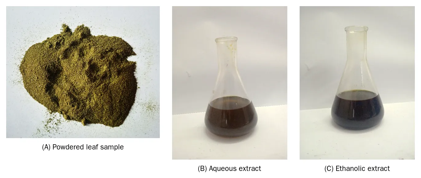
Figure 1. Plant material and extracts of Caesalpinia bonducella. (A) Shade-dried, powdered leaf sample. (B) Aqueous extract. (C) Ethanolic (Soxhlet) extract.
Qualitative phytochemical screening
Qualitative phytochemical screening (Table 1, Figure 2) revealed a clear solvent-dependent difference in the phytoconstituent profile of the two extracts. The ethanolic extract tested positive for seven of the ten classes screened: alkaloids, carbohydrates, glycosides, saponins, phenolic compounds, flavonoids and terpenoids, whereas the aqueous extract tested positive for only four classes (alkaloids, phenolic compounds, flavonoids and terpenoids) and was negative for carbohydrates, glycosides and saponins. Proteins, amino acids and steroids were not detected in either extract. This pattern is consistent with previous phytochemical surveys of Caesalpinia bonducella, in which alkaloids, flavonoids, saponins, phenolics and terpenoids have repeatedly been identified as the dominant constituent classes across plant parts and extraction solvents.¹⁻⁴,⁶ The comparatively richer profile of the ethanolic extract reflects the broader polarity range accessible to ethanol, which can co-extract both polar phenolics/glycosides and moderately lipophilic terpenoids, whereas water preferentially extracts highly polar constituents and excludes glycosides and saponins that may be less soluble or structurally bound in this particular tissue.
Table 1. Qualitative Phytochemical Screening Of Aqueous And Ethanolic Leaf Extracts Of C. Bonducella
|
S. No. |
Phytoconstituent |
Aqueous extract |
Ethanolic extract |
|
1 |
Alkaloids |
+ |
+ |
|
2 |
Carbohydrates |
− |
+ |
|
3 |
Glycosides |
− |
+ |
|
4 |
Saponins |
− |
+ |
|
5 |
Proteins |
− |
− |
|
6 |
Amino acids |
− |
− |
|
7 |
Phenolic compounds |
+ |
+ |
|
8 |
Flavonoids |
+ |
+ |
|
9 |
Terpenoids |
+ |
+ |
|
10 |
Steroid |
− |
− |
“+” = present; “−” = absent.
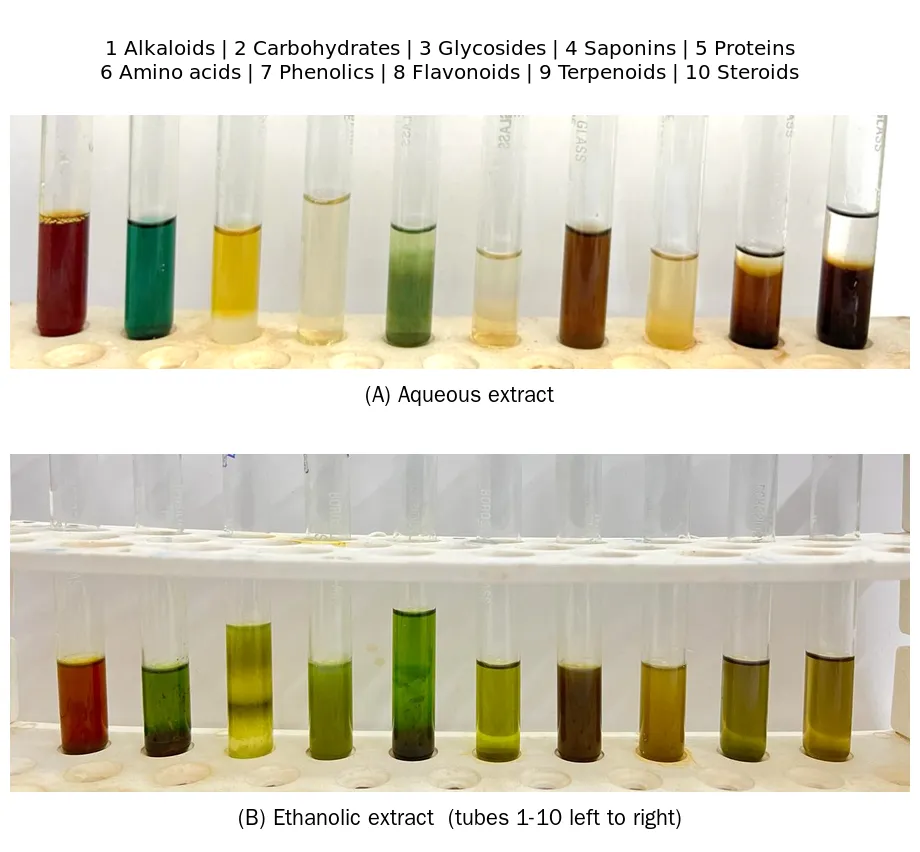
Figure 2. Colour reactions observed during qualitative phytochemical screening. (A) Aqueous extract (tubes 1–10 correspond to alkaloids, carbohydrates, glycosides, saponins, proteins, amino acids, phenolic compounds, flavonoids, terpenoids and steroids, respectively). (B) Ethanolic extract, tested in the same sequence.
DPPH radical scavenging activity
Both extracts scavenged the DPPH radical in a volume-dependent manner, evidenced by progressive bleaching of the characteristic purple DPPH chromogen to pale yellow (Figure 3A, B; Table 2). However, the two extracts differed markedly in potency. The ethanolic extract showed strong scavenging activity even at the lowest volume tested (73.18% inhibition at 20 µL), plateauing at 82.47% inhibition at 100 µL, whereas the aqueous extract showed weak, near-linear scavenging that reached only 28.95% inhibition at 100 µL (Table 2, Figure 3C). The markedly superior antioxidant activity of the ethanolic extract parallels its richer phenolic, flavonoid and terpenoid content on qualitative screening (Table 1), consistent with the well-established structure–activity relationship linking phenolic hydroxyl groups and conjugated flavonoid systems to hydrogen-atom-donating, DPPH-quenching capacity.³,⁴ This finding is in line with previous reports that ethanolic/methanolic extracts of Caesalpinia bonduc leaves and seeds consistently outperform aqueous extracts in DPPH and related free-radical scavenging assays.³,⁶
Table 2. Dpph Radical Scavenging Activity Of Aqueous And Ethanolic Leaf Extracts
|
Extract volume (µL) |
Aqueous – OD (517 nm) |
Aqueous – Inhibition (%) |
Ethanol – OD (517 nm) |
Ethanol – Inhibition (%) |
|
Control (0) |
0.936 |
– |
0.936 |
– |
|
20 |
0.920 |
1.70 |
0.251 |
73.18 |
|
40 |
0.901 |
3.73 |
0.236 |
74.78 |
|
60 |
0.843 |
9.93 |
0.202 |
78.41 |
|
80 |
0.796 |
14.95 |
0.185 |
80.23 |
|
100 |
0.665 |
28.95 |
0.164 |
82.47 |
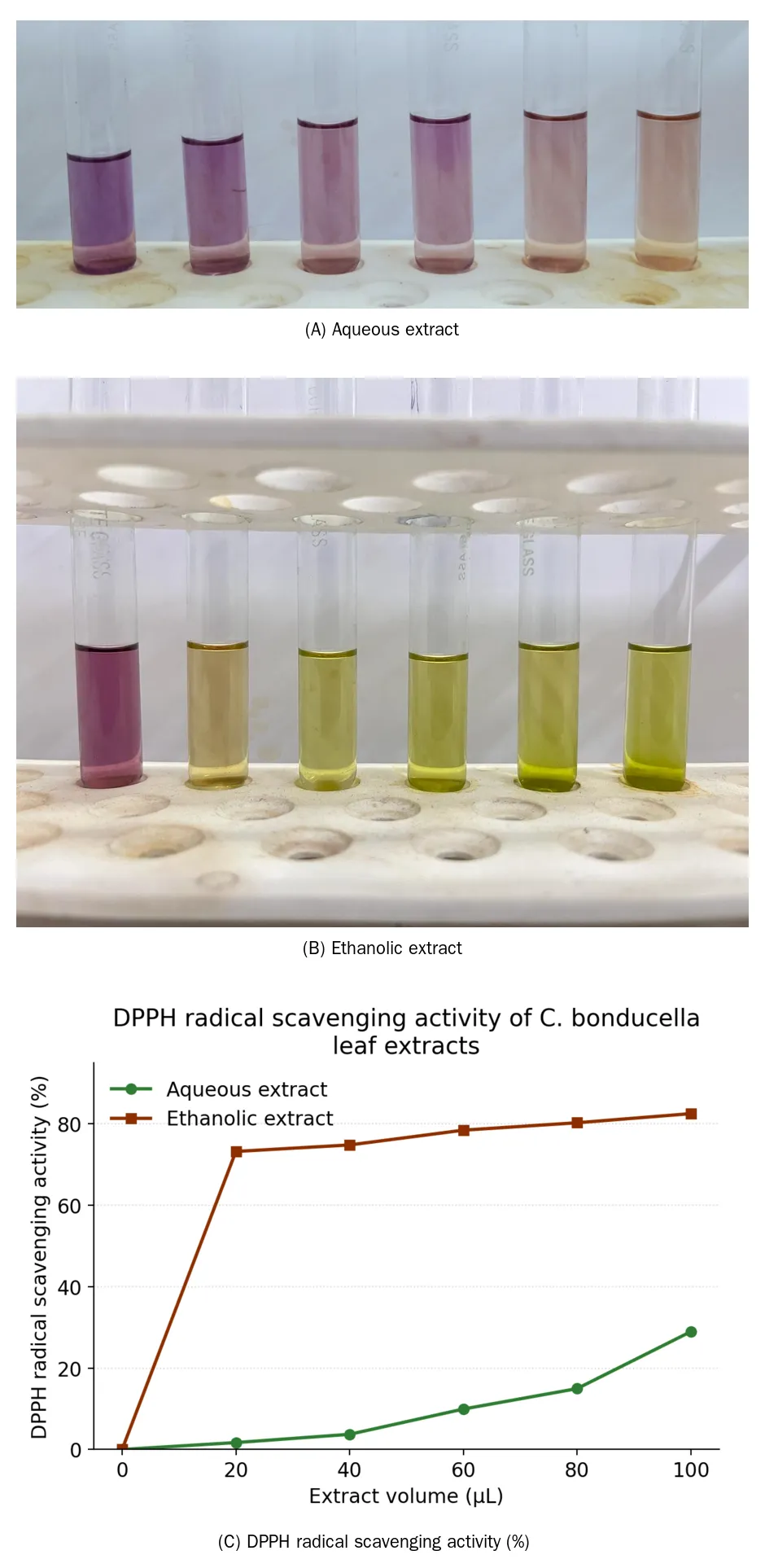
Figure 3. DPPH radical scavenging assay. (A) Colour change of the DPPH chromogen with increasing volume of aqueous extract (C = control; 20–100 µL). (B) Colour change with increasing volume of ethanolic extract. (C) Percentage DPPH radical scavenging activity of aqueous and ethanolic extracts as a function of extract volume.
Cytotoxic effect on Vero and HT-29 cell lines
The ethanolic extract was evaluated for cytotoxicity against the non-cancerous Vero cell line and the HT-29 human colorectal adenocarcinoma cell line by the MTT assay (Tables 3 and 4; Figure 4). Vero cell viability declined only modestly across the tested concentration range, from 100% (control) to 76.18% at 100 µg/mL, indicating good tolerability of the extract by normal epithelial cells; the IC₅₀ was not attained within the tested range (i.e., IC₅₀ > 100 µg/mL). In marked contrast, HT-29 viability fell steeply and dose-dependently, from 100% (control) to 12.72% at 100 µg/mL, with the IC₅₀ estimated by linear interpolation at approximately 47.4 µg/mL. The resulting estimated selectivity index (IC₅₀ Vero / IC₅₀ HT-29) exceeds 2.1, indicating preferential cytotoxicity toward the malignant cell line over the normal cell line at the concentrations tested.
Table 3. Mtt Cytotoxicity Of Ethanolic Extract On Vero (Normal) Cell Line
|
Concentration (µg/mL) |
Absorbance I |
Absorbance II |
Mean Abs. |
Cell viability (%) |
|
Control |
0.668 |
0.663 |
0.6655 |
100.00 |
|
20 |
0.651 |
0.652 |
0.6515 |
97.90 |
|
40 |
0.638 |
0.633 |
0.6355 |
95.49 |
|
60 |
0.597 |
0.601 |
0.5990 |
90.01 |
|
80 |
0.558 |
0.554 |
0.5560 |
83.55 |
|
100 |
0.511 |
0.503 |
0.5070 |
76.18 |
Table 4. Mtt Cytotoxicity Of Ethanolic Extract On Ht-29 (Colorectal Adenocarcinoma) Cell Line
|
Concentration (µg/mL) |
Absorbance I |
Absorbance II |
Mean Abs. |
Cell viability (%) |
|
Control |
0.743 |
0.751 |
0.7470 |
100.00 |
|
20 |
0.641 |
0.634 |
0.6375 |
85.34 |
|
40 |
0.454 |
0.443 |
0.4485 |
60.04 |
|
60 |
0.253 |
0.241 |
0.2470 |
33.07 |
|
80 |
0.126 |
0.123 |
0.1245 |
16.67 |
|
100 |
0.098 |
0.092 |
0.0950 |
12.72 |
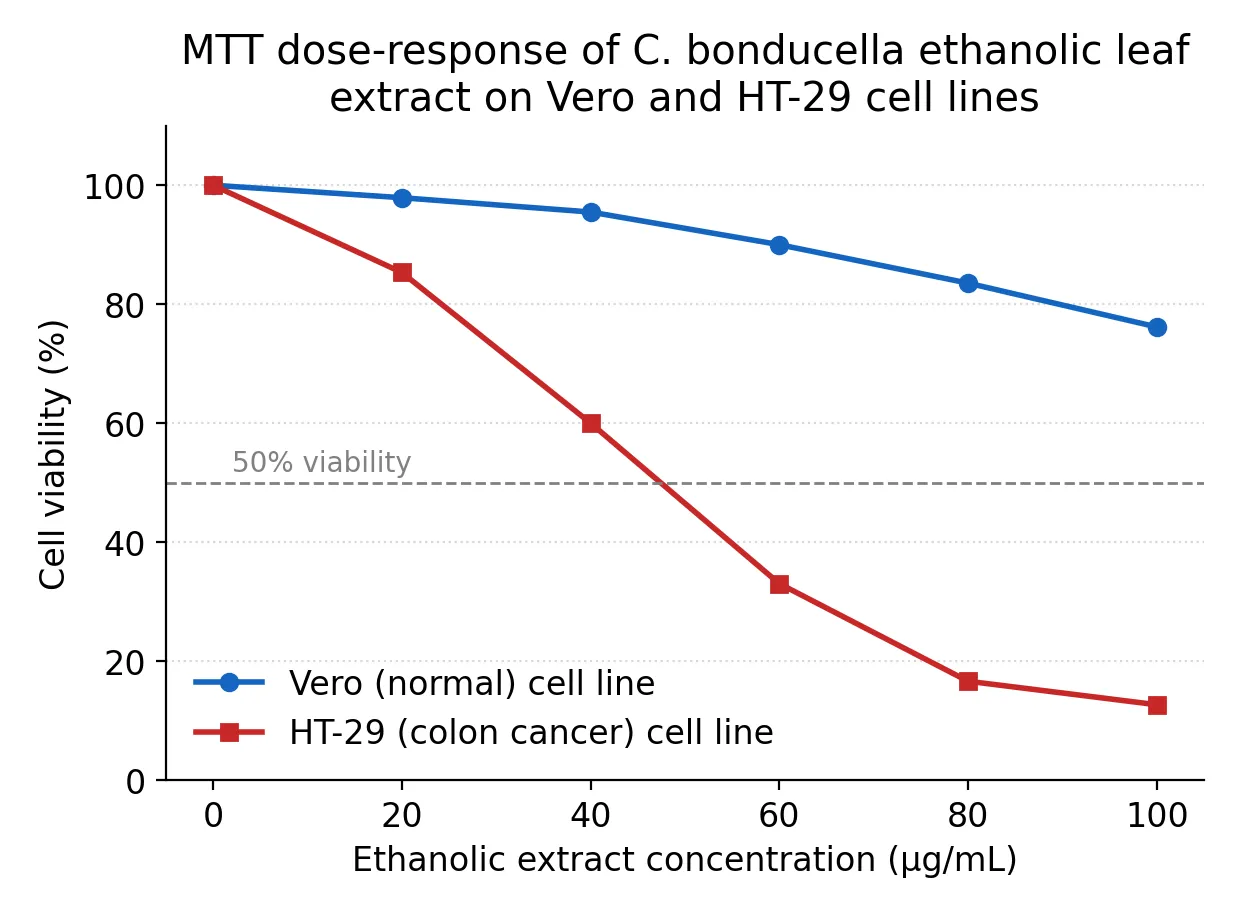
Figure 4. Dose-response curves showing percentage cell viability of Vero (normal) and HT-29 (colorectal adenocarcinoma) cell lines following 24 h treatment with the ethanolic leaf extract of C. bonducella (MTT assay). The dashed line indicates 50% viability
Photomicrographic examination corroborated the absorbance-based viability data. HT-29 cells showed progressive loss of confluent monolayer architecture, cell rounding, membrane blebbing and detachment with increasing extract concentration (Figure 5), changes that were most pronounced at 80-100 µg/mL. Vero cells, by contrast, retained their characteristic spindle-shaped, confluent monolayer morphology across the same concentration range, with only minor evidence of cell rounding at the highest concentration tested (Figure 6). These morphological observations are consistent with the selective, concentration-dependent cytotoxic action of the extract toward the malignant cell line, and align with previous reports describing selective in vitro cytotoxicity of Caesalpinia bonducella extracts and related plant extracts against colorectal and other cancer cell lines while sparing normal cell lines.¹,²
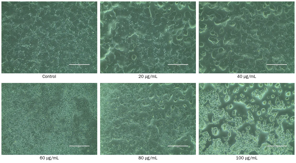
Figure 5. Phase-contrast photomicrographs (20×) of HT-29 cells following 24 h treatment with the ethanolic leaf extract of C. bonducella at 0 (control), 20, 40, 60, 80 and 100 µg/mL, showing progressive loss of monolayer confluence, cell rounding and membrane blebbing with increasing concentration. Scale bar, 200 µm.
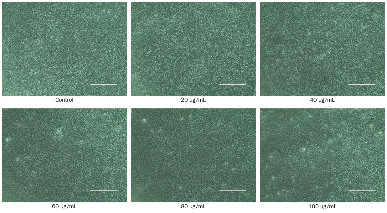
Figure 6. Phase-contrast photomicrographs (20×) of Vero cells following 24 h treatment with the ethanolic leaf extract of C. bonducella at 0 (control), 20, 40, 60, 80 and 100 µg/mL, showing largely preserved monolayer morphology across the tested concentration range. Scale bar, 200 µm.
Induction of apoptosis (AO&PI dual staining)
To determine whether the extract-induced loss of HT-29 viability proceeded through an apoptotic mechanism, treated and untreated cells were examined by AO/PI dual fluorescence staining (Figure 7). Untreated control cells displayed uniform, bright green nuclear fluorescence with AO and negligible PI uptake, consistent with an intact plasma membrane and viable cell population. Extract-treated HT-29 cells, in contrast, showed a marked reduction in green (AO-only) fluorescence and a corresponding increase in red/orange PI fluorescence; the merged AO/PI image revealed numerous nuclei with condensed, fragmented chromatin and yellow-to-orange staining, hallmarks of cells in early-to-late apoptosis. A subpopulation of cells showed uniform bright red/orange staining with pronounced nuclear condensation, indicative of late apoptotic or secondary necrotic cells. These morphological features are classical hallmarks of apoptotic cell death and corroborate the MTT-based cytotoxicity findings, together indicating that the ethanolic leaf extract of C. bonducella inhibits HT-29 cell proliferation, at least in part, through induction of apoptosis rather than purely necrotic mechanisms.
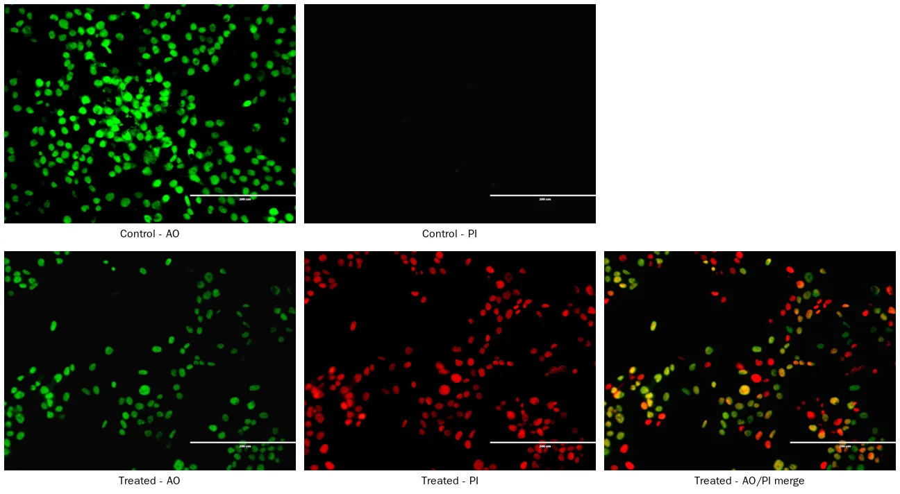
Figure 7. Acridine orange/propidium iodide (AO/PI) dual fluorescence staining of HT-29 cells. Top row: untreated control cells stained with AO (left) and PI (right), showing uniform green nuclear fluorescence and negligible PI uptake. Bottom row: extract-treated cells stained with AO (left), PI (centre) and the AO/PI merge (right), showing reduced green fluorescence, increased red/orange PI uptake, and condensed, fragmented nuclei consistent with early and late apoptosis. Scale bar, 200 µm.
Taken together, the phytochemical, antioxidant and cytotoxicity/apoptosis data indicate that the phenolic-, flavonoid- and terpenoid-rich ethanolic leaf extract of C. bonducella combines meaningful free-radical scavenging capacity with selective, apoptosis-associated cytotoxicity toward HT-29 colorectal cancer cells, while showing comparatively limited toxicity toward the non-cancerous Vero cell line at the same concentrations. Phenolic and flavonoid compounds are well recognised for their dual capacity to scavenge free radicals and to modulate apoptotic signalling pathways (e.g., via mitochondrial membrane depolarisation and caspase activation) in malignant cells, and it is plausible that the constituents responsible for the strong DPPH scavenging activity of the ethanolic extract also contribute to its pro-apoptotic action on HT-29 cells; however, bioassay-guided fractionation and mechanistic (caspase activity, mitochondrial membrane potential, gene expression) studies would be required to establish this link directly.
CONCLUSION
The present study demonstrates that the ethanolic leaf extract of Caesalpinia bonducella is richer in alkaloids, carbohydrates, glycosides, saponins, phenolic compounds, flavonoids and terpenoids than the corresponding aqueous extract, and correspondingly exhibits substantially stronger DPPH radical scavenging activity. In vitro cytotoxicity screening further showed that the ethanolic extract selectively reduced the viability of HT-29 colorectal adenocarcinoma cells (IC₅₀ ≈ 47.4 µg/mL) while largely sparing the non-cancerous Vero cell line, and AO/PI dual staining confirmed that this cytotoxicity was accompanied by classical morphological features of apoptosis. These findings support the traditional use of C. bonducella and warrant further bioassay-guided isolation of the active constituents, together with mechanistic studies (caspase activation, mitochondrial membrane potential, cell cycle analysis) and in vivo validation, to fully establish its potential as a source of antioxidant and colorectal-cancer-selective cytotoxic agents.
ACKNOWLEDGEMENT
The authors thank the Department of Microbiology and the Department of Bio-Engineering, Vels Institute of Science, Technology and Advanced Studies (VISTAS), Chennai, for laboratory facilities and support, and the National Centre for Cell Science (NCCS), Pune, for supply of the Vero and HT-29 cell lines.
ETHICAL STATEMENT
This study did not involve any human participants or live animal experimentation. The Vero and HT-29 cell lines used are established, commercially procured cell lines obtained from the National Centre for Cell Science (NCCS), Pune, India, and their use does not require institutional ethics committee approval.
CONFLICTS OF INTEREST
The authors declare that they have no conflicts of interest relevant to the content of this article.
REFERENCES


V. Renu, S. Eswari, Pravin kumar*, Phytochemical Screening, In Vitro Antioxidant And Apoptotic Potential Of Aqueous And Ethanolic Leaf Extracts Of Caesalpinia Bonducella (L.) Roxb, Int. J. of Pharm. Sci., 2026, Vol 4, Issue 8, 3867-3878. https://doi.org/ 10.5281/zenodo.22078493
 10.5281/zenodo.22078493