

We use cookies to ensure our website works properly and to personalise your experience. Cookies policy
Maharashtra Institute of Pharmacy, Betala, Bramhapuri, Chandrapur.
Diabetic wounds are one of the most challenging complications of diabetes mellitus because of impaired angiogenesis, prolonged inflammation, oxidative stress, microbial infection, and delayed tissue regeneration. Although conventional wound therapies are widely used, their effectiveness is often limited by poor drug penetration, frequent application, and inadequate healing. Consequently, there is growing interest in herbal medicines and nanotechnology-based drug delivery systems as alternative approaches for improving wound management.Moringa oleifera and Mimosa pudica are medicinal plants rich in flavonoids, phenolic compounds, alkaloids, and tannins that possess antioxidant, anti-inflammatory, antimicrobial, and wound healing properties. Incorporation of these phytoconstituents into nano-herbal formulations enhances their stability, skin penetration, bioavailability, and sustained drug release, thereby improving therapeutic efficacy. Nano-herbal gels have emerged as promising topical delivery systems because they provide prolonged drug retention at the wound site while promoting collagen synthesis, angiogenesis, fibroblast proliferation, and re-epithelialization.This review summarizes the pathophysiology of diabetic wound healing, the therapeutic potential of Moringa oleifera and Mimosa pudica, recent advances in nano-herbal drug delivery systems, and their applications in diabetic wound management. It also highlights current challenges and future perspectives for the clinical translation of nano-herbal formulations. Overall, nano-herbal gels represent a promising strategy for enhancing diabetic wound healing and may offer a safe, effective, and patient-friendly alternative to conventional wound therapies.
1.1 Diabetes Mellitus and Diabetic Wounds
Diabetes mellitus (DM) is a chronic metabolic disorder characterized by persistent hyperglycemia resulting from impaired insulin secretion, insulin action, or both.[1] It is one of the fastest-growing non-communicable diseases worldwide and is associated with several long-term microvascular and macrovascular complications. [2] Among these complications, diabetic wounds, particularly diabetic foot ulcers (DFUs), represent one of the most severe and difficult-to-treat conditions. [4] Persistent hyperglycemia impairs normal cellular metabolism, weakens the immune response, reduces blood circulation, and delays tissue regeneration, thereby significantly affecting the wound healing process. Diabetic wounds frequently become chronic because of prolonged inflammation, recurrent infections, impaired angiogenesis, and defective collagen synthesis. If left untreated, these wounds may progress to severe infection, tissue necrosis, gangrene, and ultimately lower-limb amputation. [4] Therefore, the development of safe, effective, and affordable therapeutic strategies for diabetic wound healing has become a major focus of current pharmaceutical and biomedical research. [5]
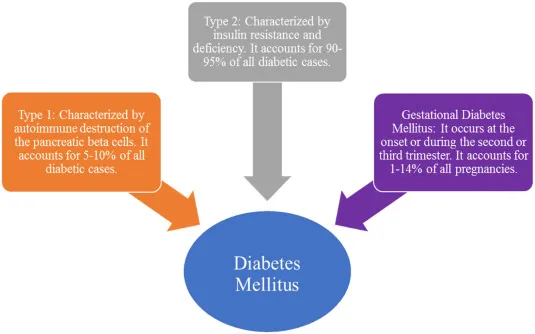
Figure 1: Diabetes mellitus
1.2 Burden of Chronic Wounds
Chronic wounds constitute a major healthcare challenge because of their prolonged healing period, high recurrence rate, and substantial economic burden. [6] The increasing global prevalence of diabetes has resulted in a corresponding rise in diabetic foot ulcers and other chronic wounds. [7] Patients suffering from chronic wounds often experience persistent pain, reduced mobility, frequent hospitalization, and diminished quality of life. [8] The financial burden associated with wound management is also considerable due to repeated clinical visits, prolonged medication use, surgical interventions, and long-term rehabilitation. [9] In addition to physical complications, chronic wounds frequently affect the psychological well-being of patients by causing anxiety, depression, and social isolation. Consequently, improving wound management remains an important healthcare priority worldwide. [10]
1.3 Limitations of Conventional Therapies
Current treatment strategies for diabetic wound management include wound debridement, antibiotics, antiseptics, growth factors, skin substitutes, negative pressure wound therapy, and advanced wound dressings. Although these approaches have improved clinical outcomes to some extent, several limitations remain. Conventional therapies are often associated with high treatment costs, limited availability, frequent dressing changes, poor patient compliance, and the emergence of antimicrobial resistance due to prolonged antibiotic use. [11] Moreover, many synthetic agents primarily target a single pathological pathway and fail to address the multifactorial nature of diabetic wounds, including oxidative stress, inflammation, impaired angiogenesis, and delayed collagen synthesis. These limitations have encouraged researchers to investigate alternative therapeutic approaches that are more effective, safer, and capable of promoting comprehensive tissue regeneration. [12]
Table 1: Limitations of Conventional Therapies
|
Conventional Therapy |
Limitation |
|
Antibiotics |
Antimicrobial resistance |
|
Conventional dressings |
Frequent replacement required |
|
Topical creams/ointments |
Poor skin penetration |
|
Growth factor therapy |
High cost and instability |
|
Skin grafting |
Limited donor tissue availability |
|
Debridement |
Painful and requires skilled professionals |
|
Systemic drugs |
Risk of systemic side effects |
|
Overall therapy |
Delayed healing in chronic diabetic wounds |
1.4 Importance of Herbal Medicines
Medicinal plants have been used for centuries in traditional healthcare systems for the treatment of wounds and skin disorders. Herbal medicines are rich sources of biologically active phytochemicals such as flavonoids, phenolic acids, tannins, alkaloids, terpenoids, and saponins, which possess antioxidant, anti-inflammatory, antimicrobial, and tissue regenerative properties. [13] These bioactive constituents act through multiple biological pathways to reduce oxidative stress, inhibit microbial growth, stimulate fibroblast proliferation, enhance collagen synthesis, promote angiogenesis, and accelerate re-epithelialization. Among various medicinal plants, Moringa oleifera and Mimosa pudica have received considerable scientific attention due to their well-documented pharmacological activities and excellent safety profiles. Their combined use may produce synergistic therapeutic effects, making them promising candidates for the development of novel phytopharmaceutical formulations for diabetic wound healing. [14]
1.5 Role of Nanotechnology
Nanotechnology has revolutionized modern drug delivery by providing innovative systems capable of improving the therapeutic performance of conventional and herbal medicines. Nano-sized carriers such as nanoemulsions, nanoparticles, liposomes, and nanogels enhance the solubility, stability, permeability, and controlled release of bioactive compounds. In topical drug delivery, nano-herbal gels facilitate deeper penetration of phytoconstituents into the skin while protecting them from premature degradation. The increased surface area of nanoparticles improves interaction with biological tissues and promotes sustained drug release at the wound site, thereby reducing dosing frequency and enhancing patient compliance. Consequently, nano-herbal formulations have emerged as promising therapeutic systems for chronic wound management and tissue regeneration. [15]
Table 2: Role of Nanotechnology in Topical Drug Delivery
|
Feature |
Benefit in Topical Drug Delivery |
|
Nano-sized Particles |
Enhance skin penetration and drug absorption. |
|
Increased Surface Area |
Improves drug solubility and bioavailability. |
|
Controlled Drug Release |
Provides prolonged therapeutic action. |
|
Targeted Drug Delivery |
Delivers drugs directly to the affected site. |
|
Enhanced Stability |
Protects active constituents from degradation. |
|
Improved Retention |
Increases residence time at the wound site. |
|
Reduced Dosing Frequency |
Improves patient compliance and convenience. |
1.6 Aim of the Review
The present review aims to comprehensively summarize the current understanding of diabetic wound healing, the major pathological factors responsible for delayed tissue repair, and recent advances in phytopharmaceutical and nanotechnology-based therapeutic approaches. Particular emphasis is given to the wound healing potential of Moringa oleifera and Mimosa pudica, the role of nano-herbal gel formulations in enhancing topical drug delivery, and future opportunities for clinical translation and commercialization of herbal nanomedicines for diabetic wound management.
2. Pathophysiology of Diabetic Wound Healing
2.1 Normal Wound Healing
Wound healing is a dynamic and highly regulated biological process that restores the structural and functional integrity of injured tissue. It involves coordinated interactions among inflammatory cells, fibroblasts, keratinocytes, endothelial cells, growth factors, cytokines, and extracellular matrix proteins. Under normal physiological conditions, wound healing progresses through four overlapping phases: hemostasis, inflammation, proliferation, and remodeling. Successful completion of each phase is essential for effective tissue repair and restoration of normal skin architecture. Any disturbance in these sequential events may result in delayed healing or chronic wound formation. [16]
2.2 Hemostasis
Hemostasis is the immediate response following tissue injury and serves to prevent excessive blood loss. During this phase, blood vessels constrict, and platelets rapidly aggregate at the injury site to form a stable fibrin clot. This clot acts as a temporary extracellular matrix that supports the migration of inflammatory cells and fibroblasts into the wound. Activated platelets release several growth factors, including platelet-derived growth factor (PDGF), transforming growth factor-beta (TGF-β), and vascular endothelial growth factor (VEGF), which initiate subsequent phases of tissue repair and promote cellular recruitment. [17]
2.3 Inflammation
The inflammatory phase begins shortly after hemostasis and plays a critical role in protecting the wound from microbial invasion and removing damaged tissue. Neutrophils are the first inflammatory cells to infiltrate the wound, followed by macrophages that phagocytose cellular debris and pathogens. Macrophages also secrete cytokines, chemokines, and growth factors that regulate fibroblast proliferation, angiogenesis, and extracellular matrix synthesis. Although inflammation is essential for wound healing, excessive or prolonged inflammation results in continuous tissue damage and significantly delays wound repair. [18]
2.4 Proliferation
The proliferative phase is characterized by active tissue regeneration and wound closure. Fibroblasts migrate into the wound bed and synthesize collagen, fibronectin, and other extracellular matrix proteins that form granulation tissue. Simultaneously, endothelial cells promote angiogenesis to establish a new vascular network capable of supplying oxygen and nutrients to regenerating tissue. Keratinocytes proliferate and migrate across the wound surface to restore the epidermal barrier through re-epithelialization. Coordinated cellular activity during this phase is essential for successful wound contraction and tissue regeneration. [19]
2.5 Remodeling
Remodeling represents the final stage of wound healing and involves gradual maturation of newly formed tissue. During this phase, type III collagen is progressively replaced by stronger type I collagen, resulting in increased tensile strength of the repaired tissue. Matrix metalloproteinases regulate extracellular matrix remodeling by balancing collagen synthesis and degradation. Scar tissue becomes more organized and functionally stable over time, although it rarely regains the full strength of uninjured skin. Remodeling may continue for several months following complete wound closure. [20]
2.6 Impaired Healing in Diabetes
Diabetes mellitus profoundly disrupts every stage of the wound healing process. Persistent hyperglycemia increases oxidative stress, impairs immune cell function, decreases growth factor production, inhibits fibroblast proliferation, and suppresses collagen synthesis. Endothelial dysfunction reduces angiogenesis and compromises blood supply to the wound, while prolonged inflammation causes excessive tissue destruction. Furthermore, diabetic neuropathy and vascular insufficiency increase susceptibility to repeated injury and infection. These pathological alterations create a chronic inflammatory environment that prevents timely progression through the normal stages of wound healing, ultimately leading to chronic non-healing ulcers. [21]
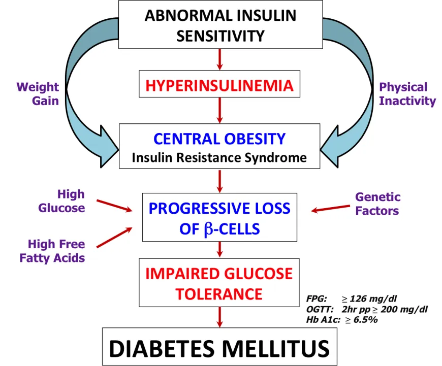
Figure 2: Diabetes mellitus pathophysiology
3. Factors Responsible for Delayed Wound Healing
3.1 Hyperglycemia
Persistent hyperglycemia is the principal pathological factor responsible for impaired diabetic wound healing. Elevated blood glucose promotes the formation of advanced glycation end products (AGEs), which alter cellular signaling pathways, impair fibroblast activity, inhibit collagen synthesis, and reduce the regenerative capacity of skin tissue. Hyperglycemia also compromises immune cell function, thereby increasing susceptibility to infection. [22]
3.2 Oxidative Stress
Oxidative stress results from excessive production of reactive oxygen species (ROS) that exceeds the body's antioxidant defense capacity. High ROS levels damage cellular proteins, membrane lipids, nucleic acids, and mitochondria, leading to apoptosis of fibroblasts and endothelial cells. This oxidative damage delays angiogenesis, collagen deposition, and epithelial regeneration, thereby prolonging wound healing. [23]
3.3 Chronic Inflammation
Diabetic wounds are characterized by persistent activation of inflammatory pathways and continuous release of pro-inflammatory cytokines such as TNF-α, IL-1β, and IL-6. Sustained inflammation delays the transition from the inflammatory phase to the proliferative phase, resulting in excessive tissue destruction and impaired wound closure.
3.4 Infection
Reduced immune competence and impaired leukocyte function increase the susceptibility of diabetic wounds to bacterial infection. Microbial colonization and biofilm formation prolong inflammation, damage healthy tissue, and reduce the effectiveness of antimicrobial therapy, thereby significantly delaying wound healing. [24]
3.5 Poor Angiogenesis
Angiogenesis is essential for supplying oxygen and nutrients to regenerating tissues. In diabetes, reduced expression of VEGF and endothelial dysfunction impair the formation of new blood vessels. Consequently, inadequate vascularization delays granulation tissue formation, collagen deposition, and epithelial regeneration.
3.6 Neuropathy
Diabetic peripheral neuropathy causes loss of protective sensation in the extremities, allowing repeated trauma and excessive pressure to occur without patient awareness. Continuous mechanical injury further damages tissue, increases the risk of ulcer formation, and prolongs wound healing.
3.7 Extracellular Matrix (ECM) Degradation
The extracellular matrix provides structural support for cell migration, collagen deposition, and tissue regeneration. In diabetic wounds, excessive activity of matrix metalloproteinases accelerates degradation of extracellular matrix proteins while reducing collagen accumulation. This imbalance weakens tissue integrity, delays wound contraction, and contributes to the persistence of chronic non-healing wounds. [25]
4. Herbal Medicines in Diabetic Wound Healing
4.1 Moringa oleifera
Moringa oleifera is one of the most extensively investigated medicinal plants for wound healing because of its rich phytochemical composition and broad pharmacological activities. [26] The leaves contain flavonoids, phenolic compounds, tannins, saponins, alkaloids, vitamins, and essential minerals that collectively promote tissue repair. [27] These bioactive constituents exhibit potent antioxidant properties that neutralize reactive oxygen species, thereby reducing oxidative stress associated with diabetic wounds. [28] In addition, Moringa oleifera possesses significant anti-inflammatory activity by suppressing pro-inflammatory cytokines, while its antimicrobial properties help prevent wound infection. Experimental studies have demonstrated that Moringa oleifera accelerates fibroblast proliferation, collagen deposition, angiogenesis, and re-epithelialization, resulting in faster wound contraction and improved tissue regeneration. [29] Owing to its excellent safety profile and multiple therapeutic mechanisms, Moringa oleifera is considered a promising phytopharmaceutical candidate for diabetic wound management. [30]
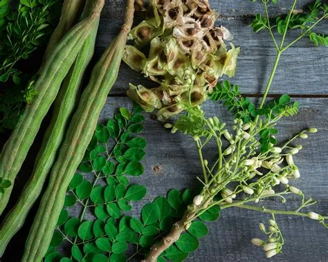
Figure 3: Moringa oleifera
Table 4: Moringa oleifera
|
Parameter |
Details |
|
Botanical Name |
Moringa oleifera Lam. |
|
Family |
Moringaceae |
|
Common Name |
Drumstick Tree |
|
Part Used |
Leaves |
|
Major Constituents |
Flavonoids, Phenolics, Vitamins |
|
Key Activities |
Antioxidant, Anti-inflammatory, Wound Healing |
4.2 Mimosa pudica
Mimosa pudica, commonly known as the sensitive plant, has long been used in traditional medicine for treating wounds, burns, ulcers, and skin infections. [31] The plant is rich in flavonoids, tannins, alkaloids, terpenoids, glycosides, and phenolic compounds that contribute to its wound healing activity. [32] These phytochemicals exhibit antioxidant and anti-inflammatory effects by reducing oxidative damage and controlling excessive inflammatory responses at the wound site. [33] The antimicrobial activity of Mimosa pudica also helps inhibit the growth of pathogenic microorganisms, thereby minimizing wound infection. [34] Several experimental studies have reported that Mimosa pudica enhances fibroblast proliferation, collagen synthesis, granulation tissue formation, and epithelial regeneration, leading to accelerated wound closure. These pharmacological properties make it a valuable herbal ingredient for developing advanced wound healing formulations. [35]
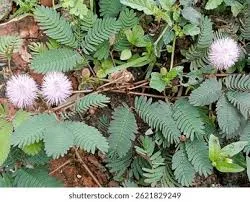
Figure 4: Mimosa pudica
Table 6: Mimosa pudica
|
Parameter |
Details |
|
Botanical Name |
Mimosa pudica L. |
|
Family |
Fabaceae (Mimosaceae) |
|
Common Name |
Touch-Me-Not Plant |
|
Part Used |
Whole Plant / Leaves |
|
Major Constituents |
Flavonoids, Tannins, Alkaloids, Glycosides |
|
Key Activities |
Antioxidant, Anti-inflammatory, Antimicrobial, Wound Healing |
4.3 Aloe vera
Aloe vera is widely recognized for its wound healing, moisturizing, and anti-inflammatory properties. The gel obtained from its leaves contains polysaccharides, glycoproteins, amino acids, vitamins, and enzymes that support tissue regeneration. Aloe vera maintains a moist wound environment, stimulates fibroblast activity, enhances collagen synthesis, and promotes epithelial cell migration. It also exhibits antimicrobial and antioxidant activities that reduce microbial contamination and oxidative stress. Owing to these therapeutic properties, Aloe vera has been extensively incorporated into topical formulations for chronic wound management. [36]
4.4 Curcumin
Curcumin, the principal bioactive constituent of Curcuma longa (turmeric), has gained significant attention for its potent antioxidant and anti-inflammatory activities. It suppresses inflammatory mediators such as TNF-α, IL-1β, and IL-6 while scavenging reactive oxygen species generated during diabetic wound healing. Curcumin also stimulates collagen deposition, fibroblast proliferation, angiogenesis, and extracellular matrix remodeling. However, its poor aqueous solubility and limited bioavailability have encouraged the development of nano-based delivery systems to enhance its therapeutic efficacy. [37]
4.5 Centella asiatica
Centella asiatica is an important medicinal herb traditionally used to accelerate wound healing. The plant contains triterpenoids such as asiaticoside and madecassoside, which stimulate fibroblast proliferation, collagen synthesis, angiogenesis, and extracellular matrix production. These bioactive compounds enhance granulation tissue formation, improve wound contraction, and increase the tensile strength of healed tissue. Its antioxidant and anti-inflammatory properties further contribute to rapid tissue regeneration and scar reduction. [38]
4.6 Neem (Azadirachta indica)
Azadirachta indica (Neem) possesses remarkable antimicrobial, antioxidant, anti-inflammatory, and immunomodulatory properties that make it beneficial for wound management. The leaves and bark contain bioactive compounds including nimbidin, azadirachtin, flavonoids, and tannins that inhibit microbial growth and reduce inflammation. Neem extracts have been reported to enhance collagen maturation, stimulate angiogenesis, and accelerate epithelialization, thereby promoting faster wound healing. Due to its broad-spectrum antimicrobial activity, neem is particularly useful in preventing wound infections in diabetic patients. [39]
4.7 Other Medicinal Plants
Several other medicinal plants have also demonstrated significant wound healing potential. Calendula officinalis promotes granulation tissue formation and collagen synthesis through its anti-inflammatory and antioxidant activities. Terminalia arjuna enhances tissue regeneration by improving collagen deposition and reducing oxidative stress. Ocimum sanctum (Tulsi) exhibits antimicrobial and immunomodulatory properties that support wound healing, while Hibiscus sabdariffa, Butea monosperma, and Catharanthus roseus have also shown promising wound healing effects in experimental studies. Collectively, these medicinal plants provide valuable sources of bioactive phytochemicals for the development of effective phytopharmaceutical formulations for diabetic wound management. [40]
5. Phytochemicals Responsible for Wound Healing
5.1 Flavonoids
Flavonoids are among the most important phytochemicals involved in wound healing due to their potent antioxidant, anti-inflammatory, and antimicrobial properties. They effectively scavenge reactive oxygen species, reduce lipid peroxidation, and protect cells from oxidative damage. Flavonoids also stimulate fibroblast proliferation, collagen synthesis, angiogenesis, and re-epithelialization, thereby accelerating tissue regeneration and wound closure. [41]
5.2 Phenolic Compounds
Phenolic compounds possess strong antioxidant activity that protects damaged tissues from oxidative stress during wound healing. They reduce inflammatory responses, inhibit microbial growth, and promote collagen maturation. Their ability to regulate cellular signaling pathways contributes significantly to tissue repair and remodeling in diabetic wounds. [42]
5.3 Tannins
Tannins exhibit astringent, antimicrobial, antioxidant, and anti-inflammatory properties that support wound healing. They promote wound contraction by forming a protective layer over damaged tissue, reduce exudate formation, and enhance collagen deposition. Tannins also inhibit bacterial growth, thereby preventing wound infection and accelerating tissue repair. [43]
5.4 Alkaloids
Alkaloids contribute to wound healing through their antimicrobial, anti-inflammatory, and analgesic activities. These compounds help reduce microbial contamination, control inflammation, and promote fibroblast proliferation. Certain alkaloids have also been reported to stimulate angiogenesis and collagen synthesis, facilitating faster tissue regeneration. [44]
5.5 Saponins
Saponins possess immunomodulatory and antioxidant properties that enhance the wound healing process. They stimulate fibroblast migration, collagen synthesis, angiogenesis, and granulation tissue formation. In addition, saponins improve epithelial regeneration and contribute to faster wound contraction. [45]
5.6 Terpenoids
Terpenoids are well known for their anti-inflammatory, antimicrobial, and antioxidant activities. They reduce inflammatory cytokine production, enhance fibroblast proliferation, stimulate collagen deposition, and improve angiogenesis. These pharmacological effects promote rapid tissue regeneration and reduce healing time in chronic wounds. [46]
5.7 Glycosides
Glycosides have been reported to facilitate wound healing by enhancing cellular proliferation, collagen formation, and extracellular matrix synthesis. They also possess antioxidant properties that minimize oxidative damage and support tissue remodeling. Their combined biological activities contribute to improved wound contraction and restoration of skin integrity. [47]
5.8 Synergistic Role of Phytochemicals
The wound healing activity of medicinal plants is generally attributed to the synergistic action of multiple phytochemicals rather than a single constituent. Flavonoids, phenolics, tannins, alkaloids, saponins, terpenoids, and glycosides act through complementary mechanisms to reduce oxidative stress, suppress inflammation, inhibit microbial growth, stimulate angiogenesis, enhance collagen synthesis, and promote epithelialization. This multi-targeted pharmacological approach makes phytopharmaceuticals highly effective candidates for the treatment of diabetic wounds and supports their incorporation into advanced nano-herbal drug delivery systems. [48]
6. Nanotechnology-Based Drug Delivery Systems
6.1 Nanoemulsions
Nanoemulsions are thermodynamically or kinetically stable colloidal systems composed of oil, water, surfactants, and co-surfactants, with droplet sizes typically ranging from 20 to 200 nm. Their small droplet size provides a large surface area, which enhances the solubility, stability, and skin permeation of poorly water-soluble phytoconstituents. In diabetic wound healing, nanoemulsions improve drug penetration into the deeper layers of the skin, protect bioactive compounds from degradation, and provide sustained drug release. These characteristics contribute to enhanced antioxidant, anti-inflammatory, and antimicrobial activities, thereby accelerating tissue regeneration and wound closure. [49]
6.2 Nanogels
Nanogels are three-dimensional cross-linked polymeric networks capable of encapsulating therapeutic agents within nano-sized structures. They possess high water content, excellent biocompatibility, and superior swelling capacity, making them suitable for topical wound applications. Nanogels maintain a moist wound environment, improve drug retention at the wound site, and provide controlled release of encapsulated phytochemicals. Their ability to enhance skin penetration and prolong therapeutic action makes them promising carriers for diabetic wound management. [50]
6.3 Liposomes
Liposomes are spherical vesicular systems composed of one or more phospholipid bilayers enclosing an aqueous core. They can encapsulate both hydrophilic and lipophilic bioactive compounds, thereby improving drug stability and bioavailability. Liposomal drug delivery enhances penetration of herbal constituents into damaged skin while minimizing systemic exposure. In diabetic wound healing, liposomes facilitate controlled drug release, reduce inflammation, promote collagen synthesis, and accelerate tissue repair, making them effective carriers for phytopharmaceutical formulations. [51]
6.4 Polymeric Nanoparticles
Polymeric nanoparticles are biodegradable colloidal carriers prepared using natural or synthetic polymers. These nanoparticles protect encapsulated herbal compounds from degradation and provide sustained and targeted drug release. Their nano-sized dimensions facilitate efficient penetration into damaged tissues and improve cellular uptake. Polymeric nanoparticles have demonstrated considerable potential in enhancing antioxidant activity, reducing inflammation, promoting angiogenesis, and improving collagen deposition during diabetic wound healing. [52]
6.5 Solid Lipid Nanoparticles
Solid lipid nanoparticles (SLNs) are submicron-sized lipid carriers composed of physiologically compatible solid lipids stabilized by surfactants. They offer high drug loading capacity, excellent physical stability, controlled drug release, and protection of encapsulated phytochemicals from chemical degradation. SLNs also improve skin hydration by forming an occlusive film on the wound surface, thereby enhancing drug penetration and accelerating tissue regeneration. These properties make them attractive carriers for topical herbal drug delivery. [53]
6.6 Nanofibers
Nanofibers are ultrafine fibrous structures generally produced by electrospinning techniques. They possess a high surface area-to-volume ratio and structural similarity to the extracellular matrix, providing an ideal environment for cell attachment and proliferation. Nanofiber-based wound dressings promote oxygen exchange, absorb wound exudates, maintain moisture, and support sustained release of incorporated herbal bioactive compounds. Their excellent mechanical strength and biocompatibility contribute to improved diabetic wound healing outcomes. [54]
6.7 Hydrogels
Hydrogels are three-dimensional hydrophilic polymeric networks capable of absorbing large amounts of water while maintaining structural integrity. They provide a moist wound environment that facilitates cell migration, granulation tissue formation, and epithelialization. Hydrogels can also serve as effective carriers for herbal extracts and nanoparticles, enabling sustained drug release directly at the wound site. Their excellent biocompatibility, cooling effect, and ability to reduce wound pain make hydrogels one of the most widely used topical delivery systems for chronic wound management. [55]
Table 6: Nanotechnology-Based Drug Delivery Systems
|
Drug Delivery System |
Key Advantage |
|
Nanoparticles |
Improved drug stability |
|
Nanoemulsion |
Enhanced skin penetration |
|
Nanogel |
Sustained drug release |
|
Liposomes |
Targeted drug delivery |
|
Solid Lipid Nanoparticles (SLNs) |
Controlled drug release |
|
Nanostructured Lipid Carriers (NLCs) |
High drug loading |
|
Polymeric Nanoparticles |
Enhanced bioavailability |
|
Nano-Herbal Gel |
Improved wound healing efficacy |
7. Nano Herbal Gel
7.1 Definition
Nano-herbal gel is an advanced topical drug delivery system in which herbal bioactive compounds are incorporated into nano-sized carriers, such as nanoemulsions or nanoparticles, and subsequently dispersed within a gel matrix. This formulation combines the pharmacological benefits of medicinal plants with the advantages of nanotechnology to improve topical drug delivery. The nano-sized particles enhance skin penetration, increase bioavailability, and provide sustained release of herbal constituents at the wound site, thereby improving therapeutic efficacy in diabetic wound healing. [56]
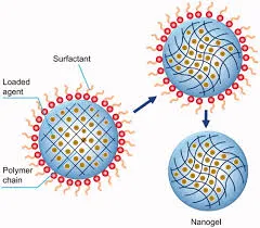
Figure 7: Nanogel structure
7.2 Composition
A nano-herbal gel generally consists of herbal extracts, oil phase, surfactants, co-surfactants, gelling agents, preservatives, pH-adjusting agents, and purified water. In the present context, Moringa oleifera and Mimosa pudica extracts serve as the active herbal ingredients, while Carbopol 940 acts as the gelling agent. Tween 80 functions as the surfactant, propylene glycol serves as a co-surfactant and penetration enhancer, triethanolamine adjusts the pH, and methyl paraben is incorporated as a preservative. The combination of these ingredients ensures formulation stability, uniformity, and effective topical delivery. [57]
7.3 Preparation Methods
Nano-herbal gels are generally prepared by first developing a stable nanoemulsion containing the herbal extracts through methods such as high-speed homogenization, ultrasonication, spontaneous emulsification, or high-pressure homogenization. The optimized nanoemulsion is then gradually incorporated into a hydrated gel base under continuous stirring to obtain a homogeneous nano-herbal gel. The pH of the formulation is adjusted to match the physiological skin pH, ensuring patient comfort and minimizing irritation. [58]
7.4 Mechanism of Action
Nano-herbal gels promote wound healing through multiple complementary mechanisms. The nano-sized droplets enhance penetration of phytoconstituents into deeper skin layers and provide sustained drug release at the wound site. Herbal bioactive compounds reduce oxidative stress, suppress inflammatory cytokines, inhibit microbial growth, stimulate fibroblast proliferation, promote collagen synthesis, enhance angiogenesis, and accelerate epithelialization. Together, these actions improve tissue regeneration and facilitate rapid wound closure in diabetic patients. [59]
7.5 Advantages
Nano-herbal gels offer several advantages over conventional topical formulations. They improve drug solubility, enhance skin penetration, provide sustained drug release, increase bioavailability, and reduce dosing frequency. Their non-invasive nature improves patient compliance, while the incorporation of herbal extracts minimizes adverse effects associated with synthetic drugs. Furthermore, nano-herbal gels provide better formulation stability and greater therapeutic efficacy in the treatment of chronic diabetic wounds. [60]
7.6 Limitations
Despite their numerous advantages, nano-herbal gels also have certain limitations. Formulation development requires specialized equipment and technical expertise, increasing production costs. The long-term physical stability of nanoformulations may be affected by aggregation or phase separation if not properly optimized. Standardization of herbal extracts and regulatory approval remain significant challenges due to variability in phytochemical composition. Additionally, large-scale manufacturing and clinical validation are necessary before widespread commercialization of nano-herbal wound healing formulations. [61]
Table 8: Nano Herbal Gel
|
Parameter |
Description |
|
Dosage Form |
Topical nano-sized gel |
|
Particle Size |
20–200 nm |
|
Drug Carrier |
Nanoemulsion/Nanoparticles |
|
Drug Release |
Sustained and controlled |
|
Skin Penetration |
Enhanced |
|
Bioavailability |
Improved |
|
Therapeutic Effect |
Accelerated wound healing |
|
Advantages |
Stable, biocompatible, and patient-friendly |
8. Evaluation of Nano-Herbal Gel
8.1 Particle Size
Particle size is one of the most important parameters influencing the therapeutic performance of nano-herbal formulations. It affects skin penetration, drug diffusion, formulation stability, and bioavailability of herbal phytoconstituents. Nano-sized droplets provide a larger surface area, facilitating enhanced permeation through the skin and improved drug retention at the wound site. Particle size is commonly determined using Dynamic Light Scattering (DLS), and formulations with particle sizes below 200 nm generally exhibit superior topical performance. Smaller particle sizes also reduce sedimentation and aggregation, thereby improving the physical stability of the formulation. [62]
8.2 Polydispersity Index (PDI)
The Polydispersity Index (PDI) is used to evaluate the uniformity of particle size distribution within the nano-herbal gel. A lower PDI value indicates a narrow and homogeneous particle size distribution, reflecting better formulation consistency and stability. Generally, PDI values below 0.30 indicate monodispersed systems with excellent physical stability. Uniform particle distribution minimizes particle aggregation and contributes to reproducible drug release and enhanced therapeutic efficacy. [63]
8.3 Zeta Potential
Zeta potential measures the electrical surface charge of nano-sized particles and serves as an important indicator of colloidal stability. Higher positive or negative zeta potential values generate stronger electrostatic repulsive forces between particles, preventing aggregation during storage. Nano-herbal formulations with zeta potential values greater than ±30 mV are generally considered physically stable. Adequate zeta potential enhances formulation stability, prolongs shelf life, and maintains the uniform dispersion of herbal bioactive constituents. [65]
8.4 Entrapment Efficiency
Entrapment efficiency represents the percentage of herbal bioactive compounds successfully encapsulated within the nano-carriers. High entrapment efficiency ensures maximum drug loading, minimizes drug loss during formulation, and provides sustained release of phytoconstituents at the wound site. Efficient encapsulation also protects sensitive herbal compounds from degradation and enhances their stability, resulting in improved therapeutic effectiveness during diabetic wound healing. [66]
8.5 Drug Content
Drug content analysis is performed to determine the uniform distribution of herbal extracts throughout the nano-herbal gel formulation. Uniform drug content ensures accurate dosing, batch-to-batch consistency, and reproducible therapeutic performance. The drug content is generally estimated using UV–Visible spectrophotometry based on a previously established calibration curve. Drug content values within the acceptable pharmacopeial limits indicate proper formulation homogeneity and quality. [68]
8.6 pH
The pH of the nano-herbal gel is measured to ensure compatibility with the physiological pH of human skin. A formulation with an appropriate pH minimizes skin irritation while maintaining the stability of herbal bioactive constituents. Generally, topical gels with pH values between 5.5 and 7.0 are considered suitable for dermal application and provide better patient comfort. [69]
8.7 Viscosity
Viscosity determines the flow behavior and consistency of the nano-herbal gel. Appropriate viscosity ensures easy application, prolonged retention at the wound site, and controlled drug release. Highly viscous formulations may be difficult to spread, whereas very low viscosity may reduce residence time on the skin. Therefore, optimum viscosity is essential for effective topical drug delivery. [70]
8.8 Spreadability
Spreadability indicates the ease with which the nano-herbal gel can be uniformly applied over the wound surface. Good spreadability improves patient convenience, facilitates uniform drug distribution, and enhances contact between the formulation and damaged tissue. It also contributes to better therapeutic efficacy by ensuring complete coverage of the wound area. [71]
8.9 Extrudability
Extrudability is evaluated to determine the ease with which the nano-herbal gel can be removed from collapsible tubes or containers. Good extrudability ensures convenient dispensing without excessive force and allows accurate application of the required quantity of gel. This parameter directly influences patient acceptability and formulation usability. [72]
8.10 In Vitro Drug Release
In vitro drug release studies are conducted to evaluate the release profile of herbal bioactive constituents from the nano-herbal gel. The study is generally performed using a Franz diffusion cell with phosphate buffer as the dissolution medium. Controlled and sustained drug release improves drug availability at the wound site, enhances therapeutic efficacy, and reduces the frequency of application. [73]
8.11 Stability Studies
Stability studies are performed to evaluate the physical, chemical, and microbiological stability of the nano-herbal gel during storage. Parameters such as appearance, pH, viscosity, drug content, particle size, and entrapment efficiency are periodically monitored under specified storage conditions according to ICH guidelines. Stable formulations should show no significant changes in their physicochemical characteristics throughout the study period. [74]
Table 9: Evaluation of Nano-Herbal Gel
|
Evaluation Parameter |
Purpose |
|
Physical Appearance |
Visual assessment |
|
pH |
Skin compatibility |
|
Viscosity |
Flow behavior |
|
Spreadability |
Ease of application |
|
Extrudability |
Ease of gel extrusion |
|
Drug Content |
Uniformity of drug distribution |
|
In-vitro Drug Release |
Drug release profile |
|
Stability Study |
Physical and chemical stability |
9. Mechanism of Wound Healing by Herbal Nanoformulations
9.1 Antioxidant Mechanism
Herbal nanoformulations contain phytochemicals such as flavonoids and phenolic compounds that effectively scavenge reactive oxygen species generated in diabetic wounds. Reduction of oxidative stress protects cells from damage, promotes fibroblast survival, and accelerates tissue regeneration. The nano-sized delivery system further enhances the availability of these antioxidants at the wound site.
9.2 Anti-inflammatory Mechanism
Nano-herbal formulations suppress excessive inflammatory responses by reducing the production of pro-inflammatory cytokines, including TNF-α, IL-1β, and IL-6. Controlled inflammation facilitates the transition from the inflammatory phase to the proliferative phase of wound healing, thereby promoting faster tissue repair and reducing chronic wound formation.
9.3 Antimicrobial Activity
Many herbal phytoconstituents exhibit broad-spectrum antimicrobial activity against wound pathogens. Nano-based delivery improves the penetration of these bioactive compounds into infected tissues, inhibits microbial growth, prevents biofilm formation, and reduces the risk of wound infection, thereby creating a favorable environment for healing.
9.4 Angiogenesis
Herbal nanoformulations stimulate angiogenesis by enhancing the expression of vascular endothelial growth factor (VEGF) and promoting endothelial cell proliferation. Increased formation of new blood vessels improves oxygen and nutrient supply to damaged tissues, thereby accelerating granulation tissue formation and wound closure.
9.5 Fibroblast Proliferation
Fibroblasts play a central role in extracellular matrix production and tissue regeneration. Herbal bioactive compounds stimulate fibroblast migration and proliferation, leading to increased synthesis of collagen and other structural proteins. Nano-sized carriers improve cellular uptake of these compounds and enhance their biological activity.
9.6 Collagen Synthesis
Collagen provides structural support to newly formed tissue and is essential for wound strength. Herbal nanoformulations stimulate collagen deposition while regulating collagen maturation and extracellular matrix remodeling. Increased collagen synthesis contributes to faster wound contraction and improved tensile strength of healed tissue.
9.7 Re-epithelialization
Re-epithelialization is the final stage of wound closure, during which keratinocytes migrate and proliferate to restore the epidermal barrier. Nano-herbal formulations enhance keratinocyte activity, maintain a moist wound environment, and provide sustained release of phytoconstituents, thereby accelerating epithelial regeneration and complete wound healing.
10. Recent Advances (2020–2025)
In recent years, significant advances in nanotechnology have transformed the field of diabetic wound management. Smart nanogels and stimuli-responsive drug delivery systems have been developed to provide controlled and targeted release of therapeutic agents in response to changes in the wound environment, such as pH or glucose levels. Growth factor-loaded nanocarriers have shown the ability to enhance angiogenesis, collagen synthesis, and tissue regeneration. Green nanotechnology has also gained considerable attention by utilizing plant extracts for eco-friendly nanoparticle synthesis with improved biocompatibility. Moreover, the integration of artificial intelligence and machine learning has simplified formulation optimization, stability prediction, and quality assessment, while emerging technologies such as stem cell therapy and three-dimensional (3D) bioprinting offer promising opportunities for future wound care applications.
11. Clinical Studies
Clinical investigations have demonstrated that herbal nanoformulations possess considerable potential for the treatment of diabetic wounds. Several studies have reported improved wound contraction, enhanced collagen deposition, accelerated epithelialization, reduced inflammation, and faster healing compared with conventional topical therapies. Nano-based formulations also provide better skin penetration, sustained drug release, and improved bioavailability of herbal phytoconstituents. Although a number of nanotechnology-based wound dressings are commercially available, herbal nanoformulations are still undergoing clinical evaluation. Therefore, larger multicenter clinical trials are required to establish their long-term safety, efficacy, and therapeutic benefits.
12. Challenges and Limitations
Despite encouraging research outcomes, several challenges restrict the clinical translation of nano-herbal formulations. Maintaining long-term physical and chemical stability, ensuring batch-to-batch consistency, and achieving large-scale manufacturing remain major concerns. Standardization of herbal extracts is also difficult because phytochemical composition varies according to geographical location, harvesting season, and extraction methods. Furthermore, comprehensive toxicity evaluation, regulatory approval, and cost-effective production are essential before these formulations can be widely adopted in clinical practice.
13. Future Perspectives
Future research should focus on developing personalized wound care strategies based on patient-specific clinical conditions. Artificial intelligence-assisted formulation design, polyherbal nanoformulations, and advanced tissue engineering approaches such as 3D bioprinting are expected to improve therapeutic outcomes. Greater emphasis should also be placed on standardized manufacturing processes, extensive preclinical and clinical studies, and collaborations between researchers, clinicians, and pharmaceutical industries to facilitate successful commercialization of nano-herbal wound healing products.
CONCLUSION
Diabetic wound healing remains a significant healthcare challenge because of persistent inflammation, oxidative stress, impaired angiogenesis, and delayed tissue regeneration. Herbal medicines such as Moringa oleifera and Mimosa pudica possess diverse phytoconstituents that exhibit antioxidant, anti-inflammatory, antimicrobial, and wound healing properties. Incorporation of these bioactive compounds into nanotechnology-based drug delivery systems significantly improves their stability, skin penetration, bioavailability, and sustained release. Consequently, nano-herbal gels have emerged as promising topical formulations for the effective management of diabetic wounds. Although challenges related to standardization, regulatory approval, and large-scale production still exist, continued advances in nanotechnology and pharmaceutical research are expected to support the future development of safe, effective, and commercially viable nano-herbal therapies.
REFERENCES


Manaswee Nandagawli, Dr. Suhas Sakarkar, Savita Shende, Dr. Sachin Dudhe, Phytopharmaceutical Development and Evaluation of Nano Herbal Gel Containing Moringa oleifera and Mimosa pudica for Diabetic Patients, Int. J. of Pharm. Sci., 2026, Vol 4, Issue 7, https://doi.org/10.5281/zenodo.21672117
 10.5281/zenodo.21672117