View Article

  • Phytosomal Carriers For Improved Phytoconstituent Delivery In Wound Healing: A Review

  • Department of Pharmacognosy, Bharati Vidyapeeth Deemed to be University, Poona College of Pharmacy, Erandwane, Pune, Maharashtra 411038, India.

Abstract

Wound healing is a complex and tightly regulated biological process involving the sequential phases of hemostasis, inflammation, proliferation, and tissue remodeling. Although plant-derived bioactive compounds have demonstrated significant therapeutic potential in wound management due to their anti-inflammatory, antioxidant, and regenerative properties, their clinical effectiveness is often limited by poor solubility, instability, low permeability, and inadequate bioavailability. Phytosomes, lipid-based complexes formed through the interaction of phytoconstituents with phospholipids, have emerged as an advanced delivery system capable of overcoming these limitations by enhancing stability, skin penetration, and systemic bioavailability of plant-derived compounds. This review critically examines the role of phytosomal formulations in enhancing wound healing outcomes by integrating current knowledge on wound pathophysiology, phytosome preparation techniques, and their mechanisms of action in tissue repair. Particular emphasis is placed on the ability of phytosome-based systems to improve dermal delivery and therapeutic efficacy of phytochemicals involved in modulating inflammation, reducing oxidative stress, promoting fibroblast proliferation, stimulating collagen synthesis, and accelerating tissue regeneration. In addition, recent preclinical investigations demonstrating improved wound healing performance of phytosome-based formulations are summarized and analyzed. Furthermore, key formulation parameters influencing therapeutic efficiency, along with current challenges, limitations, and future research perspectives for the clinical translation of phytosomal therapies in wound management, are discussed. By bridging advances in phytochemistry and nanocarrier-based drug delivery, this review highlights phytosomes as a promising platform for improving the therapeutic potential of herbal medicines in modern wound care.

Keywords

Wound Repair, Phytosomes, Natural Therapeutics, Anti-Inflammatory, Antioxidant

Introduction

× Popup Image

Globally, wounds represent a critical health concern, leading to massive patient morbidity, disability and death [1]. Diabetes amplifies the burden, affecting 500 million individuals across the world. 1 in 3-5 are affected with diabetic foot ulcers (DFU), which recur in 40% of cases per year and 65% over 5 years, driving higher long-term mortality [2]. DFUs in the United States are associated with a 31% 5-year mortality rate, comparable to that of several malignancies, serving as a life-threatening condition [3]. Worldwide, the prevalence of wounds is steadily increasing. Data from the United Kingdom and Denmark show 4 cases per 1000 individuals, with nearly 15% showing non-healing wound outcomes [4]. A study from Varanasi in India reported an incidence of 1.89 per 1000, a high magnitude in rural areas [5]. Acute wound infection rates can vary between 5.6% and 26% due to microbial group and tissue damage. Pathogens such as Staphylococcus aureus and Escherichia coli are responsible for these kinds of infections[6]. There are different types of acute wounds, including surgical sites, burns and trauma. Burn injuries, being severe, account for about 75% of mortality worldwide, which is around 180,000 deaths per year[7]. The 50% of localised wounds that show no indication of infection pose a challenge for accurate diagnostic assessment[8]. Around 70% of bacteria involved in wound infection are resistant to antibiotics[9]. Despite rising disease incidence, the clinical utility of conventional pharmacotherapies remains constrained by therapeutic limitations, driving significant clinical interest in botanical bioactives for their favourable toxicological profiles and reduced adverse effects[10]. However, the clinical translation of many phytochemicals is frequently constrained by poor membrane permeability, low lipophilicity, and structural complexity, which collectively impair their systemic bioavailability and attenuate their therapeutic efficacy.

 To circumvent these biopharmaceutical limitations, phytosomes, amphiphilic phospholipid bioactive complexes, have emerged as an advanced delivery platform engineered to augment the physicochemical stability, membrane permeability, and cellular internalisation of phytochemicals [11]-[14].

In contrast to previous reviews, which broadly discussed the applications of phytosomes, the current work focuses specifically on the use of phytosomes in wound care. Moreover, it discusses formulation hurdles and delivery limitations, with different context-specific strategies. The discussion not only enriches current knowledge but also highlights the continuous development of phytosomal preparations and their action in modern wound management.

Wound Healing Pathophysiology

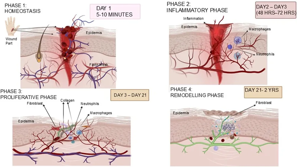
The healing of a wound is a biological process that gets activated through a cascade of events following an injury Table 1. The diagrammatic representation of wound healing phases is given in Figure 1.

Table 1: Phases of Wound Healing

Phase

Phase illustration

Duration

References

Phase:

Homeostasis

During the early stage of an injury, the blood clotting cascade mechanism starts to prevent fluid loss. As a result, platelets get activated and aggregate near the collagen. Followed by these, there is activation of the thrombin enzyme, which leads to the formation of fibrin, which aggregates the platelets to a stable clot.

5-10 minutes

[12], [15]

Phase: Inflammatory

After homeostasis, neutrophils become the first immune cells to respond. They produce their anti-inflammatory substances and secrete pro-inflammatory cytokines with degradative enzymes. They infiltrate the lesion and phagocytose cellular debris to secrete growth factors important for tissue regeneration.

24-78 hours

[16]

Phase:

Proliferative

The phase is followed by activation of fibroblasts, which initiates extracellular matrix deposition, forms granulation tissue, promotes angiogenesis, and promotes reepithelialisation.

3-21 days

[17]

Phase: Remodelling

The fibroblasts, along with matrix metalloproteinase, mediate collagen remodelling, leading to maturation of granulation tissue to scar tissue. Myofibroblasts promote wound contraction, Keratinocytes induce TGF-β essential in the development of scar tissue.

21 days- 2 years

[18]


Figure 1 schematic representation of wound healing stages

Phytosomes

Wounds display an impaired cellular response, with continued inflammation and poor diffusion of medicine into the dermal layers. As a result, it is necessary to develop novel delivery systems to overcome these constraints. Phytosomes, being one of them, enhance wound healing; as a result, the study of their structure and characteristics is necessary. Phytosomes are made by chemically bonding a small number of molecules together [19]. Phospholipids exhibit a cohesive force on polyphenolic compounds to form a supramolecular adduct, forming a stable complex [10]. Phosphatidylcholine is an amphiphilic lipid composed of a hydrophilic phosphatidyl head group and a lipophilic fatty acid tail domain. Due to this amphipathic architecture, Phosphotidylcholine acts as an interfacial mediator between polar and non-polar molecules. During phytosome complexation, hydrophilic phytoconstituents associate with the polar head via non-covalent interactions (such as hydrogen bonding), while the lipophilic acyl chains encapsulate and shield the bioactive molecule. This unique structural arrangement protects the phytoconstituents from degradation, thereby enhancing both their physicochemical stability and bioavailability [20], [21].

Methods of preparation

Understanding the nature of these structural interactions is essential for choosing suitable preparation techniques (Figure 2), as the effectiveness and stability of the resulting phyto-phospholipid complexes are significantly influenced by the degree of integration of these elements during the formulation process. Some of the methods of preparation are

Solvent evaporation method: The method encompasses mixing of plant material and the phospholipid, followed by the addition of acetone to precipitate phytosomes [22].

Mechanical dispersion: In this technique, lipids solubilised in an organic solvent are combined with an aqueous solution containing drugs. The solvent is recovered under reduced pressure, forming a phyto-phospholipid complex. Recently, more sophisticated techniques, such as supercritical fluid methods including Gas Anti-Solvent technique (GAS), Compressed Anti-Solvent Process (PCA), and Supercritical Anti Solvent Method (SAS), have been utilised for the preparation of phospholipid complexes[23].

Lyophilisation methods: The lyophilisation method involves mixing the plant extract and phospholipids in suitable solvents to form a complex and then isolating the resulting phytosomes through a freeze-drying process. This technique improves the stability and bioavailability of bioactives[24].

Thin Film Hydration Method: Surfactants, as well as cholesterol, are solubilised in solvents, like chloroform and diethyl ether. Using a rotary evaporator, solvents are evaporated, forming a thin lipid film adhering to the flask. This lipid film is rehydrated with PBS (Phosphate-Buffered Saline) and subjected to sonication, generating MLVs (Multilamellar Vesicles). For the preparation of enzymosomes, enzymes pre-dissolved in a phosphate buffer at an acidic pH were introduced into the liposomal suspension to promote their encapsulation within the vesicular structure [19], [25].

Ether injection: Initially, the drug and lipid are dissolved in a suitable solvent, which is further incorporated into a hot aqueous solvent to promote vesicle formation. The behaviour of the amphiphilic molecule changes with its concentration. At lower concentrations, they remain as monomers, whereas at higher concentrations they can assemble into an organic structure such as spherical, cylindrical, disc-like, or sometimes cubic or hexagonal morphologies [14], [26].

Salting Out Process: In this process, the plant extracts are solubilised in an inorganic solvent which subsequent addition of phospholipids. Further addition of n-hexane results in precipitation and yields a phospholipid-extract complex [27].

Rotary evaporation method: A definite amount of herbal extract and phospholipids is dissolved in a water-miscible organic solvent like acetone and agitated for several hours at an elevated temperature using a Rotary evaporator. As a result of the mixing step, a thin film is treated with an anti-solvent like n-hexane, which results in precipitation leading to phytosome formation. The phytosomal preparations are stored in amber coloured vials with protected temperature and humidity conditions. Using an alternative method, phospholipids are dissolved in ether and incorporated into a solution that contains phytoconstituents. As a result, was the solvent evaporates, there is a formation of vesicles. The structural organisation of phytosomes is affected by the concentration of the amphiphiles. At lower concentrations, they exist as monomers, whereas as concentrations increase, they yield intricate vesicle structures like spherical, cylindrical, disc-like, cubic or hexagonal formations [23], [28].

Anti-solvent precipitation technique: Numerous investigations have examined anti-solvent methodologies, including the application of n-hexane to precipitate pharmaceutical substances from mixtures of phospholipids and organic solvents. One patented approach utilised n-hexane as the anti-solvent with dichloromethane as the reaction medium to form a complex of andrographolide and phytophospholipid, followed by solvent removal and subsequent vacuum drying. In another research effort, anhydrous cosolvent lyophilisation was employed to formulate a rutin-phospholipid complex, where each component was initially dissolved in methanol. Upon mechanical mixing and evaporation of the solvent, the resulting complex exhibited an amorphous structure rather than a crystalline one, with the most favourable outcomes observed at a drug-to-phospholipid ratio[26], [29].
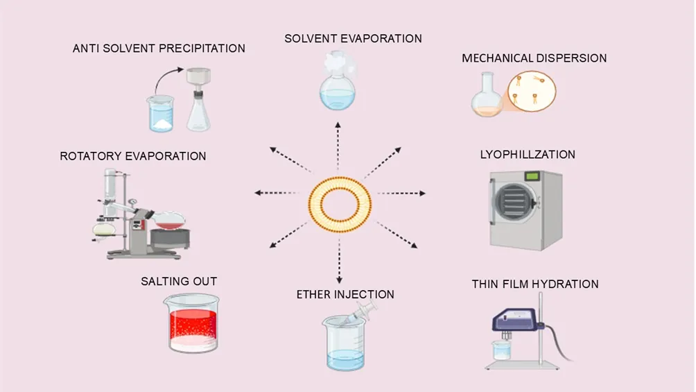
 

Figure 2 various strategies used in the formulation of phytosomes

Phytosomes: the smart structure for better herbal drug delivery

A range of preparation methods, which comprises thin-film hydration, solvent evaporation and anti-solvent precipitation, has been utilised to create stable and efficient phytosomal formulations. Driven by advancements in drug delivery, significant research focus has been shifted towards characterising the structural dynamics of phytosomes. Specifically, the phospholipid-based architecture plays a pivotal role in optimising the transdermal absorption, membrane permeability and targeted delivery of essential bioactive phyto constituents during tissue regeneration and wound repair. Maximizing the full therapeutic potential of plant-derived bioactive fundamentally hinges upon the integration of phytosomal delivery systems [30]. Phytosomes minimise the risk of unfavourable immunological effects and exhibit biochemical stability[31]. The favourable safety and biocompatibility profiles of phytosomal delivery systems render them highly effective for targeted localised delivery. By insulating sensitive phytochemicals from environmental destabilisers such as light, molecular oxygen, and ambient pH fluctuations, phytosomal complexation prevents premature degradation, resulting in improved bioaccessibility, structural integrity, and sustained radical-scavenging activity[32], which are vital for the pathway of wound healing and facilitate enhanced absorption within the dermal layers.[33], [34] The overall absorption profile is fundamentally depends upon optimal lipophilic-hydrophilic partitioning inherent to both the plant bioactive and the phytosomal carrier.[35] The existence of a large functional group, such as a glycoside and hydroxyl in their structural configuration, hinders the absorption of phytonutrients. The substantial molecular size of these compounds leads to poor membrane permeability, restricting the bioavailability of the phytochemicals such as flavonoids, phenolics, and glycosidic aglycons. [36], [37]

Encapsulating these bioactives within phytosomal matrices facilitates transcellular transport, significantly enhances dermal penetration, and amplifies topical therapeutic efficacy. Consequently, optimising phytosomal formulation parameters is paramount for maintaining a sustained, therapeutically effective intradermal concentration over an extended residence time. Owing to their structural homology with biological cell membranes, phospholipid-based delivery platforms seamlessly permeate lipophilic epidermal barriers without eliciting cytotoxicity or compromising cellular integrity. [38]

Essential oils possess considerable antibacterial potential in wound management; however, their high volatility can compromise their stability and therapeutic utility. Incorporation of essential oils into phytosomal complexes can minimize their evaporation, enhance chemical stability, and preserve the integrity of their bioactive constituents without altering their fundamental properties[39]. Such encapsulation may consequently improve their therapeutic performance, particularly their antimicrobial activity. Furthermore, nanosized phytosomal systems can facilitate enhanced tissue penetration by reducing diffusion barriers, thereby promoting more effective delivery of essential oils to the wound site [40].

Working Principle of Phytosomes

Phytosomes facilitate wound repair through multi-faceted synergistic mechanisms. By significantly improving the bio accessibility of complex phytoconstituents, these platforms promote efficient localised cellular uptake and tissue absorption within the injury site [41]. In addition to this, they facilitate the liberation of anti- inflammatory cytokine IL-10 and can inhibit the synthesis of pro-inflammatory cytokines such as IL-4 and IL-7, which will significantly reduce inflammation. Furthermore, it also contributes by displaying its antibacterial, antioxidant, and collagen-boosting properties as studied in carvacrol-loaded phytosomes[42]. Phospholipid complexation significantly enhances both the transmembrane permeation and bioavailability of phytochemicals, thereby facilitating their passive diffusion across semipermeable biological membranes [43]. Collectively, these physicochemical attributes enable phytosomes to penetrate target lesion sites more efficiently, thereby accelerating tissue regeneration and wound healing dynamics [44]. These delivery platforms play a pivotal role in encapsulating and translocating key bioactive phytochemicals, specifically tannins, terpenoids, and flavonoids, which constitute the primary therapeutic constituents in numerous botanical medicines [43]

Furthermore, phytosomes aid in tissue remodelling, encouraging the genesis of newer blood vessels, also termed angiogenesis (Figure 3). It also enhances the formation of cells, increasing fibroblast activity [45]. Angiogenesis as well as collagen synthesis will accelerate the process of wound healing [46]. Moreover, anti-microbial and antioxidative activity will suppress the growth of bacteria during the process of wound healing [47]. Phytosomes are suitable for the targeted delivery of the active phytoconstituent and prolonged remedial effects [48].   The mechanistic overview of phytosomes in wound healing is illustrated in Figure 3
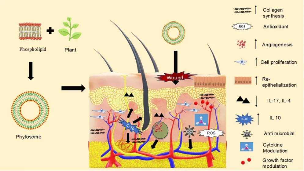
 

Figure 3: Mechanistic overview of phytosome-mediated wound healing

Contributions of phytosomes in wound restoration

Multiple studies have been conducted to explore the antibacterial, antioxidant, anti-inflammatory and consequences of phytoconstituents on the production of procollagen with respect to wound restoration. All these actions can be achieved by complexing the natural phytochemicals into the phytosome[42]. Oregano essential oil consists of a natural phytoconstituent, carvacrol (CAR), which plays a major role in wound repair [49]. It modulates the immune response at the inflammatory stage by inhibiting the formation of pro-inflammatory cytokines, which accounts for redness and swelling and increases the production of IL- 10 and other anti-inflammatory cytokines. It also counters the progression of effects generated by inflammatory mediators, namely IL-1, IL-4, IL-17 and (Cyclooxygenase) COX-2, depleting the inflammation[50] CAR speeds up the process of angiogenesis, collagen deposition and re-epithelialization in the proliferative phase, which are also aided by the increased level of TGF-beta and VEGF (Vascular Endothelial Growth Factor). Additionally, CAR exhibited anti-bacterial property against pathogens like Staphylococcus aureus and Staphylococcus epidermidis [42].

The antibiotic free hydrogel encapsulated in phytosomes and chitosomes such as Hydrogel-Eugenol (HG-Eu), HydrogelPhytosome@Eugenol (meaning eugenol encapsulated into phytosomes within a hydrogel) (HGPhyt@Eu), and Hydrogel-Phytosome/Chitosome@Eugenol (meaning eugenol encapsulated into both phytosomes and chitosomes within a hydrogel) (HG-Phy/Chit@Eu) were administered topically to the infected wounds. Their application led to a sustained release of the natural phytochemical, which further magnified their antibacterial action against both Gram-negative and Gram-positive bacteria. HG-Phyt/Chit@Eu displayed efficient wound restoration by depleting the bacteria which in turn decreases the amounts of inflammatory biomarkers namely (4-Hydroxynonenal) 4-HNE, COX-2, (Inducible Nitric Oxide Synthase) iNOS and (Nuclear Factor Kappa light-chain enhancer of activated B cells) NF-κB, and will enhance the expression of (Collagen Type I Alpha) COL-1A and (Nuclear Factor Erythroid 2 Related Factor 2) which are essential for tissue regeneration. All these effects suggest that HG-Phyt/Chit@Eu is an effective and safer replacement for antibiotics like mupirocin for treating infected wounds [47]

Moreover, leucoselect phytosome, alpha-lipoic acid and Ginkgo Biloba have been thoroughly examined for wound healing properties [51]. These formulations restored diabetic wounds, reducing wound size, promoting tissue repair over time, and alleviating pain.

Additionally, Sinigrin-Loaded phytosomes demonstrate improved stability and promote accelerated wound closure, achieving a remarkable 71% closure within 42 hours during tests on skin cells and clinical trials, without any evidence of toxicity [12].

Furthermore, crocetin derived from Nyctanthes arbor tristis phytosomal formulation bolstered wound healing, thereby reinforcing the integrity of the repaired skin [52].

Lim A-W et al investigated the wound-repairing efficacy of a phytosomal formulation derived from Moringa Olifera leaf extract, achieving 94.8% wound closure within 24 hours at an optimal dosage. Phytosomes potentiated the topical delivery of the extract[53]

Curcumin-loaded phytosomes have demonstrated significant anti-inflammatory effects primarily in rat models, by inhibiting NF-κB activation.

Moreover, phytosome-based wound healing formulations incorporating diverse botanical extracts are discussed in Table 2. Certain formulations have shown better bioavailability and sustained release properties, thus contributing to faster wound contraction and less scarring. In general, nanophytosomal delivery systems have proven to be effective applications to enhance wound healing properties

Table 2: Phytosomal preparations for wound healing

Sr. no.

Formulation

Key Property

Reference

  1.  

Rhodiola rosea Phytosomal Gel

improved wound healing, complete tissue regeneration and collagen deposition

[54]

  1.  

Phytosomal gel of Annona squamosa and Cinnamomum tamala leaves ethanolic extracts

Antimicrobial and antioxidant properties

[55]

  1.  

Pumpkin Seed Oil Nano Phytosome Loaded Lidocaine

Enhanced vascularity and fortified collagen fibers. Stimulated the formation and migration of fibroblasts and mitogenesis, improved re-epithelialization

[56]

  1.  

Curcuma longa L. and Plumbago zeylanica L.

Improved wound contraction and decreased time for epithelialization

[57]

  1.  

Naphthoquinone-Rich root bark of Onosma echioides

enhanced contraction and tensile strength, decrease in lipid peroxidation, and increased catalytic activity

[58]

  1.  

White Ginger Phytosome Gel

Development of granulation tissue and epithelialization.

[59]

  1.  

Polysiloxane-based gel loaded with resveratrol and quercetin

Prevented dehydration and scar formation

[60]

  1.  

Moringa oleifera leaf extract–loaded phytophospholipid complex

Improved NHDF (Normal Human Dermal Fibroblast) cell migration without cytotoxic effect.

[55]

  1.  

PEGylated (polyethylene glycol) Nanophytosomes Loaded with 6-Gingerol


Reduced inflammatory markers and cytokines, and improved wound healing

[61]

  1.  

Geraniol nanophytosomes loaded into polyvinyl alcohol

Decrease bacterial colonization, reduced inflammation, promoted angiogenesis, and improved the expression of COL1A.

[62]

  1.  

Novel Mangiferin-Infused Phytosomal Gel

Enhanced bioavailability, regulated release, and accelerated wound contraction within 14 days

[63]

  1.  

Gold nanoparticle loaded phytosomal systems

demonstrated antioxidant

capabilities and enhanced wound healing effects

[28]

  1.  

Spirulina platensis Nanophytosomes

reduced inflammatory markers and apoptotic processes, downregulated HMGB1(High Mobility Group Box 1)

[64]

Challenges and limitations

Despite their therapeutic potential, phytosomal formulations encounter several biopharmaceutical and commercial constraints. Physicochemical susceptibility to environmental stressors such as photooxidation, thermal degradation, and atmospheric oxidation demands specialised primary packaging. Furthermore, high supply cost for pharmaceutical-grade phospholipids, supply chain scarcity of standardised botanical extracts, and complex compounding parameters for topical hydrogel incorporation pose significant hurdles. Clinically and industrially, potential cutaneous hyperreactivity, stringent regulatory landscape, batch-to-batch quality control challenges, and the requirement for specialised scalable manufacturing processes collectively impede their commercial translation[65]. A significant challenge in advancing research is the paucity of available clinical trial data in wound healing, which impedes the optimisation of future investigations [44]. Post formulation safety evaluation remains a formidable bottleneck in the clinical translation of phytosomal delivery platforms. Despite the biological inertness of constituent phospholipids, rigorous toxicological and pharmacokinetic profiling specifically evaluating biocompatibility alongside in vivo absorption, distribution, metabolism, and excretion dynamics is indispensable. Furthermore, advanced pH responsive phytosomal architectures engineered to optimize site - specific absorption introduces added complexity, presenting heightened regulatory and toxicological challenges regulatory and toxicological challenges [43].

Future aspects to be considered

Despite the expandability and legal obstacles, phytosomes put forward a constructive modern remedy serving healthcare and patients[44]. (Epigallocatechin-3-gallate) EGCG-loaded phytosomes obtained from green tea have anti-inflammatory and antioxidant properties, helping tissue repair, but two factors, namely clinical evidence and stability, limit their use[66]. Phytosomes represent an advanced drug delivery platform that holds distinct therapeutic promise across various pathological conditions, including chronic diabetic wounds, by successfully mitigating biopharmaceutical challenges related to physicochemical stability, aqueous solubility, membrane absorption, and systemic bioavailability [67]. Studies specify that their pharmacokinetic profile is preferable to other delivery systems like liposomes [48]. Modern analytical techniques such as HPLC are regularly implemented for standardisation and efficacy testing of phytosomal formulations[65]. Overall, phytosomes serve a salient function of contributing a more efficacious, safer, and targeted approach to wound healing [44]. Furthermore, the spectrum of phytosomes is extensive when the focus is on elucidating the mechanisms accountable for phytosomes' effective delivery to improve their therapeutic efficiency in wound restoration by restricting their side effects [47].

CONCLUSION

In summary, phytosomes represent a promising phospholipid-based delivery platform for enhancing the therapeutic potential of plant-derived bioactives in wound management. By improving the stability, solubility, dermal penetration, and bioavailability of phytochemicals, phytosomal formulations may potentiate their anti-inflammatory, antioxidant, pro-angiogenic, fibroblast-promoting, and collagen-regulating activities across the different phases of wound repair. Their ability to improve the delivery and therapeutic performance of poorly bioavailable phytoconstituents provides an important bridge between traditional phytotherapy and contemporary drug-delivery technologies. However, despite encouraging preclinical evidence, the clinical translation of phytosomal wound-healing systems remains limited by challenges related to formulation optimization, manufacturing scalability, quality control, stability, regulatory requirements, and the paucity of well-designed long-term clinical studies. Future research should therefore focus on establishing robust structure–activity and formulation–performance relationships, standardizing manufacturing processes, and generating comprehensive clinical evidence to determine the safety, efficacy, and therapeutic value of phytosomes in advanced wound care.

ABBREVIATIONS

  • DFUs: Diabetic Foot Ulcers
  • TGF-β: Transforming Growth Factor Beta
  • GAS: Gas Anti-Solvent Technique
  • PCA: Compressed Anti-Solvent Process
  • SAS: Supercritical Anti-Solvent Method
  • PBS: Phosphate-Buffered Saline
  • MLVs: Multilamellar Vesicles
  • IL: Interleukins
  • CAR: Carvacrol
  • COX: Cyclooxygenase
  • VEGF: Vascular Endothelial Growth Factor
  • 4-HNE: 4-Hydroxynonenal
  • iNOS: Inducible Nitric Oxide Synthase
  • NF-κB: Nuclear Factor Kappa
  • COL-1A: Collagen Type I Alpha
  • Nrf2: Nuclear Factor Erythroid 2–Related Factor 2
  • NHDF: Normal Human Dermal Fibroblast Cells
  • HMGB: High Mobility Group Box 1
  • EGCG: Epigallocatechin-3 Gallate

CONFLICTS OF INTEREST

The authors declare no conflicts of interest.

ACKNOWLEDGEMENTS

The authors are thankful to the Principal, Bharati Vidyapeeth’s Poona College of Pharmacy, Pune, for providing infrastructure and all necessary facilities.

REFERENCES

  1. Olsson M, Jarbrink K, Divakar U, et al. The humanistic and economic burden of chronic wounds: A systematic review. Wound Repair and Regeneration. 2018, 27(1):114–125. https://doi.org/10.1111/wrr.12683
  2. Burgess JL, Wyant WA, Abdo Abujamra B, Kirsner RS, Jozic I. Diabetic Wound-Healing Science. Medicina. 2021, 57(10):1072.  https://doi.org/10.3390/medicina57101072 
  3. Sen CK. Human Wound and Its Burden: Updated 2020 Compendium of Estimates. Advances in Wound Care. 2021;10:281–92 https://doi.org/10.1089/wound.2021.0026
  4. Ksenchyna K, Ksenchyn O, Dmytriiev D, Nazarchuk O. Wound infection and pain one month after trauma: an underestimated threat. Frontiers in Pain Research. 2025; 6 https://doi.org/10.3389/fpain.2025.1647785
  5. Sharma A, Srivastava V, Shankar R, Yadav AK, Ansari MA, Gupta SK .Epidemiological Profile of Chronic Wounds in an Indian Population: A Community-Based Cross-Sectional Observational Study. Cures Journal of Medical Science. 2024;16(9)  https://doi.org/10.7759/cureus.68684
  6. Ding X, Tang Q, Xu Z, Xu Y, Zhang H, Zheng D, Wang S, Tan Q, Maitz J, Maitz PK, Yin S, Wang Y, Chen J .Challenges and innovations in treating chronic and acute wound infections: from basic science to clinical practice. Burns and Trauma. 2022; 10 https://doi.org/10.1093/burnst/tkac014
  7. Sganga G, Pea F, Aloj D, Corcione S, Pierangeli M, Stefani S, Rossolini GM, Francesco M. Acute wound infections management: the ‘Don’ts’ from a multidisciplinary expert panel. Expert Review of Anti-infective Therapy. 2020;18:231-240 https://doi.org/10.1080/14787210.2020.1726740
  8. Thet NT, Mercer-Chalmers J, Greenwood RJ. SPaCE Swab: Point-of-Care Sensor for Simple and Rapid Detection of Acute Wound Infection. ACS Sensors. 2020; 5(8):2652–2657 https://doi.org/10.1021/acssensors.0c01265
  9. Liu YF, Ni PW, Huang Y, Xie T. Therapeutic strategies for chronic wound infection. Chinese Journal of Traumatology. 2022; 25(1):11-16 https://doi.org/10.1016/j.cjtee.2021.07.004
  10. Barani M, Sangiovanni E, Angarano M, Rajizadeh MA, Mehrabani M, Piazza S, Gangadharappa HV, Pardakhty A, Mehrbani M, Dell’Agli M, Nematollahi MH. Phyto-phospholipid complexes (phytosomes): A novel strategy to improve the bioavailability of active constituents. International Journal of Nanomedicine. 2021;(16):6983–7022 https://doi.org/10.2147/IJN.S318416
  11. Mei Lu, Qiujun Qiu, Xiang Luo, Xinrong Liu, Jing Sun, Cunyang Wang, Xiangyun Lin, Yihui Deng, Yanzhi Song. A novel strategy to improve the bioavailability of active constituents. Asian Journal of Pharmaceutical Sciences. 2019; 14(3):265–274 https://doi.org/10.1016/j.ajps.2018.05.011
  12. Kumar M, Keshwania P, Chopra S, Mahmood S, Bhatia A. Therapeutic Potential of Nanocarrier-Mediated Delivery of Phytoconstituents for Wound Healing: Their Current Status and Future Perspective. AAPS PharmSciTech. 2023; 24:155 https://doi.org/10.1208/s12249-023-02616-6
  13. Koppula S, Shaik B, Maddi S. Phytosomes as a New Frontier and Emerging Nanotechnology Platform for Phytopharmaceuticals: Therapeutic and Clinical Applications. Phytotherapy Research.2025; 39(5):2217–49  https://doi.org/10.1002/ptr.8465
  14. Dhoke PS, Shelke PG, Bakal RL, Hatwar PR. Phytosomes: A promising approach for enhancing herbal medicinal efficacy. International Journal of Herbal Medicine. 2025; 13(2):01-08 https://www.doi.org/10.22271/flora.2025.v13.i2a.972
  15. Ayadi AEl, Jay JW, Prasai A. Current Approaches Targeting the Wound Healing Phases to Attenuate Fibrosis and Scarring. International Journal of Molecular Sciences. 2020; 21(3):1105 https://doi.org/10.3390/ijms21031105
  16. Xu Z, Han S, Gu Z, Wu J. Advances and Impact of Antioxidant Hydrogel in Chronic Wound Healing. Advanced Healthcare Materials. 2020; 9(5) https://doi.org/10.1002/adhm.201901502
  17. Jatoi AW, Ogasawara H, Kim IS, Ni QQ. Polyvinyl alcohol nanofiber based three phase wound dressings for sustained wound healing applications. Materials Letters. 2020; 241:168–71 https://doi.org/10.1016/j.matlet.2019.01.084
  18. Dehkordi AN, Babaheydari FM, Chehelgerdi M, Dehkordi SR. Skin tissue engineering: wound healing based on stem-cell-based therapeutic strategies. Stem Cell Research and Therapy.2019 ;10(2019):111 https://doi.org/10.1186/s13287-019-1212-2
  19. Pawar HA, Bhangale BD. Phytosome as a Novel Biomedicine: A Microencapsulated Drug Delivery System. Journal Of Bioanalysis and biomedicine. 2015;7 10.4172/1948593X.1000116
  20. Jain N, Gupta BP, Thakur N, et al. Phytosome: A Novel Drug Delivery System for Herbal Medicine. International Journal of Pharmaceutical Sciences and Drug Research. 2010; 2(4):224–228 https://doi.org/10.25004/IJPSDR.2010.020401
  21. Kumar S, Baldi A, Sharma DK. A Modernistic Approach for Novel Herbal Drug Delivery: Enhancing Bioavailability and Revealing Endless Frontier of Phytopharmaceuticals. Journal of Developing Drugs.2020; 9:2 10.4172/2329-6631. 1000195
  22. Modak A, Bhakat SP, Kar B, Roy S. Innovative Approaches in Drug Delivery: Exploring Nanosuspension as Phytosome. International Journal of Pharmaceutical Research and Applications. 2024; 9(3):1286-1298 10.35629/4494-090312861298
  23. Gaurav V, Paliwal S, Singh A, Pandey S, Aqil M. Phytosomes: Preparation, Evaluation and Application. International Journal of Engineering Science. 2021 9(2):35-39  https://doi.org/10.48175/ijarsct-29240
  24. Grange LL, Wang M, Watkins R, et al. Protective effects of the flavonoid mixture, silymarin, on fetal rat brain and liver. Journal Of Ethnopharmacology. 1999; 65(1):53–61 https://doi.org/10.1016/S0378-8741(98)00144-5
  25. Kusuma Priya M D, Kumar V, Damini VK, Eswar Ka, Kadiri RR, Brito Raj, sucharitha P. somes: A Review on Composition, Formulation Methods and Evaluations of Different Types of “Somes” Drug Delivery System. International Journal of Applied Sciences. 2020;12(6):7-18
  26. Dwivedi J, Sachan P, Wal P, Kosey S, Khan U. Progressive Journey of Phytosomes: Preparation, Characterization, Patents, Clinical trials & Commercial products. Journal of Research in Pharmacy.2023; 27(5):1687–733 10.29228/jrp.457
  27. Khan S, Kotkar P. Phytosomes in Nanovesicle Herbal Formulation: A Contemporary Advancement in Natural Medicine. Journal Of Pharmaceutical Advanced Research. 2023; (10): 1940-1947https://www.researchgate.net/publication/380493331_Phytosomes_in_Nanovesicle_Herbal_Formulation_A_Contemporary_Advancement_in_Natural_Medicine
  28. Demir B, Barlas FB, Guler E, et al. Gold nanoparticle loaded phytosomal systems: synthesis, characterization and in vitro investigations. RSC Advances. 2014;4:34687–34695 https://xlink.rsc.org/?DOI=C4RA05108D
  29. Maryana W, Rachmawati H, Mudhakir D.Formation of Phytosome Containing Silymarin Using Thin Layer-Hydration Technique Aimed for Oral Delivery. Materials today: Proceedings. 2016;3:855–866 https://doi.org/10.1016/j.matpr.2016.02.019
  30. Mota AH, Prazeres I, Mestre H, et al. A Newfangled Collagenase Inhibitor Topical Formulation Based on Ethosomes with Sambucus nigra L. Extract. Pharmaceuticals. 2021;14(5):467 https://doi.org/10.3390/ph14050467
  31. Tallam AK, Sahithi A, Nuli MV. A review on phytosomes as innovative delivery systems for phytochemicals. International Journal of Pharmacognosy and Chemistry. 2023; 4(1):1–8. https://doi.org/10.46796/ijpc.v4i1.416
  32. Jahanfar S, Gahavami M, Khosravi-Darani K, Jahadi M, Mozafari MR. Entrapment of rosemary extract by liposomes formulated by Mozafari method: physicochemical characterization and optimization. Heliyon. 2021; 7(12) https://doi.org/10.1016/j.heliyon.2021.e08632
  33. Manconi M, Marongiu F, Castangia I, et al. Polymer-associated liposomes for the oral delivery of grape pomace extract. Colloids and Surfaces B: Biointerfaces. 2016;146:910–917 https://doi.org/10.1016/j.colsurfb.2016.07.043
  34. Mehta G, Rani R, Singh AP, Singh AP. Phytosomes:An Overview. International Journal of Pharmaceutics and Drug Analysis. 2024; 12(1):65-71 https://doi.org/10.47957/ijpda.v12i1.578
  35. Alharbi WS, Almughem FA, Almehmady AM, et al. Phytosomes as an Emerging Nanotechnology Platform for the Topical Delivery of Bioactive Phytochemicals. Pharmaceutics. 2021;13(9):1475 https://doi.org/10.3390/pharmaceutics13091475
  36. Ali M, Abdel Motaal A, Ahmed MA, Alsayari A, El-Gazayerly. An in vivo study of Hypericum perforatum in a niosomal topical drug delivery system. Drug Delivery. 2018;25:417–425. https://doi.org/10.1080/10717544.2018.1431977
  37. Un RN, Barlas FB, Yavuz M, et al. Phyto-Niosomes: In Vitro Assessment of the Novel Nanovesicles Containing Marigold Extract. International Journal of Polymeric Materials and Polymeric Biomaterials. 2015; 64(17):927-937 https://doi.org/10.1080/00914037.2015.1030663
  38. Patzelt A, Lademann J. Recent advances in follicular drug delivery of nanoparticles. Expert Opinion on Drug Delivery. 2020;17(1):49-60 https://doi.org/10.1080/17425247.2020.1700226
  39. Usach I, Margarucci E, Manca ML, et al. Comparison between Citral and Pompia Essential Oil Loaded in Phospholipid Vesicles for the Treatment of Skin and Mucosal Infections. Nanomaterials. 2020; 10(2):286  https://doi.org/10.3390/nano10020286
  40. Safta DA, Bogdan C, Moldovan ML. Vesicular Nanocarriers for Phytocompounds in Wound Care: Preparation and Characterization. Pharmaceutics. 2022;14(5):991  https://doi.org/10.3390/pharmaceutics14050991
  41. Vishwakarma DK, Mishra NJ, Shukla AK, Singh AP. Phytosomes as a Novel Approach to Drug Delivery System. Smart Drug Delivery Systems - Futuristic Window in Cancer Therapy. 2024  http://dx.doi.org/10.5772/intechopen.113914
  42. Tafish AM, El-Sherbiny M, Al-Karmalawy AA, Soliman OAEA, Saleh NM.Carvacrol-Loaded Phytosomes for Enhanced Wound Healing: Molecular Docking, Formulation, DoE-Aided Optimization, and in vitro/in vivo Evaluation. International Journal of Nanomedicine.2023 ;(18):5749—5780  https://doi.org/10.2147/IJN.S421617
  43. Sakure K, Patel A, Pradhan M, Badwaik HR. Recent Trends and Future Prospects of Phytosomes: A Concise Review. Indian Journal of Pharmaceutical Sciences. 2024; 86(3):772-790  https://doi.org/10.36468/pharmaceutical-sciences.1334
  44. Kakurte S, Shinde K, Rascal P. Application Of Phytosome In Wound Healing: A Comprehensive Review. International Journal of Pharmaceutical Sciences. 2024; 2(11):600-611  10.5281/zenodo.14151452
  45. Agrawal D, Singh MP, Sharma GK. Pharmacodynamic approaches of phytoconstitutents in wound healing mechanisms. Pharmacological Research - Natural Products. 2024;5 https://doi.org/10.1016/j.prenap.2024.100119
  46. Suhag D. Skin and Wound Healing Biomaterials. Handbook of Biomaterials for Medical Applications. 2024  2:281–320 https://doi.org/10.1007/978-981-97-5906-4_9
  47. Dashti A, Karamibonari AR, Farahpour MR, Tabatabaei ZG. Topical effectiveness of eugenol phytosome / chitosome hydrogels on the healing process of infected excision wounds. Colloids and Surfaces A: Physicochemical and Engineering Aspects. 2024;687 https://doi.org/10.1016/j.colsurfa.2024.133482
  48. Talebi M, Shahbazi K, Dakkali MS, et al. Phytosomes: A promising nanocarrier system for enhanced bioavailability and therapeutic efficacy of herbal products. Phytomedicine Plus. 2025; 5(2)  https://doi.org/10.1016/j.phyplu.2025.100779
  49. Costa MF, Durço AO, Rabelo TK, Barreto RDSS, Guimarães AG. Effects of Carvacrol, Thymol and essential oils containing such monoterpenes on wound healing: a systematic review. Journal of Pharmacy and Pharmacology. 2019; 71(2):141-155 https://doi.org/10.1111/jphp.13054
  50. Marinelli L, Di Stefano A, Cacciatore I. Carvacrol and its derivatives as antibacterial agents. Phytochemistry Reviews. 2018;17:903–921 https://doi.org/10.1007/s11101-018-9569-x
  51. Poudyal A, Subba B, Bhattarai A, Regmi D, Tiwari B, Pradhan A (2022) A Comprehensive Review on Phytosomes: A Novel Drug Delivery System of Phytoconstituents. International Journal of Life science and Pharma Research 12(5):143-161 https://doi.org/10.22376/ijpbs/lpr.2022.12.5.p143-161
  52. Varadkar M, Gadgoli C. Preparation and evaluation of wound healing activity of phytosomes of crocetin from Nyctanthes arbor-tristis in rats. Journal of Traditional and Complementary Medicine. 2022;12(4):354-360  https://doi.org/10.1016/j.jtcme.2021.10.002
  53. Lim AW, Ng PY, Chieng N, Ng SF. Moringa oleifera leaf extract–loaded phytophospholipid complex for potential application as wound dressing. Journal of Drug Delivery Science and Technology. 2019; 54  https://doi.org/10.1016/j.jddst.2019.101329
  54. Kalusalingam A, Khan A, Kanakal MM, et al. Enhanced Wound Healing with Rhodiola rosea Phytosomal Gel: Extraction, Formulation, and In vivo Evaluation. Current Pharmaceutical Design. 2025  31(33):2651-2665 https://doi.org/10.2174/0113816128359150250211053431
  55. Khan AD, Singh MK, Lavhale PM, Yasir M, Singh L. Exploring the wound healing activity of phytosomal gel of Annona squamosa and Cinnamomum tamala leaves ethanolic extracts with antioxidant and antimicrobial activities in S aureus infected excision wound model. Journal of Biomaterials Science. 2024; Polymer Edition 35 (16):2447-2468  https://doi.org/10.1080/09205063.2024.2382540
  56. Abduwhab WA, Hasan WA, AL-Bayati MA. Burned Wound Healing Effect of Prepared Pumpkin Seed Oil Nano Phytosome Loaded Lidocaine in Rabbit. Advances in Animal and Veterinary Sciences. 2024;12(4):723-731 https://dx.doi.org/10.17582/journal.aavs/2024/12.4.723.731
  57. Shukla B, Kushwaha P, Saxena S, Gupta A, Panjwani D, Kumar S. Development and efficacy assessment of polyherbal phytosomal gel for accelerated wound healing. Journal of biomaterials Science. Polymer. 2024;35(11):1684-1705 https://doi.org/10.1080/09205063.2024.2346400
  58. Pananchery J, Gadgoli C. Phytosomes of Naphthoquinone Enriched Extract of Root bark of Onosma echioides Exhibit Wound Healing Activity in Rats. Indonesian Journal of Pharmacy. 2021; 32(4):474-483 https://doi.org/10.22146/ijp.2351
  59. Auliah N, Sartini, Djawad K, et al. Effects of White Ginger Phytosome Gel on Wound Healing in Mice: Studies on TGF-β1, VEGF, and Tissue Growth. Pakistan Journal of Life and Social Sciences. 2025;23(1):5933-5950 https://doi.org/10.57239/PJLSS-2025-23.1.00463
  60. Naseriyeh T, Kahrizi D, Arkan E, Moradi S, Kahrizi F. Development and characterization of polysiloxane-based gel loaded with phytoingredients encapsulated in phytosomes for scar management. Cellular and Molecular Biology. 2024;70(8):235-243  https://doi.org/10.14715/cmb/2024.70.8.32
  61. Al-Samydai A, Qaraleh MA, Alshaer W, et al. Preparation, Characterization, Wound Healing, and Cytotoxicity Assay of PEGylated Nanophytosomes Loaded with 6-Gingerol. Nutrients. 2022 ; 14(23): 5170 https://doi.org/10.3390/nu14235170
  62. Babaei P, Farahpour MR, Tabatabaei ZG. Fabrication of geraniol nanophytosomes loaded into polyvinyl alcohol: A new product for the treatment of wounds infected with methicillin-resistant Staphylococcus aureus. Journal of Tissue Viability. 2024;33(1):116-125  https://doi.org/10.1016/j.jtv.2023.11.002
  63. Mankar SD, Siddheshwar SS, Dighe SB, Wani A. Novel Mangiferin-Infused Phytosomal Gel for Wound Healing: Development, Characterization and In Vivo Assessment. Indian Drugs. 2025;62(1):35-47 https://doi.org/10.53879/id.62.01.14914
  64. Refai H, El-Gazar AA, Ragab GM, et al. Enhanced Wound Healing Potential of Spirulina platensis Nanophytosomes: Metabolomic Profiling, Molecular Networking, and Modulation of HMGB-1 in an Excisional Wound Rat Model. Marine drugs. 2023;21(3):149  https://doi.org/10.3390/md21030149
  65. Mohare A, Meti V, Guddad A, Harish KH, Dasankoppa F. Herbal Phytosomal Gels: Benefits, challenges, and future directions: A review. IP International Journal of Comprehensive and Advanced Pharmacology. 2025;10(1):3-15 https://doi.org/10.18231/j.ijcaap.2025.002
  66. Shariare MH, Afnan K, Iqbal F, et al. Development and Optimization of Epigallocatechin-3-Gallate (EGCG) Nano Phytosome Using Design of Experiment (DoE) and Their In Vivo Anti-Inflammatory Studies.  Molecules. 2020; 25(22):5453  https://doi.org/10.3390/molecules25225453
  67. Mardiana L, Milanda T, Hadisaputri YE, Chaerunisaa AY. Phytosome-Enhanced Secondary Metabolites for Improved Anticancer Efficacy: Mechanisms and Bioavailability Review. Drug Design Development and Therapy. 2025; 19:201-218 AY.10.2147/DDDT.S483

Reference

  1. Olsson M, Jarbrink K, Divakar U, et al. The humanistic and economic burden of chronic wounds: A systematic review. Wound Repair and Regeneration. 2018, 27(1):114–125. https://doi.org/10.1111/wrr.12683
  2. Burgess JL, Wyant WA, Abdo Abujamra B, Kirsner RS, Jozic I. Diabetic Wound-Healing Science. Medicina. 2021, 57(10):1072.  https://doi.org/10.3390/medicina57101072 
  3. Sen CK. Human Wound and Its Burden: Updated 2020 Compendium of Estimates. Advances in Wound Care. 2021;10:281–92 https://doi.org/10.1089/wound.2021.0026
  4. Ksenchyna K, Ksenchyn O, Dmytriiev D, Nazarchuk O. Wound infection and pain one month after trauma: an underestimated threat. Frontiers in Pain Research. 2025; 6 https://doi.org/10.3389/fpain.2025.1647785
  5. Sharma A, Srivastava V, Shankar R, Yadav AK, Ansari MA, Gupta SK .Epidemiological Profile of Chronic Wounds in an Indian Population: A Community-Based Cross-Sectional Observational Study. Cures Journal of Medical Science. 2024;16(9)  https://doi.org/10.7759/cureus.68684
  6. Ding X, Tang Q, Xu Z, Xu Y, Zhang H, Zheng D, Wang S, Tan Q, Maitz J, Maitz PK, Yin S, Wang Y, Chen J .Challenges and innovations in treating chronic and acute wound infections: from basic science to clinical practice. Burns and Trauma. 2022; 10 https://doi.org/10.1093/burnst/tkac014
  7. Sganga G, Pea F, Aloj D, Corcione S, Pierangeli M, Stefani S, Rossolini GM, Francesco M. Acute wound infections management: the ‘Don’ts’ from a multidisciplinary expert panel. Expert Review of Anti-infective Therapy. 2020;18:231-240 https://doi.org/10.1080/14787210.2020.1726740
  8. Thet NT, Mercer-Chalmers J, Greenwood RJ. SPaCE Swab: Point-of-Care Sensor for Simple and Rapid Detection of Acute Wound Infection. ACS Sensors. 2020; 5(8):2652–2657 https://doi.org/10.1021/acssensors.0c01265
  9. Liu YF, Ni PW, Huang Y, Xie T. Therapeutic strategies for chronic wound infection. Chinese Journal of Traumatology. 2022; 25(1):11-16 https://doi.org/10.1016/j.cjtee.2021.07.004
  10. Barani M, Sangiovanni E, Angarano M, Rajizadeh MA, Mehrabani M, Piazza S, Gangadharappa HV, Pardakhty A, Mehrbani M, Dell’Agli M, Nematollahi MH. Phyto-phospholipid complexes (phytosomes): A novel strategy to improve the bioavailability of active constituents. International Journal of Nanomedicine. 2021;(16):6983–7022 https://doi.org/10.2147/IJN.S318416
  11. Mei Lu, Qiujun Qiu, Xiang Luo, Xinrong Liu, Jing Sun, Cunyang Wang, Xiangyun Lin, Yihui Deng, Yanzhi Song. A novel strategy to improve the bioavailability of active constituents. Asian Journal of Pharmaceutical Sciences. 2019; 14(3):265–274 https://doi.org/10.1016/j.ajps.2018.05.011
  12. Kumar M, Keshwania P, Chopra S, Mahmood S, Bhatia A. Therapeutic Potential of Nanocarrier-Mediated Delivery of Phytoconstituents for Wound Healing: Their Current Status and Future Perspective. AAPS PharmSciTech. 2023; 24:155 https://doi.org/10.1208/s12249-023-02616-6
  13. Koppula S, Shaik B, Maddi S. Phytosomes as a New Frontier and Emerging Nanotechnology Platform for Phytopharmaceuticals: Therapeutic and Clinical Applications. Phytotherapy Research.2025; 39(5):2217–49  https://doi.org/10.1002/ptr.8465
  14. Dhoke PS, Shelke PG, Bakal RL, Hatwar PR. Phytosomes: A promising approach for enhancing herbal medicinal efficacy. International Journal of Herbal Medicine. 2025; 13(2):01-08 https://www.doi.org/10.22271/flora.2025.v13.i2a.972
  15. Ayadi AEl, Jay JW, Prasai A. Current Approaches Targeting the Wound Healing Phases to Attenuate Fibrosis and Scarring. International Journal of Molecular Sciences. 2020; 21(3):1105 https://doi.org/10.3390/ijms21031105
  16. Xu Z, Han S, Gu Z, Wu J. Advances and Impact of Antioxidant Hydrogel in Chronic Wound Healing. Advanced Healthcare Materials. 2020; 9(5) https://doi.org/10.1002/adhm.201901502
  17. Jatoi AW, Ogasawara H, Kim IS, Ni QQ. Polyvinyl alcohol nanofiber based three phase wound dressings for sustained wound healing applications. Materials Letters. 2020; 241:168–71 https://doi.org/10.1016/j.matlet.2019.01.084
  18. Dehkordi AN, Babaheydari FM, Chehelgerdi M, Dehkordi SR. Skin tissue engineering: wound healing based on stem-cell-based therapeutic strategies. Stem Cell Research and Therapy.2019 ;10(2019):111 https://doi.org/10.1186/s13287-019-1212-2
  19. Pawar HA, Bhangale BD. Phytosome as a Novel Biomedicine: A Microencapsulated Drug Delivery System. Journal Of Bioanalysis and biomedicine. 2015;7 10.4172/1948593X.1000116
  20. Jain N, Gupta BP, Thakur N, et al. Phytosome: A Novel Drug Delivery System for Herbal Medicine. International Journal of Pharmaceutical Sciences and Drug Research. 2010; 2(4):224–228 https://doi.org/10.25004/IJPSDR.2010.020401
  21. Kumar S, Baldi A, Sharma DK. A Modernistic Approach for Novel Herbal Drug Delivery: Enhancing Bioavailability and Revealing Endless Frontier of Phytopharmaceuticals. Journal of Developing Drugs.2020; 9:2 10.4172/2329-6631. 1000195
  22. Modak A, Bhakat SP, Kar B, Roy S. Innovative Approaches in Drug Delivery: Exploring Nanosuspension as Phytosome. International Journal of Pharmaceutical Research and Applications. 2024; 9(3):1286-1298 10.35629/4494-090312861298
  23. Gaurav V, Paliwal S, Singh A, Pandey S, Aqil M. Phytosomes: Preparation, Evaluation and Application. International Journal of Engineering Science. 2021 9(2):35-39  https://doi.org/10.48175/ijarsct-29240
  24. Grange LL, Wang M, Watkins R, et al. Protective effects of the flavonoid mixture, silymarin, on fetal rat brain and liver. Journal Of Ethnopharmacology. 1999; 65(1):53–61 https://doi.org/10.1016/S0378-8741(98)00144-5
  25. Kusuma Priya M D, Kumar V, Damini VK, Eswar Ka, Kadiri RR, Brito Raj, sucharitha P. somes: A Review on Composition, Formulation Methods and Evaluations of Different Types of “Somes” Drug Delivery System. International Journal of Applied Sciences. 2020;12(6):7-18
  26. Dwivedi J, Sachan P, Wal P, Kosey S, Khan U. Progressive Journey of Phytosomes: Preparation, Characterization, Patents, Clinical trials & Commercial products. Journal of Research in Pharmacy.2023; 27(5):1687–733 10.29228/jrp.457
  27. Khan S, Kotkar P. Phytosomes in Nanovesicle Herbal Formulation: A Contemporary Advancement in Natural Medicine. Journal Of Pharmaceutical Advanced Research. 2023; (10): 1940-1947https://www.researchgate.net/publication/380493331_Phytosomes_in_Nanovesicle_Herbal_Formulation_A_Contemporary_Advancement_in_Natural_Medicine
  28. Demir B, Barlas FB, Guler E, et al. Gold nanoparticle loaded phytosomal systems: synthesis, characterization and in vitro investigations. RSC Advances. 2014;4:34687–34695 https://xlink.rsc.org/?DOI=C4RA05108D
  29. Maryana W, Rachmawati H, Mudhakir D.Formation of Phytosome Containing Silymarin Using Thin Layer-Hydration Technique Aimed for Oral Delivery. Materials today: Proceedings. 2016;3:855–866 https://doi.org/10.1016/j.matpr.2016.02.019
  30. Mota AH, Prazeres I, Mestre H, et al. A Newfangled Collagenase Inhibitor Topical Formulation Based on Ethosomes with Sambucus nigra L. Extract. Pharmaceuticals. 2021;14(5):467 https://doi.org/10.3390/ph14050467
  31. Tallam AK, Sahithi A, Nuli MV. A review on phytosomes as innovative delivery systems for phytochemicals. International Journal of Pharmacognosy and Chemistry. 2023; 4(1):1–8. https://doi.org/10.46796/ijpc.v4i1.416
  32. Jahanfar S, Gahavami M, Khosravi-Darani K, Jahadi M, Mozafari MR. Entrapment of rosemary extract by liposomes formulated by Mozafari method: physicochemical characterization and optimization. Heliyon. 2021; 7(12) https://doi.org/10.1016/j.heliyon.2021.e08632
  33. Manconi M, Marongiu F, Castangia I, et al. Polymer-associated liposomes for the oral delivery of grape pomace extract. Colloids and Surfaces B: Biointerfaces. 2016;146:910–917 https://doi.org/10.1016/j.colsurfb.2016.07.043
  34. Mehta G, Rani R, Singh AP, Singh AP. Phytosomes:An Overview. International Journal of Pharmaceutics and Drug Analysis. 2024; 12(1):65-71 https://doi.org/10.47957/ijpda.v12i1.578
  35. Alharbi WS, Almughem FA, Almehmady AM, et al. Phytosomes as an Emerging Nanotechnology Platform for the Topical Delivery of Bioactive Phytochemicals. Pharmaceutics. 2021;13(9):1475 https://doi.org/10.3390/pharmaceutics13091475
  36. Ali M, Abdel Motaal A, Ahmed MA, Alsayari A, El-Gazayerly. An in vivo study of Hypericum perforatum in a niosomal topical drug delivery system. Drug Delivery. 2018;25:417–425. https://doi.org/10.1080/10717544.2018.1431977
  37. Un RN, Barlas FB, Yavuz M, et al. Phyto-Niosomes: In Vitro Assessment of the Novel Nanovesicles Containing Marigold Extract. International Journal of Polymeric Materials and Polymeric Biomaterials. 2015; 64(17):927-937 https://doi.org/10.1080/00914037.2015.1030663
  38. Patzelt A, Lademann J. Recent advances in follicular drug delivery of nanoparticles. Expert Opinion on Drug Delivery. 2020;17(1):49-60 https://doi.org/10.1080/17425247.2020.1700226
  39. Usach I, Margarucci E, Manca ML, et al. Comparison between Citral and Pompia Essential Oil Loaded in Phospholipid Vesicles for the Treatment of Skin and Mucosal Infections. Nanomaterials. 2020; 10(2):286  https://doi.org/10.3390/nano10020286
  40. Safta DA, Bogdan C, Moldovan ML. Vesicular Nanocarriers for Phytocompounds in Wound Care: Preparation and Characterization. Pharmaceutics. 2022;14(5):991  https://doi.org/10.3390/pharmaceutics14050991
  41. Vishwakarma DK, Mishra NJ, Shukla AK, Singh AP. Phytosomes as a Novel Approach to Drug Delivery System. Smart Drug Delivery Systems - Futuristic Window in Cancer Therapy. 2024  http://dx.doi.org/10.5772/intechopen.113914
  42. Tafish AM, El-Sherbiny M, Al-Karmalawy AA, Soliman OAEA, Saleh NM.Carvacrol-Loaded Phytosomes for Enhanced Wound Healing: Molecular Docking, Formulation, DoE-Aided Optimization, and in vitro/in vivo Evaluation. International Journal of Nanomedicine.2023 ;(18):5749—5780  https://doi.org/10.2147/IJN.S421617
  43. Sakure K, Patel A, Pradhan M, Badwaik HR. Recent Trends and Future Prospects of Phytosomes: A Concise Review. Indian Journal of Pharmaceutical Sciences. 2024; 86(3):772-790  https://doi.org/10.36468/pharmaceutical-sciences.1334
  44. Kakurte S, Shinde K, Rascal P. Application Of Phytosome In Wound Healing: A Comprehensive Review. International Journal of Pharmaceutical Sciences. 2024; 2(11):600-611  10.5281/zenodo.14151452
  45. Agrawal D, Singh MP, Sharma GK. Pharmacodynamic approaches of phytoconstitutents in wound healing mechanisms. Pharmacological Research - Natural Products. 2024;5 https://doi.org/10.1016/j.prenap.2024.100119
  46. Suhag D. Skin and Wound Healing Biomaterials. Handbook of Biomaterials for Medical Applications. 2024  2:281–320 https://doi.org/10.1007/978-981-97-5906-4_9
  47. Dashti A, Karamibonari AR, Farahpour MR, Tabatabaei ZG. Topical effectiveness of eugenol phytosome / chitosome hydrogels on the healing process of infected excision wounds. Colloids and Surfaces A: Physicochemical and Engineering Aspects. 2024;687 https://doi.org/10.1016/j.colsurfa.2024.133482
  48. Talebi M, Shahbazi K, Dakkali MS, et al. Phytosomes: A promising nanocarrier system for enhanced bioavailability and therapeutic efficacy of herbal products. Phytomedicine Plus. 2025; 5(2)  https://doi.org/10.1016/j.phyplu.2025.100779
  49. Costa MF, Durço AO, Rabelo TK, Barreto RDSS, Guimarães AG. Effects of Carvacrol, Thymol and essential oils containing such monoterpenes on wound healing: a systematic review. Journal of Pharmacy and Pharmacology. 2019; 71(2):141-155 https://doi.org/10.1111/jphp.13054
  50. Marinelli L, Di Stefano A, Cacciatore I. Carvacrol and its derivatives as antibacterial agents. Phytochemistry Reviews. 2018;17:903–921 https://doi.org/10.1007/s11101-018-9569-x
  51. Poudyal A, Subba B, Bhattarai A, Regmi D, Tiwari B, Pradhan A (2022) A Comprehensive Review on Phytosomes: A Novel Drug Delivery System of Phytoconstituents. International Journal of Life science and Pharma Research 12(5):143-161 https://doi.org/10.22376/ijpbs/lpr.2022.12.5.p143-161
  52. Varadkar M, Gadgoli C. Preparation and evaluation of wound healing activity of phytosomes of crocetin from Nyctanthes arbor-tristis in rats. Journal of Traditional and Complementary Medicine. 2022;12(4):354-360  https://doi.org/10.1016/j.jtcme.2021.10.002
  53. Lim AW, Ng PY, Chieng N, Ng SF. Moringa oleifera leaf extract–loaded phytophospholipid complex for potential application as wound dressing. Journal of Drug Delivery Science and Technology. 2019; 54  https://doi.org/10.1016/j.jddst.2019.101329
  54. Kalusalingam A, Khan A, Kanakal MM, et al. Enhanced Wound Healing with Rhodiola rosea Phytosomal Gel: Extraction, Formulation, and In vivo Evaluation. Current Pharmaceutical Design. 2025  31(33):2651-2665 https://doi.org/10.2174/0113816128359150250211053431
  55. Khan AD, Singh MK, Lavhale PM, Yasir M, Singh L. Exploring the wound healing activity of phytosomal gel of Annona squamosa and Cinnamomum tamala leaves ethanolic extracts with antioxidant and antimicrobial activities in S aureus infected excision wound model. Journal of Biomaterials Science. 2024; Polymer Edition 35 (16):2447-2468  https://doi.org/10.1080/09205063.2024.2382540
  56. Abduwhab WA, Hasan WA, AL-Bayati MA. Burned Wound Healing Effect of Prepared Pumpkin Seed Oil Nano Phytosome Loaded Lidocaine in Rabbit. Advances in Animal and Veterinary Sciences. 2024;12(4):723-731 https://dx.doi.org/10.17582/journal.aavs/2024/12.4.723.731
  57. Shukla B, Kushwaha P, Saxena S, Gupta A, Panjwani D, Kumar S. Development and efficacy assessment of polyherbal phytosomal gel for accelerated wound healing. Journal of biomaterials Science. Polymer. 2024;35(11):1684-1705 https://doi.org/10.1080/09205063.2024.2346400
  58. Pananchery J, Gadgoli C. Phytosomes of Naphthoquinone Enriched Extract of Root bark of Onosma echioides Exhibit Wound Healing Activity in Rats. Indonesian Journal of Pharmacy. 2021; 32(4):474-483 https://doi.org/10.22146/ijp.2351
  59. Auliah N, Sartini, Djawad K, et al. Effects of White Ginger Phytosome Gel on Wound Healing in Mice: Studies on TGF-β1, VEGF, and Tissue Growth. Pakistan Journal of Life and Social Sciences. 2025;23(1):5933-5950 https://doi.org/10.57239/PJLSS-2025-23.1.00463
  60. Naseriyeh T, Kahrizi D, Arkan E, Moradi S, Kahrizi F. Development and characterization of polysiloxane-based gel loaded with phytoingredients encapsulated in phytosomes for scar management. Cellular and Molecular Biology. 2024;70(8):235-243  https://doi.org/10.14715/cmb/2024.70.8.32
  61. Al-Samydai A, Qaraleh MA, Alshaer W, et al. Preparation, Characterization, Wound Healing, and Cytotoxicity Assay of PEGylated Nanophytosomes Loaded with 6-Gingerol. Nutrients. 2022 ; 14(23): 5170 https://doi.org/10.3390/nu14235170
  62. Babaei P, Farahpour MR, Tabatabaei ZG. Fabrication of geraniol nanophytosomes loaded into polyvinyl alcohol: A new product for the treatment of wounds infected with methicillin-resistant Staphylococcus aureus. Journal of Tissue Viability. 2024;33(1):116-125  https://doi.org/10.1016/j.jtv.2023.11.002
  63. Mankar SD, Siddheshwar SS, Dighe SB, Wani A. Novel Mangiferin-Infused Phytosomal Gel for Wound Healing: Development, Characterization and In Vivo Assessment. Indian Drugs. 2025;62(1):35-47 https://doi.org/10.53879/id.62.01.14914
  64. Refai H, El-Gazar AA, Ragab GM, et al. Enhanced Wound Healing Potential of Spirulina platensis Nanophytosomes: Metabolomic Profiling, Molecular Networking, and Modulation of HMGB-1 in an Excisional Wound Rat Model. Marine drugs. 2023;21(3):149  https://doi.org/10.3390/md21030149
  65. Mohare A, Meti V, Guddad A, Harish KH, Dasankoppa F. Herbal Phytosomal Gels: Benefits, challenges, and future directions: A review. IP International Journal of Comprehensive and Advanced Pharmacology. 2025;10(1):3-15 https://doi.org/10.18231/j.ijcaap.2025.002
  66. Shariare MH, Afnan K, Iqbal F, et al. Development and Optimization of Epigallocatechin-3-Gallate (EGCG) Nano Phytosome Using Design of Experiment (DoE) and Their In Vivo Anti-Inflammatory Studies.  Molecules. 2020; 25(22):5453  https://doi.org/10.3390/molecules25225453
  67. Mardiana L, Milanda T, Hadisaputri YE, Chaerunisaa AY. Phytosome-Enhanced Secondary Metabolites for Improved Anticancer Efficacy: Mechanisms and Bioavailability Review. Drug Design Development and Therapy. 2025; 19:201-218 AY.10.2147/DDDT.S483

Photo
Dr. Kaumudee Bodas
Corresponding author

Department of Pharmacognosy, Bharati Vidyapeeth Deemed to be University, Poona College of Pharmacy, Erandwane, Pune, Maharashtra 411038, India

Photo
Apurva Upadhye
Co-author

Department of Pharmacognosy, Bharati Vidyapeeth Deemed to be University, Poona College of Pharmacy, Erandwane, Pune, Maharashtra 411038, India.

Photo
Neha Shah
Co-author

Department of Pharmacognosy, Bharati Vidyapeeth Deemed to be University, Poona College of Pharmacy, Erandwane, Pune, Maharashtra 411038, India.

: Kaumudee Bodas*, Apurva Upadhye, Neha Shah, Phytosomal Carriers For Improved Phytoconstituent Delivery In Wound Healing: A Review, Int. J. of Pharm. Sci., 2026, Vol 4, Issue 8, 3879-3894. https://doi.org/ 10.5281/zenodo.22078963

More related articles
Proton Beam Therapy in Clinical Oncology: Indicati...
Shrinath Satpute, Sakshi Humane, Preeti Kulkarni...
Hirschsprung's Disease Contingency for Novel Thera...
Sakshi Humane, Shrinath Satpute, Preeti Kulkarni...
Asian Common Technical Document: An Overview of Re...
Akash Alandikar, Shruti Phadke, Shivakumar Baradol, Bhagate Naksh...
Related Articles
Advances In Vesicular Drug Delivery. Novasomes As Next Generation...
Keerthana C., Vineeth Chandy, J. Adlin Jino Nesalin, Ganesh N. S. ...
Exploration of Fenugreek Seed Mucilage as a Natural Polymer for the Formulation ...
Dinesh Pawar, Suhas Awati, Siddhant Sonavale, Dr.S.S.Mathapati...
A Review on Oral Drug Delivery of Insulin...
Mohit Wadhwani , Nidhi Dhanbhar, Vedika Gawai, Vedant Chandan, Sushant Padmane, Yash Purswani, Anu...
More related articles
Hirschsprung's Disease Contingency for Novel Therapies and Diagnosis...
Sakshi Humane, Shrinath Satpute, Preeti Kulkarni...
Asian Common Technical Document: An Overview of Regulatory Harmonization in ASEA...
Akash Alandikar, Shruti Phadke, Shivakumar Baradol, Bhagate Nakshtra, Achhegav Rajkumar, Dharmanna S...
Hirschsprung's Disease Contingency for Novel Therapies and Diagnosis...
Sakshi Humane, Shrinath Satpute, Preeti Kulkarni...
Asian Common Technical Document: An Overview of Regulatory Harmonization in ASEA...
Akash Alandikar, Shruti Phadke, Shivakumar Baradol, Bhagate Nakshtra, Achhegav Rajkumar, Dharmanna S...