

We use cookies to ensure our website works properly and to personalise your experience. Cookies policy
Department of Pharmaceutical Chemistry, The Erode College of Pharmacy-Erode, Tamil Nadu, India.
Colorectal cancer (CRC) remains a major worldwide health problem and requires better strategies for selective therapy, drug delivery and disease monitoring. Nanomedicine is an important research area since nanoscale systems can alter the solubility, stability, biodistribution, cellular uptake and release of drugs. Silver nanoparticles (AgNPs) have attracted particular interest because of the inherent biological activity of silver and the capability to engineer the nanoscale surface for therapeutic, delivery and diagnostic applications. The plant-mediated or green synthesis is an alternative approach to the traditional physical and chemical methods for nanoparticle synthesis. Plant extracts are known to contain a wealth of phenolics, flavonoids, terpenoids, proteins, sugars and other metabolites that can be involved in silver-ion reduction and surface stabilization. The formed phytochemical corona can affect particle formation, stability and biological interactions.This review critically examines plant-mediated AgNP synthesis, phytochemical participation, physicochemical characterization, colon-cancer biology, molecular mechanisms, selective cytotoxicity, colon-targeted delivery, combination therapy, imaging and theranostic potential, nanotoxicology, reproducibility, regulatory considerations and future research priorities. Emphasis is placed on the distinction between promising in-vitro evidence and clinically relevant tumor targeting. Future progress depends on standardized botanical starting materials, quantitative nanoparticle characterization, mechanistic validation, advanced tumor models, pharmacokinetic and biodistribution studies, and rigorous safety assessment
Colorectal cancer arises from a multistep process featuring genetic and epigenetic changes, loss of epithelial homeostasis and interactions with the tumor microenvironment. GLOBOCAN 2022 estimated more than 1.9 million new colorectal cancer cases and approximately 900,000 deaths worldwide when colon and rectal sites are considered together [8]. Colon cancer alone accounted for more than 1.14 million new cases and more than 538,000 deaths [9]. These figures underline the continuing need for therapeutic approaches that improve efficacy while reducing treatment-associated toxicity.
Current CRC management is determined by stage and molecular characteristics and may include surgery, fluoropyrimidine-based chemotherapy, oxaliplatin- or irinotecan-containing regimens, anti-VEGF or anti-EGFR therapy in selected patients, BRAF-directed treatment, HER2-directed treatment and immune-checkpoint inhibition in appropriate molecular subgroups. Although these approaches have improved outcomes, advanced disease remains difficult to control because of metastasis, clonal heterogeneity and acquired resistance. Nanomedicine is therefore being explored as a means of modifying the delivery and biological behaviour of established and emerging anticancer agents [10–12].
Nanoparticles may improve therapeutic performance through improved solubility, protection from degradation, controlled release, altered circulation and cellular uptake. In colon-targeted systems, pH-sensitive, enzyme-responsive, mucoadhesive and receptor-mediated strategies have been investigated to improve localization within the gastrointestinal tract or tumor tissue [11]. However, nanoparticle performance is determined by composition, surface chemistry, size distribution and biological identity rather than size alone.
1.1Rationale for silver nanoparticles:
Silver has long been used in biomedical and antimicrobial contexts, but nanoscale silver exhibits physicochemical properties that differ from bulk silver. AgNPs possess a high surface-to-volume ratio, surface plasmon resonance and surfaces capable of binding biological molecules. Their biological effects can involve direct nanoparticle-cell interactions, release of silver ions and changes in intracellular redox balance [13,14].
For oncology, the attraction is twofold. AgNPs can act as active therapeutic materials rather than merely passive carriers, and their surfaces can potentially be functionalized with polymers, targeting ligands, imaging molecules or other therapeutic agents. Reviews consistently identify oxidative stress, mitochondrial dysfunction, DNA damage, cell-cycle arrest and apoptosis among major AgNP responses [14,15].
1.2Why green synthesis?
Conventional AgNP synthesis may use strong reducing agents, organic solvents, high energy input or synthetic stabilizers. Plant-mediated synthesis replaces some of these components with biomolecules present in extracts and can often be performed under comparatively mild conditions. Plants are abundant sources of chemically diverse reducing and capping molecules [1–4].
Nevertheless, green should be considered a synthesis strategy rather than a guarantee of safety. A plant-derived nanoparticle can still release silver ions, generate oxidative stress or interact adversely with normal tissues. The term should therefore not be used as evidence of clinical safety without experimental validation.
2. PLANT EXTRACT MEDIATED GREEN SYNTHESIS OF SILVER NANOPARTICLES:
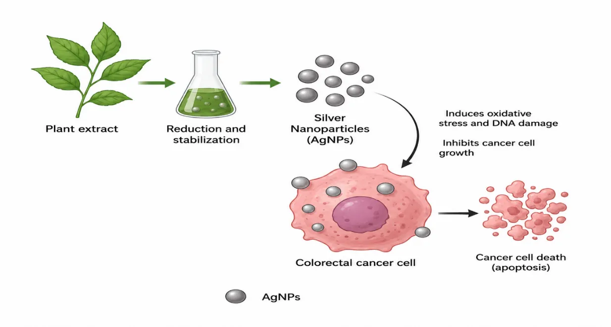
Plant mediated synthesis involves the contact of a silver precursor (usually silver nitrate) with an aqueous or organic plant extract. Reduction of Ag+ to metallic Ag0 initiates nucleation, growth and stabilisation. Phytochemicals can donate electrons, coordinate Ag atoms or adsorb on the surface of the growing particle. The process is usually accompanied by a colour change due to the surface plasmon resonance of nanoscale silver [1-4].The chemistry is more complicated than a simple one-step reduction. Various metabolites have different reducing power and binding affinity. Phenolic acids, flavonoids, proteins, sugars and terpenoid structures can participate in reduction or surface binding. Thus, the composition of extract influences nucleation rate, particle size distribution and surface chemistry. Recent reviews highlight how final characteristics can be modified by pH, temperature, precursor concentration, extract concentration and reaction time [2–4].
2.1 Phytochemicals role
Phenolic acids and flavonoids are often discussed as reducing molecules, since oxidation of their functional groups can facilitate electron transfer. Proteins and polysaccharides may assist in stabilisation by adsorption at the particle surface. Other metabolites, such as terpenoids, might also be involved, depending on the species and the extraction conditions. Crucially, phytochemicals associated with the surface may persist in biological activity post-synthesis, creating a hybrid material in which silver and botanical constituents contribute to an overall response [1,3].
This feature can be advantageous but creates a reproducibility problem. A whole-plant extract is not a chemically defined reagent. The abundance of a particular flavonoid or phenolic compound can change with cultivar, geography, season, plant age, harvesting conditions and extraction solvent. Two batches from the same nominal plant can therefore generate nanoparticles with different surface chemistry and biological behaviour.
2.2 Factor controlling nanoparticle formulation
pH influences phytochemical ionization and reduction capacity and can affect nucleation and aggregation. Temperature change’s reaction kinetics and may degrade heat-sensitive metabolites. Silver precursor concentration affects nucleation density and particle growth. The extract-to-precursor ratio controls the relative abundance of reducing and capping molecules. Reaction time can change particle growth and aggregation, while light can influence photochemical processes in some systems.
These variables should be treated as critical process parameters. A high-quality publication should report them sufficiently to permit replication [2,3].
2.3 Advantages and limitations
Plant‑mediated synthesis brings good points. It uses biological materials it lessens the need for dangerous synthetic chemicals it is easy to process, and it can add useful bioactive molecules to the nanoparticle corona [1–4]. Plant‑mediated synthesis also has some downsides. The extracts can vary the reaction mechanisms not fully known batches differ from one another it is hard to keep particle uniform, and we do not know which phytochemicals stay attached to the final product. Green synthesis in short is a sustainable way to make materials but it still needs the same strict quality control that the pharmaceutical industry uses.
3. PHYSICOCHEMICAL CHARACTERIZATION AND STRUCTURE–ACTIVITY RELATIONSHIPS:
To understand how plant‑mediated nanoparticles work good characterization is essential. Biological activity depends on size, shape, surface charge, how particles stick together crystal quality, how they dissolve and the biomolecules on their surface. Simply giving a spectrum and a rough size number is not enough for serious research [2,3].
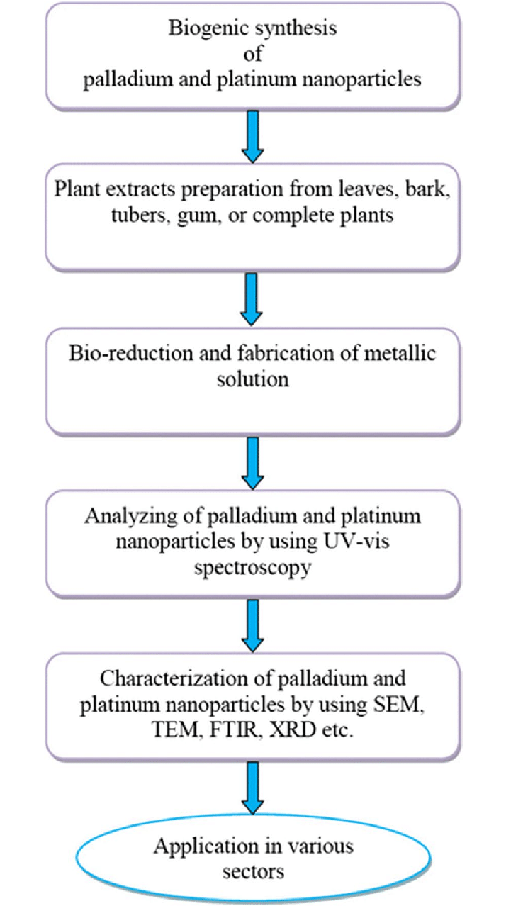
UV‑visible spectroscopy watches the surface plasmon resonance of nanoparticles. A shift or broadening of the peak can show a change in size spread or in particle clumping. Uv‑visible data alone does not reveal shape. Transmission electron microscopy (TEM) and scanning electron microscopy (SEM) give pictures of the particles. Dynamic light scattering (DLS) tells the diameter while the particles are, in liquid. Fourier‑transform infrared spectroscopy (FTIR) can spot groups that come from plant material. X‑ray diffraction (XRD) confirms the presence of metallic silver. Energy‑dispersive X‑ray spectroscopy (EDX) verifies the elements. Zeta potential measures the surface charge and helps judge whether the particles will stay dispersed.
3.1 Size, shape and surface chemistry
The size of nanoparticles plays a role in how cells take them in how deeply they reach into tissues how fast they are cleared from the body and how they interact with proteins. Shape also matters because it changes how nanoparticles interact with cell membranes and how they move inside cells. Surface chemistry is especially important. When nanoparticles enter fluids they get covered quickly by proteins and other molecules. This creates what is known as an identity, which can change how the body sees the nanoparticle. Green silver nanoparticles or AgNPs often come with a layer from plants. This plant-derived coating may change how the biological identity forms [2,13].
A wide review of plant-based AgNPs shows that the way they are made strongly affects their size and shape. This means researchers should make sure that the physical properties of the nanoparticles are linked directly to their effects [3]. There is also evidence that smaller nanoparticles can be more toxic to cells in some cases. But this is not always true results vary depending on the system being studied [16].
3.2 Dissolution and silver-ion contribution
The effects of nanoparticles on biological systems are not always due to the particles themselves. These particles can break down in environments through a process called oxidative dissolution. When this happens silver ions (Ag+) are released into the surrounding fluid. These ions can bind to proteins and enzymes that contain sulfur groups, which can disrupt cellular functions [13–15]. The balance between the effects of particles and the effects of released silver ions depends on several factors. These include the size of the nanoparticles the material used to coat them the composition of the surrounding fluid, the pH level and the specific biological conditions.
3.3 Minimum characterization package
To build an anticancer study researchers should provide a full description of the plant material used including how it was grown and authenticated. They should also report the method used to extract compounds from the plant. A detailed phytochemical fingerprint helps identify ingredients. Information about the precursor and the concentration of the plant extract is essential. The pH and temperature during synthesis must be recorded.
Key characterization tools include UV- spectroscopy to track nanoparticle formation and electron microscopy (TEM or SEM) to measure size and shape. Dynamic light scattering (DLS) gives size distribution data in solution. Zeta potential tells us about surface charge and stability. FTIR helps identify groups on the nanoparticle surface. XRD gives crystal structure details. Elemental analysis shows the chemical makeup. Colloidal stability over time is important to assess. If silver ions are released this should be. Reported.
Linking all these chemical properties to biological results—such as cell death or tumor shrinkage—builds a structure–activity framework. This provides insight than just describing how the nanoparticles were made.
4. COLON CANCER BIOLOGY RELEVANT TO NANOPARTICLE THERAPY:
Colorectal cancer (CRC) is made up of different types. Tumors can vary widely in how they grow how well they are supplied with blood the makeup of the surrounding tissue how many immune cells are present and the specific genetic changes they carry. Important pathways involved in CRC include Wnt/beta-catenin, MAPK, PI3K/AKT, TGF-beta, DNA damage repair and apoptosis. Mutations in genes like APC, KRAS, TP53 and SMAD4 are common. Their presence and frequency can differ between tumors.
Because of this diversity a nanoparticle that works on one type of colon cancer cell line cannot be expected to work the way on all CRC subtypes. For example, HCT116, HT-29, HCT15, SW480 and Caco-2 cells have genetic makeups different levels of cell maturity and different responses to drugs. Studies should therefore use than one cell line and include noncancerous intestinal cells as controls whenever possible.
4.1 Tumor microenvironment
The tumor microenvironment is a network of cells and materials. It includes fibroblasts, blood vessel cells, immune cells and a network of proteins and molecules outside the cells. These elements affect how nanoparticles move through tissue and how they are taken up by tumor cells. A dense extracellular matrix can block nanoparticle movement. Abnormal blood vessels can lead to delivery of nanoparticles. Macrophages, which are part of the system may swallow up nanoparticles before they ever reach the tumor.
This complexity makes cell culture methods—like flat monolayers—limited in their usefulness. Advanced models, such as three-dimensional spheroids organoids and co-culture systems give a better idea of what might happen in real tumors. These models can help predict how well a nanoparticle therapy will work before testing it in animals.
4.2 Limitations of therapy and rationale for nanotechnology
Standard chemotherapy treats cancer by targeting rapidly dividing cells. This includes healthy tissues like the lining of the gut and bone marrow. This leads to effects that limit how high a dose can be given. Over time cancer cells can also develop resistance making treatment less effective.
Nanoparticles offer a way to improve treatment. They can increase drug solubility protect drugs from breaking down quickly extend how long they stay in the bloodstream and help them collect in tumor tissue. Some systems can even release their drug payload in a controlled way.
For cancer researchers are focusing on colon-targeted delivery. The gut environment changes along its length— in pH and enzyme activity. Nanoparticles can be designed to respond to these differences. For example, some coatings dissolve at a pH others break down when they meet certain enzymes and others stick to mucus or react to bacteria in the gut. A 2024 review highlights that colon-targeted nanomedicine can combine site- delivery, with receptor-based targeting and smart release triggered by internal cues [11].
5. PRECLINICAL EVIDENCE OF AgNPs AGAINST COLORECTAL CANCER:
The current evidence is mainly preclinical. It is varied enough to find repeating biological patterns. A 2026 systematic review that focused on aqueous plant-extract synthesis found 19 studies involving more than 20 plant species and showed activity against HCT-116, HT-29 Caco-2 and SW480 models. Most particles were round and between about 3.9–76 nm while the strongest in-vitro effects were linked to some Annona muricata and Plumeria alba systems. The review also pointed out reactive oxygen species generation, mitochondrial issues, DNA cuts and cell death as processes and said particle size, surface charge and plant chemicals that
cover the surface were important factors [17]. These findings back the idea that plant-made AgNPs can be biologically active against CRC. The big differences in particle features and testing situations stop direct comparisons of the different forms.
5.1 Vitex negundo-derived AgNPs
Prabhu et al. Made AgNPs from Vitex negundo leaf extract. Tested them on HCT15 human colon cancer cells. The nanoparticles stopped growth with an IC50 of 20 μg/mL after 48 hours. The study did viability tests along with looking at the shape of the nucleus DNA cuts, a comet test and cell cycle analysis. AgNP exposure made the G0/G1 and G2/M groups bigger while making the S-phase group smaller meaning it affected DNA making and how the cell divides [5]. This study is important because it linked a drop in survival with clear reasons instead of just using MTT reduction as the only proof of cancer fighting action.
5.2 Ginger–turmeric-mediated AgNPs
Venkatadri et al. Made AgNPs using water extracts from the roots of Zingiber officinale and Curcuma longa. TEM showed particles of about 20–51 nm. Sem showed round particles of about 42–61 nm. The nanoparticles had a concentration-related effect on HT-29 cells with an IC50 of 150.8 μg/mL [6]. The study shows how combining plants can give reducing and covering molecules from plant chemicals. However, the high IC50 compared to some reports also shows why numbers for how strong they are should not be compared without thinking about particle features how long the test lasted, the cell type and how the cells were exposed.
5.3 Naringenin-mediated AgNPs and molecular testing
Gurunathan et al. Looked at naringenin-made AgNPs in HCT116 cells. Went further into testing the genes. The study used UV–XRD, FTIR, DLS and TEM to look at the particles and studied how cells responded at a genetic level [7]. The work connected AgNP exposure with stress from oxygen problems with the power plants inside the cell effects on DNA and changes in how genes work. The value of this study is in the way it was done looking at the genes can find pathways and possible markers that you can't find from checking if cells are alive.
5.4 Manilkara zapota and using it with other treatments
AgNPs made using Manilkara zapota leaf extract have also been looked at in HCT116 cells. The study said the AgNPs stopped HCT116 growth and caused changes in cell death and when used with cisplatin it caused stress from oxygen, less energy in the cell and signs of cell death [18]. These studies make it possible that green AgNPs could help other treatments work better or be used with them. However real help must be shown clearly. Not just because the mix is more effective than each part on its own.
5.5 Moringa oleifera and looking at the way genes work
AgNPs made from Moringa oleifera have been tested on HCT116, SW480 and HT-29 models. One study said the AgNPs had a dose-related effect on HCT116 and SW480 cells and changes in genes linked to Wnt/βcatenin signaling, such as CTNNB1, LRP5, LRP6 and APC [19]. Since Wnt signaling is important for colon cancer these results are interesting. Still changes in
how genes work needs to be checked at the protein and how they work level before a path can be seen to treat cancer.
5.6 What the proof really shows
Overall, the research supports four points. First plant-made AgNPs can stop the growth of colon cancer cell lines. Second the usual reactions are stress from oxygen, problems with the cells power plant, DNA damage changes in the cell cycle and cell death. Third the way the particles look and the chemicals from the plant on the surface seem to change how strong they are. Fourth proof that they work in life is still low because most studies are done in the lab. So "cancer fighting ability" is okay, for studies but saying they can treat cancer or are tested in real patients is too soon [17,20].
|
Biogenic source |
CRC model |
Reported particle/response |
|
Vitex negundo leaf |
HCT15 |
IC50 ≈20 μg/mL at 48 h; cell-cycle arrest, DNA damage and apoptosis |
|
Zingiber officinale + Curcuma longa |
HT-29 |
20–51 nm by TEM; IC50 150.8 μg/mL; concentration-dependent cytotoxicity |
|
Naringenin |
HCT116 |
Biogenic AgNPs; oxidative stress, mitochondrial dysfunction, DNA effects; transcriptomic analysis |
|
Ajuga bracteosa |
HCT-116, HT-29 |
~50 nm; dose-dependent reduction in viability |
|
Manilkara zapota |
HCT116 |
Selective growth suppression; apoptosis-associated effects; combination with cisplatin investigated |
|
Moringa oleifera |
HCT116, SW480, HT-29 |
Dose-dependent activity; changes reported in Wnt/β-catenin-related genes |
6. PHARMACODYNAMIC DETERMINANTS OF AgNP ACTIVITY:
The biological response to AgNPs is governed by a combination of dose, exposure duration, particle number, surface area, aggregation state, dissolution and intracellular localization. Reporting dose only as μg/mL of suspension can obscure differences between preparations. Two formulations containing the mass concentration may contain different numbers of particles and different available surface areas. Future studies should therefore report concentration together with size distribution and where feasible particle number or surface-area-related parameters.
6.1 Protein corona and biological identity
When nanoparticles enter fluids, proteins, lipids and other biomolecules adsorb onto their surfaces. This "protein corona" can alter colloidal stability, cellular recognition and uptake. Green AgNPs enter systems with a pre-existing phytochemical layer but that layer can be remodeled by serum proteins. Consequently, characterization in water is not sufficient to predict behavior in culture medium or plasma.
6.2 Cellular uptake and intracellular trafficking
Endocytosis is a route of nanoparticle internalization. Once internalized AgNPs may traffic through lysosomal compartments. Acidic intracellular environments and oxidative conditions can influence silver-ion release. Localization therefore matters when interpreting ROS and mitochondrial effects. Fluorescence or electron microscopy combined with organelle markers can provide evidence than indirect assumptions based on cytotoxicity.
6.3 Role of ions
The intact particle and dissolved Ag+ may act together. Ion release can increase protein binding and stress while the particle surface can interact directly with membranes and intracellular structures. A rigorous study should therefore compare AgNPs with an ionic-silver control and where possible evaluate dissolution kinetics. This approach helps particle-specific effects from silver chemistry.
7. MOLECULAR TARGETS AND SIGNALING NETWORKS IN CRC:
CRC involves pathways rather than a single therapeutic target. This is relevant to AgNPs because oxidative stress can influence multiple signaling cascades
7.1 Wnt/β-catenin signaling
Wnt/β-catenin signaling regulates stem-cell maintenance and epithelial proliferation. Aberrant activation, frequently associated with APC alterations is a feature of CRC. Green AgNP studies involving Moringa oleifera have reported changes in genes related to this pathway [19]. Future work should determine whether AgNP exposure produces suppression of β-catenin transcriptional activity or merely secondary changes caused by cellular stress.
7.2 PI3K/AKT and MAPK pathways
PI3K/AKT signaling supports cell survival and growth while MAPK pathways regulate proliferation and stress responses. AgNP-induced oxidative stress can modify kinase activity indirectly through redox- proteins. Because these pathways are also involved in normal-cell survival pathway inhibition alone does not guarantee cancer selectivity.
7.3 P53, BCL-2 family proteins and caspases
DNA damage and cellular stress can activate p53-associated responses. Shift the balance between pro- and anti-apoptotic BCL-2 family proteins. Mitochondrial outer membrane permeabilization can then lead to caspase activation. Demonstrating changes in p53, BAX, BCL-2, caspase-3 and PARP provides a mechanistic argument for apoptosis than morphological observation alone.
7.4 Autophagy and alternative cell-death pathways
Autophagy may function as a destructive response depending on context. AgNPs can alter lysosomal and oxidative pathways making autophagy relevant to treatment response. Future studies should distinguish flux from simple accumulation of autophagy-associated proteins. Other regulated cell-death mechanisms, including ferroptosis may also become relevant as research progresses. These should be demonstrated using pathway-specific assays rather, than inferred from ROS alone.
8. EXPERIMENTAL DESIGN FOR HIGH-QUALITY ANTICANCER STUDIES:
The methodological quality of AgNP research strongly influences how confidently findings can be interpreted.
8.1 Controls
At minimum experiments should include cells, vehicle controls, plant extract alone AgNPs and a relevant positive-control anticancer drug. Where feasible chemically synthesized or differently capped AgNPs can help determine whether the green surface chemistry contributes to activity. Ionic silver controls are useful for evaluating the role of Ag+.
8.2 Dose–response and time-course design
A single concentration is insufficient to establish behaviour. Multiple concentrations and exposure times should be used to generate concentration–response and time–response relationships. IC50 values should be accompanied by confidence intervals or suitable statistical estimates when possible. Exposure should be expressed consistently. Linked to particle characterization.
8.3 Orthogonal viability assays
MTT or related metabolic assays are screening tools but can be affected by nanoparticle optical properties, adsorption and interference with assay reagents. Confirmation using a method—such as live/dead imaging ATP-based viability membrane-integrity testing or clonogenic assays—reduces the risk of assay-specific artifacts.
8.4 Selectivity index
Cancer-cell cytotoxicity should be interpreted alongside normal-cell toxicity. A useful framework is to calculate a selectivity index based on the ratio of concentrations in normal versus malignant cells. The exact calculation should be reported clearly. A formulation that kills cancer cells efficiently but influences normal intestinal cells may have limited therapeutic value.
8.5 Statistical rigor and reproducibility
Experiments should include replicates, technical replicates where appropriate predefined statistical tests and transparent reporting of variability. Nanoparticle synthesis should be independently repeated across batches. Biological conclusions should ideally be reproduced with than one batch of nanoparticles.
9. COLON-SPECIFIC DELIVERY STRATEGIES:
Targeting the colon is not equivalent to targeting a tumor cell. An administered system must first survive gastric and small-intestinal conditions interact with mucus and microbiota and release its payload at an appropriate location.
9.1 Responsive systems
The pH changes along the gastrointestinal tract can be exploited using enteric or pH-responsive polymers. A suitable formulation can remain relatively protected in the stomach and release effectively in intestinal or colonic conditions. However, pH varies among individuals and disease states so pH responsiveness should be validated under realistic conditions.
9.2 Enzyme- and microbiota-responsive systems
Colon microbiotas produce enzymes of degrading selected polysaccharides and other substrates. This provides a route to colon- release. Green AgNPs could potentially be embedded within. Coated by biodegradable materials that respond to microbial enzymes. Such designs require assessment of whether the formulation alters microbiome composition or function.
9.3 Mucoadhesion and mucus penetration
Mucoadhesive systems can increase residence time at the surface whereas mucus-penetrating designs aim to move through the mucus layer toward epithelial cells. These strategies can have objectives. The desired behaviour should therefore be selected according to whether local luminal release or epithelial/tumor-cell interaction's the primary goal.
10. ACTIVE TARGETING OF COLORECTAL CANCER:
Active targeting uses ligands that recognize receptors or surface markers enriched in tumor tissue. A recent review specifically addressing AgNPs and CRC highlighted overexpressed receptors and the potential for multi-ligand functionalization [21].
10.1 Folate-related targeting
Folate receptors can be exploited in selected tumor models because ligand-mediated uptake may increase association. However, receptor expression must be confirmed in the chosen model and in the intended population.
10.2 Peptide and antibody targeting
Peptides can offer size and easier synthesis than full antibodies while antibodies can provide high-affinity recognition. Both approaches can increase formulation complexity and immunological considerations. Ligand density must be optimized because more ligand is not necessarily better.
10.3 multi-ligand targeting
CRC heterogeneity creates a rationale, for combining two recognition ligands. A multi-ligand nanoparticle may theoretically address receptor expression, but it also introduces additional variables. Such designs should be validated against ligand and non-targeted controls.
11. NANOPARTICLE ENABLED DRUG DELIVERY:
Silver nanoparticles can be used as carriers for chemotherapy medicines or active ingredients. Loading can happen through sticking to the surface attaching to the surface or being placed inside a polymer or mixed material.
11.1 Advantages of drug loading
Drug loading can possibly make things more stable help cells take in the drug and have an effect in the right place while exposing healthy parts less. If silver nanoparticles are harmful themselves the mixture might have two ways to work. However more activity is not always proof that the delivery's better.
11.2 Release timing
Release should be checked under conditions that're like the bodys pH and salt levels and for drinks under conditions that are like the stomach and intestines. Mathematical models can be used to explain how the drug comes out through diffusion dissolving or breaking down. The model should be picked based on the way it works of just the one that fits best.
11.3 Use together with known CRC drugs
5-fluorouracil, oxaliplatin and irinotecan are known parts of CRC treatment. A smart silver nanoparticle mix should be based on working and tested with formal combination-index or surface response methods. The goal should be to kill cancer cells with less drug in the body instead of just making the total harm more.
12. THERANOSTIC AND IMAGING USES:
The light properties of nanoparticles offer chances for sensing and making signals stronger. A theranostic system can mix a treatment part with a way to see or sense allowing to check where it goes and treat it all in one system.
12.1 Surface enhanced spectroscopic methods
Silver nanostructures can greatly improve fields near their surfaces supporting strong Raman spectroscopy and similar techniques. In CRC such systems could be made for finding markers. However, being able to see things in a controlled sample does not always mean it works well in body fluids.
12.2 Imaging guided treatment
Imaging can show where the nanoparticles are and where the tumor is. A strong study with imaging should link the image to how much's in the body and how the treatment works.
12.3 Theranostic design problems
Adding a way to see makes the mix more complicated needs steps to make and needs more safety checks. A system like this should have a purpose like finding where the tumor is or checking how the treatment is working.
13. NANOTOXICITY, DISTRIBUTION AND SAFETY:
Developing nanoparticles needs to look at both how well they work and how safe they are. A mix that is used times must be checked beyond just short-term cell death.
13.1 Blood compatibility
For mixes that go into the blood how they work with blood cells, platelets and blood proteins should be checked. Breaking blood cells making clots and activating parts of the system can affect if it works in real life.
13.2 Liver and kidney effects
The liver and spleen often take out nanoparticles while the kidneys may get rid of ones or dissolved silver. Measuring how much silver is in the tissue where possible is important. Examining the tissue and chemical signs should add to checking the elements.
13.3 Stomach safety
Mixes that go into the stomach and target the colon need checking for how they work with the inside of the stomach inflammation and how they mix with the bacteria. A mix that works in one place should be checked for how it builds up and goes away after doses.
13.4 Damage to DNA
Because DNA damage is a way cancer is treated healthy tissue damage from DNA needs attention. A treatment should harm cancer cells more than ones and not cause lasting damage to normal cells.
14. GAPS IN RESEARCH AND WHAT COMES NEXT:
The main gap in this area is the difference between making in the lab and using in life. There are already examples of making nanoparticles and their effect on cancer cells. Future work should instead focus on using plant parts checking how they work, better models, where they go and if they are safe.
14.1 Standard plant parts
Checking the mix of chemicals and specific parts could make things more consistent. Using plants that are known to be real and growing in controlled conditions can make things more the same.
14.2 Cancer models
3D groups cells taken from humans and mixing different types can give more real signs than flat layers. These models should be used with how the drug moves and where it goes in the body.
14.3 Artificial intelligence and smart medicine
Computer ways may one day link how to make the particles the plant parts and what they do with how they work in the body. These models will need clear data. AI should help not replace, tests.
14.4 Moving toward custom CRC treatment
The future of targeting nanoparticles may involve matching the parts that stick to the cancer and the drugs with signs of the cancer. This needs combining nanotechnology with how the cancer's known and tests that go with it. The 2025 review, about targeted colon cancer says this move is important but also says real tests are still needed [20].
|
Development stage |
Essential evidence |
Common weakness |
|
Synthesis |
Authentication, extraction conditions, reaction parameters |
Incomplete reporting of plant source and process variables |
|
Characterization |
TEM/SEM, DLS, zeta potential, FTIR, XRD, elemental analysis |
Only UV–visible spectra or nominal size reported |
|
In-vitro efficacy |
Dose-response, time-course, multiple CRC lines |
Single cell line and single viability assay |
|
Selectivity |
Normal intestinal controls and selectivity index |
Cancer-only cytotoxicity interpreted as selectivity |
|
Mechanism |
ROS, mitochondria, DNA, cell cycle, apoptosis with orthogonal validation |
Mechanisms inferred from one assay |
|
Targeting |
Ligand/receptor validation, uptake, competition, biodistribution |
Targeting claimed from increased cytotoxicity |
|
Translation |
PK, biodistribution, efficacy, repeated-dose toxicity |
Predominantly in-vitro evidence |
CONCLUSION
Plant-mediated silver nanoparticles offer a path at the intersection of green chemistry, phytochemistry and cancer nanomedicine. Their appeal comes from the idea that plant compounds can help form the nanoparticles while also providing an active surface layer. In cancer models green silver nanoparticles have consistently shown the ability to stop cancer cell growth. These effects are linked to oxidative stress, mitochondrial damage, DNA harm, cell-cycle blockage and programmed cell death.
Now the strongest evidence comes from preclinical studies. Research using extracts from plants like Vitex negundo, Zingiber officinale, Curcuma longa, naringenin and Moringa oleifera shows that green silver nanoparticles can affect colorectal cancer cell lines. However, the strength of the effects varies a lot between studies. This variation is not a problem to overlook. It is a key scientific question. Factors such as particle size, clumping, surface chemistry, release of silver ions the specific plant compounds used, and the types of models all influence the results.
For therapy to be truly targeted the next major step is to go beyond claims about cell death. A targeted silver nanoparticle must show that it can recognize specific receptors get inside cells focus on tumors have better pharmacokinetics and offer a higher safety-to-effect ratio. For systems meant to deliver treatment to the colon the nanoparticles must stay stable in the gut. Release their payload locally. For systems imaging data must match up with where the particles go in the body and how they respond to treatment.
Green synthesis should not be seen as the goal but as a tool that enables further progress. Its long-term value depends on whether scientists can turn plant extracts into reliable nanomedicines. These medicines must have a known chemical makeup, controlled physical and chemical traits, proven mechanisms of action predictable movement through the body and safe profiles. If these challenges are tackled in a way plant-mediated silver nanoparticles could move from being a growing area of preclinical research to becoming a more credible option, in targeted colorectal cancer nanomedicine.
REFERENCES

Hemnath R, Praveenkumar S, Rajalakshmi P, Dhanushya T, Akalya N,P Nirmala Plant-Mediated, Green Synthesized Silver Nanoparticles for Colon Cancer: From Phytochemical-Driven Nanofabrication to Target Thearpy and Theranostic Potential, Int. J. of Pharm. Sci., 2026, Vol 4, Issue 9, 2271-2286, https://doi.org/10.5281/zenodo.22847049
 10.5281/zenodo.22847049