
We use cookies to ensure our website works properly and to personalise your experience. Cookies policy
Department of Pharmaceutical Quality Assurance, Vidya Niketan Institute of Pharmacy and Research Center Bota, Tal- Sangamner, Dist- A.Nagar, Pin Code-422602.
Oral bioavailability enhancement of poorly water-soluble drugs remains one of the most persistent and pharmacoeconomically significant challenges in pharmaceutical development, with estimates indicating that up to 90% of new molecular entities belong to BCS Classes II and IV. Mesoporous silica nanoparticles have emerged as structurally distinctive inorganic carriers capable of stabilizing crystalline drugs in an amorphous state through nanopore confinement, substantially improving dissolution rate and apparent solubility. However, bare silica surfaces lack the biological responsiveness, mucoadhesive capacity, and stimulus-controlled drug gating required to fully exploit the pharmacokinetic potential of this platform in the gastrointestinal environment. Polysaccharide surface functionalization principally through chitosan, alginate, hyaluronic acid, and their derivatives addresses these limitations by introducing pH-responsive, enzyme-triggered, and redox-activated release mechanisms while simultaneously enabling reversible tight junction modulation and mucin-mediated adhesion at the intestinal epithelium. This review examines the structural diversity of mesoporous silica architectures relevant to oral delivery, the chemistry and stability consequences of three principal coating strategies, the sequential gastrointestinal barrier evasion mechanisms operative in polysaccharide-coated systems, in vivo pharmacokinetic evidence across BCS drug classes, integration into solid oral dosage forms, and the safety and regulatory considerations governing clinical translation. Collectively, the evidence positions polysaccharide-coated mesoporous silica nanoparticles as a modular, biologically responsive oral drug delivery platform whose translational realization depends on convergence between reproducible manufacturing, mechanistic pharmacokinetic characterization, and regulatory-aligned safety frameworks
Oral drug delivery is the most prescribed and preferred systemically delivered drug delivery form and the attrition rate of orally delivered drug candidates is still a challenge for pharmaceutical scientists in their drug discovery and development process to clinical stage [1]. The estimates are consistently that around 40% of all approved drugs and as high as 90% of new molecular entities in development are in BCS Classes II and IV respectively, which are the classes characterized as having low aqueous solubility with good permeability and the combination of poor aqueous solubility and poor membrane permeability [2]. The implication of this distribution in practice is that, for most modern drug candidates, dissolution in gastrointestinal fluid rather than the passage of the drug across the epithelium is the key rate-limiting step in the attainment of systemic exposure [3]. All conventional pharmaceutical approaches to solving this problem have given incremental improvements in dissolution rate, but each with its own limitation on drug loading capacity, physical stability during storage, manufacturing scalability, or lack of ability to provide controlled, site-specific release other than through promoting solubility [4].
In this context, the use of nanoparticles as drug delivery vehicles has gained significant and sustained scientific attention as a single delivery system that can overcome multiple solubility, permeation and controlled release limitations [5]. Mesoporous silica nanoparticles (MSNs) have proven to be a structurally unique and highly promising platform to deliver poorly soluble drugs orally due to their distinct functionality. The internal pore structure of these is generally ordered, and surface areas are routinely in the range of 700-1200 m²/g, allowing a drug to be loaded at concentrations that are significantly higher than those seen with soft organic carriers [6]. The ability of MSN mesopores to keep crystalline drugs in a molecularly dispersed amorphous state, which is induced by the spatial confinement resulting from the size (2–20 nm) of the channels, is critical for BCS Class II drugs, where the supersaturation at the absorption site directly determines the bioavailability [7]. One of the most cited proofs of concept for the oral MSN platform is a clinical study with 12 human subjects (IVC-01) that showed an ordered formulation of fenofibrate resulted in 54% greater oral bioavailability compared to the commercially available fenofibrate (Lipanthyl®) [8].
However, the gastrointestinal tract is a highly complex organ with structural features not fully leveraged by unmodified MSNs. Bare surfaces of silica carry a high density of negatively charged silanol groups that do not interact well with the mucus layer, lack an intrinsic mechanism of stimuli-dependent drug release and do not provide any mucoadhesion or targeted epithelial transport. The polysaccharide surface functionalization overcomes these shortcomings by introducing mucoadhesive properties, pH-sensitive swelling properties, and programmable drug-gating capabilities, which are absent in bare MSNs and are introduced by the application of biopolymers like chitosan, alginate, hyaluronic acid and pectin [9].
This review focuses on the structural rationale, functionalization chemistry, gastrointestinal barrier navigating mechanisms, stimuli-responsive release characteristics, in vivo pharmacokinetic implications, dosage form integration, and translational constraints of polysaccharide coated MSN systems as integrated oral drug delivery systems. The review is based on the large volume of literature published from 2018 to 2025 and will offer a critically evaluated, and mechanistically informed, view of the current position and potential avenues in which the platform may develop.
2. MESOPOROUS SILICA NANOPARTICLES: STRUCTURAL ARCHITECTURE AND PHYSICOCHEMICAL ADVANTAGES AS AN ORAL DRUG CARRIER
Perhaps the most underutilized pharmaceutical property of the MSN family is its crystallographic diversity. MCM-41 was first reported by the researchers at Mobil Corporation in 1992, and has a uniform pore diameter of 2–3 nm and a p6m two dimensional hexagonal arrangement of pores, thus having one of the highest surface area materials investigated in pharmaceutical sciences. Synthesized by the triblock copolymer Pluronic P123 as the structure-directing agent, SBA-15 has a remarkably similar two-dimensional hexagonal structure, but significantly larger pore diameters (6–8 nm) and pore volumes (1.0 cm³/g) parameters that directly relate to drug load capacity and, in comparative studies, the drug-to-silica loading ratio of up to 1:1 by weight for ibuprofen [10]. MCM-48 with its 3D bicontinuous cubic structure, provides interconnected pore channels that allow for better drug diffusion; hollow MSN (HMSN) are characterized by an ordered mesopore wall and a large central cavity, which can
significantly increase the total amount of drug encapsulated in the central space, especially designed for macromolecular drugs [11]. These MSNs have short diffusion path length, which is evident in their shorter dissolution time for the drugs in vitro, when compared to the conventional hexagonal MSNs with radially-arranged smaller-pore channels emanating from the particle surface. The reason for this is that the architecture of the MSN is not prescribed, but can be chosen with deliberate consideration of the pore geometry to match the physicochemical properties of the drug to be loaded, as summarized comparatively in Table 1
[12].
A very high density of silanol groups (Si–OH) is the common chemical feature of all MSN variants that creates the electrostatic, hydrogen-bonding and covalent modification handles, on which all subsequent functionalization is built, of the inner surface of their pores. The surface of silica can have three types of silanol groups: isolated, vicinal and geminal, each with a different reactivity towards the organosilane coupling agents applied in surface modification. The versatility of these MSN surfaces is the basic underlying reason that they can be hydrophilized, made negatively charged or amine-functionalized and/or polysaccharide-decorated with a relatively minor change in process conditions and without compromising the integrity of the pores [13].
It is important to point out that the drug amorphization behavior of MSNs in the nanopores is of special interest from pharmaceutical point of view. If the pore size is less than 10 nm, such as in the case of MSN, the MCM-41, the spatial confinement will hinder the nucleation processes and cannot allow the formation of a crystal lattice, obtaining a molecularly dispersed or amorphous state of the drug. The change from crystalline diffraction peaks and melting endotherms to amorphous features has been verified by both powder X-ray diffraction and differential scanning calorimetry in studies of ibuprofen, fenofibrate and carbamazepine loaded into MCM-41 and SBA-15 and stored under appropriate conditions for more than three months. Consequential to this amorphization is its bioavailability: an amorphous drug has a higher free energy state than the crystalline form, and thus an apparent higher level of solubility and dissolution rate in GI fluid. When the dissolution rate of ibuprofen loaded onto mesoporous carriers and that of bulk crystalline ibuprofen were compared at a pH of 5.5 (which is more representative of the proximal small intestine), the release of ibuprofen from the carriers was found to be 86–93% after 45 minutes, compared to 25% for the bulk crystalline ibuprofen [13].
Pharmaceutical applications of synthesis of MSNs are most frequently achieved by sol-gel processing using a structure-directing surfactant template, usually of either cetyltrimethylammonium bromide (CTAB) for the synthesis of MCM-41, or Pluronic P123 for the synthesis of SBA-15, which is followed by calcination or solvent extraction to remove the template and open the mesopore network. The silica source (typically tetraethyl orthosilicate, TEOS), surfactant-to-silica molar ratio, reaction pH and hydrothermal temperature all influence the downstream properties of the resultant pore diameter, particle size distribution and surface silanol density, which are all relevant to the loading of drugs and the efficiency of the polysaccharide coating and the colloidal properties of the particles in simulated gastrointestinal fluids. The synthesis parameter that is most directly relevant to the optimization of oral delivery (pore diameter control for amorphisation) as well as control of the particle size
(mucosal interaction) or surface silanol density (functionalisation yield) forms the backbone for the polysaccharide coating strategies [14].
Table 1. Comparative Structural and Functional Properties of Major MSN Types Used in Oral Drug Delivery [15–18]
|
MSN Type |
Pore Structure |
Pore Diamet er (nm) |
BET Surfa ce Area (m²/g) |
Pore Volu me (cm³/ g) |
Key Drug Loading Feature |
Reported Drug Examples |
Notable Oral Delivery Advantage |
|
MCM-41 |
2D hexagonal (p6m) |
2–3 |
900– 1157 |
0.7– 1.0 |
High surface area; excellent drug amorphizati on at <3 nm |
Ibuprofen, carbamazep ine, haloperidol |
Strong amorphizat ion effect; sustained release |
|
SBA-15 |
2D hexagonal (p6m) |
6–8 |
600– 800 |
>1.0 |
Highest pore volume; 1:1 w/w drug load demonstrate d |
Ibuprofen (170 mg/g), fenofibrate |
High loading capacity; faster diffusion in larger pores |
|
MCM-48 |
3D bicontinu ous cubic (Ia3d) |
3.0–3.7 |
800– 1000 |
0.8– 1.1 |
Interconnect ed channels reduce diffusion resistance |
Ibuprofen, cilostazol |
Rapid, uniform drug release; improved AUC vs MCM-41 |
|
Hollow MSN (HMS N) |
Large central cavity + mesopore shell |
Shell: 2–5 nm |
300– 700 |
0.6– 1.5 |
Large encapsulatio n volume; suitable for macromolec ules |
Anticancer drugs, proteins |
High total payload; protects labile drugs in GI |
|
Dendri tic MSN |
Radial large-pore channels |
10–30 |
400– 600 |
1.2– 2.0 |
Short diffusion paths; dendritic |
Paclitaxel, curcumin |
Rapid dissolution ; highly accessible |
3. POLYSACCHARIDE SURFACE FUNCTIONALIZATION STRATEGIES: CHEMISTRY, COATING ARCHITECTURES, AND MUCOADHESIVE OUTCOMES
The surface chemistry of an MSN is the starting point for this transformation, and the complexity of the polysaccharide coating strategy used will have a significant impact on the result of the system's subsequent handling in the gastrointestinal tract. In the literature, three main coating architectures have been reported, each having different implications regarding the stability of the coating, accessibility to pores, drug retention and evolution of the zeta potential in the physiological pH range [19]. The most basic and most widely used approach is electrostatic adsorption, in which the negative charge of the silanol groups on the surface of MSN mesoporous silica (zeta potential is generally in the range of −20 to −35 mV at neutral pH) is exploited to its full potential by the protonated amino groups of cationic polysaccharides (such as chitosan) whose pKa is in the range of 6.3–6.5, which are positively charged at mildly acidic to neutral pH. In a dilute acetic acid solution (0.1–1% v/v) the spontaneous adsorption of unmodified MSNs by a chitosan solution is controlled by ionic interactions between the Si−O⁻ groups on the silica surface and the protonated −NH₃⁺ groups in chitosan. This results in a change of zeta potential to positive values (generally between +15 and +25 mV) indicating that the surfaces have been covered. Electrostatic deposition is easy and does not need any special reagents, but it can be easily lost during the changing ionic strength and/or pH conditions in the gastrointestinal tract, so more powerful covalent coating methods have been developed [20].
The covalent functionalization is mostly carried out in two steps: (3-aminopropyl)triethoxysilane (APTES) is used to introduce primary amine functionality on the MSN surface by reacting with the silanol groups present on the surface; the amine functionalized surface is then coupled with carboxylated polysaccharide derivatives using 1-ethyl-3-(3-dimethylaminopropyl) carbodiimide (EDC) chemistry. This strategy has been used to achieve the functionalization of carboxylated MSN with chitosan–folic acid conjugates, in which APTES and succinic anhydride were sequentially grafted to the surface of MSN to provide carboxylic acid functional groups, subsequently followed by covalent attachment of chitosan–folic acid conjugates through EDC-amine bond formation [20]. The covalently attached chitosan layer preserved the charge of the particles at around +17 mV upon full conjugation and showed a significantly higher EE (89%) and LC (44%) when compared to the bare MSNs, proving the functional superiority of covalently bound chitosan over merely electrostatic binding [21]. An alternative covalent pathway is the formation of imine bonds by condensation between aldehyde-functionalized MSN surfaces and the amino groups of chitosan, without the use of EDC, and possessing stable, but hydrolysable, linkages, which can be programmed as a pH-responsive release trigger in and of itself [22]. The most architecturally controllable functionalization approach is provided by layer-by-layer assembly. In the LbL strategy, polyelectrolytes (e.g., negative HA or alginate and positive chitosan) are alternately layered onto the MSN surface, which is determined by measuring a step change in the zeta
potential. This generates a multi-layered polyelectrolyte shell that is characterized by its thickness, charge and permeability, which can be controlled via the number of deposition cycles. LbL systems made with chitosan and alginate bilayers have shown that as the number of bilayers increases the improvement of the drug retention increases, and that the permeability changes upon pH changes in simulated gastric and intestinal fluids, with the alginate component being collapsed at the acidic pH of the stomach and being expanded progressively in the alkaline environment of the small intestine [23].
The selection of polysaccharide is not random. The properties of chitosan that are unique and not found together in any naturally occurring polysaccharide employed in this manner are: Cationic nature, pKa response to pH, abundant primary amine groups that are useful for chemical modification, and proven tight junction modulating activity. Hyaluronic acid, on the other hand, carries carboxyl and acetamido groups that are responsible for the receptor-targeting and high mucoadhesiveness, respectively, which would make it more suitable for colon-targeted and/or tumor-targeted applications than for applications to the proximal intestine [24]. Alginate has the pH-responsive gelation (collapse at gastric pH and swelling at intestinal pH) which complements chitosan's behavior and is the reason why chitosan–alginate dual-coating and LbL designs are the most functionally complex oral PS-MSN systems reported so far. The two most important quality parameters for the final PSMSN construct will be the magnitude and sign of the zeta potential (above +20 mV or below −20 mV are generally deemed to be adequate for colloidal stability in pharmaceutical formulations) and the coating yield, determined by thermogravimetric analysis, which quantifies the mass fraction which can be attributed to the organic polysaccharide layer by the characteristic degradation event between 200 and 400°C, which is not masked by the Si–O–Si network characteristic of silica. The complementary confirmatory technique is FTIR spectroscopy [25].
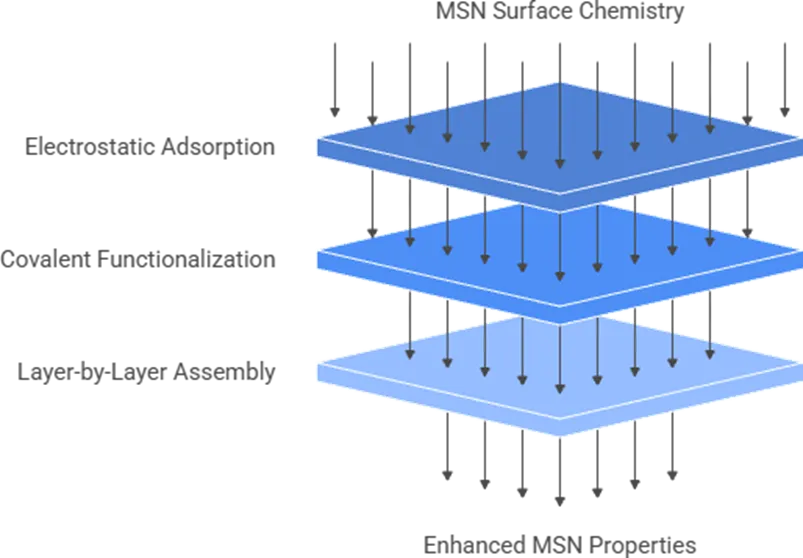
Figure: Polysaccharide Coating Strategies for MSNs
4. MECHANISMS OF GASTROINTESTINAL BARRIER EVASION BY POLYSACCHARIDE-COATED MSNs
Once taken orally, the gastrointestinal tract poses a series of successive barriers to the nanocarrier, and the efficacy of PS-MSNs in crossing these barriers is dependent on a judicious balance of their surface properties and the biology of each barrier segment. This knowledge is key to rationalize the consistent improvement in pharmacokinetic studies when comparing coated MSNs with uncoated MSNs at all these steps, as shown mechanistically in Figure 2. The first and most chemically hostile barrier is the gastric environment [26]. The structure of the encapsulated drug is protected thanks to the stability of the MSN silica framework at pH 1.2–2.0, where the dissolution of silica is limited compared to the physiological pH. A second layer of protection is provided by the polysaccharide coatings, which are protonated and water-soluble at gastric pH (pKa of 6.3-6.5), resulting in the formation of a compact surface layer of positively charged coatings around the MSN, which protects the MSN from the gastric juice and simultaneously reduce the release of the drug before reaching the target site by preventing the opening of MSN entrances, while performing electrostatic attraction with the surface of the MSN. The alginate coatings shrink to a compact gel at acidic pH, creating a second physical barrier to acid penetration into the pores. This acid response is not a simple "passive" protection, but is the first step in the sequential release design of numerous PS-MSN systems, which is based on stimuli-triggered release [26].
However, after the particle has crossed the pylorus and is in the small intestine, the mucus barrier is the predominant biophysical barrier. The thickness of the mucus gel, a hydrogel matrix consisting of mucin glycoproteins, varies from about 170 μm in the duodenum to >400 μm in the distal colon, and contains a complex network of pores and hydrophobic domains which can trap nanoparticles by steric, hydrophobic and electrostatic interactions. Chitosan coated MSNs do not just get stuck in the mucous layer, but rather have an active interaction with this layer: The positively charged surface of chitosan binds to the negatively charged sialic acid residues and sulfate groups on mucin glycoproteins, resulting in mucoadhesion and extending the residence time of particles on the intestinal epithelial surface [27]. Confocal and transmission electron microscopy studies have demonstrated that orally administered chitosan nanoparticles are able to attach to and penetrate through the mucus layer to penetrate into the mucus epithelial cells beneath, but that uncoated chitosan nanoparticles are more easily eliminated by the mucus turnover [28]. The tight junction (TJ) complex consists of a series of proteins located at the junction between adjacent enterocytes, such as the transmembrane proteins: claudin, occludin and junctional adhesion molecule (JAM-1) and the scaffolding proteins: ZO-1, ZO-2 and ZO-3, which attach the proteins to the cytoskeleton. The interaction of these TJ proteins with chitosan is a well documented and reversible positive surface charge interaction. Treatment with chitosan in Caco-2 monolayers has been shown to lead to a redistribution of JAM-1 from cell junctions to the cytosol, a decrease in the level of claudin-4 in the membrane and a reorganization of ZO-1 at cell borders, which all act to reduce transepithelial electrical resistance (TEER), and consequently, paracellular permeability in a dose- and time-dependent manner [29]. Critically, TJ opening effected by chitosan is transientand reversible: Removal of the chitosan stimulus restores TJ values to baseline, restores localization of the TJ proteins, and restores barrier integrity within hours, suggesting that this permeation-enhancing effect does not result in irreversible damage to the barrier. The TJ opening induced by chitosan is also affected by the molecular weight of chitosan, with a higher molecular weight promoting a greater and more sustained paracellular permeability increase, presumably because of the higher density of cationic amino groups and longer chain architecture of high molecular weight chitosan [30]Paracellular mechanisms are in parallel with transcellular transport pathways, such as clathrin-mediated endocytosis, macropinocytosis and caveolae-mediated uptake, to be involved in the entry of PS-MSN into enterocytes. Here, it is important to consider the size of the particles: PS-MSNs up to 200 nm are best internalized by enterocytes, whereas larger particles (300 nm and beyond) are retained more and more in the Peyer's patch M-cell compartment. The cationic surface charge of chitosan provides better interaction with the anionic apical membrane of enterocytes, increasing the chances of endocytic uptake compared to anionic and neutral particle surfaces, respectively. After internalisation, particles pass through the endolysosomal compartment to the basolateral space and finally into the systemic circulation either via the portal vein or (in the case of larger lipophilic payloads) via mesenteric lymphatic vessels. The relative contribution of each transport pathway in chitosan-MSN systems varies according to the nature of the drug and the particle size and is still under investigation, through in vitro and in situ closed loop intestinal perfusion models [31].
5. STIMULI-RESPONSIVE RELEASE MECHANISMS ENABLED BY POLYSACCHARIDE GATING
The key pharmacologic benefit of the polysaccharide coated MSN system over regular amorphous solid dispersion or nanosuspensions is its ability to release the drug on demand, depending on a specific stimulus, rather than release it in a passive manner, by way of the dissolution of the amorphous solid dispersion. When rationally engineered, the polysaccharide layer acts as a “responsive” gatekeeper, acts to manipulate the occupancy of the pore and drug egress directly in response to the physicochemical gradients experienced during gastrointestinal transit or target tissue microenvironment. The principle triggers being utilised currently in the PS-MSN literature - namely pH, enzymatic activity and redox potential, are directly linked to the well-established physiology of the gastrointestinal tract in humans, with their mechanisms clearly different from one another. The range of systems reported to date is summarized in comparative data in Table 2 [32]. The pH gradient of the gastrointestinal tract (from 1.2 in the stomach, and 6.0-7.0 in the small intestine to 7.0-7.5 in the colon) is the most naturally exploited trigger in the design of oral PS-MSN. Chitosan has a pKa between 6.3 and 6.5, protonates below that pKa (assuming a collapsed, compact conformation on the MSN surface that physically prevents the opening of pores to permit the drug to diffuse) and deprotonates above that pKa (where the polymer swells, chains separate, and the pore entrance opens to allow drug diffusion). Elegant demonstration of this mechanism was achieved in folate bioactive chitosan coated MSNs loaded with piceatannol, where only 30% of the drug was released under physiologic conditions (pH 7.4) after 24 hours, while 88% was released in only 2 hours under acidic conditions (pH 5.5) due to the collapse of the polymer. Proximal to distaldistribution of drug in the small intestine during gastric transit results in a progressive release of the drug that is retained in the pore network, while the distal small intestine provides a suitable environment for the release of payload through the polysaccharide gate, which may be opened by the increasing pH in the distal small intestine. Carboxymethyl chitosan derivatives with higher effective pKa values than the native chitosan can modify onset of release to slightly more alkaline environment in the intestinal lumen without altering MSN architecture itself, affording a means to adjust the release zone within the intestinal lumen [33].
The use of enzymes for triggering is a complementary approach and is often used in combination, especially for the delivery of drugs to the colon. Microflora in the colon produces a wide range of enzymes not found in the upper gastrointestinal tract (GI), or present in very low levels, such as azoreductases, glucosidases, pectinases, and polysaccharide lyases. This biochemical difference has been used to attach chitosan to the surface of hollow mesoporous silica nanostructures by cleavable azo linkages (–N=N–) that remain stable in the acidic and near neutral pH of the stomach and small intestine, but are broken down by bacterial azoreductases when they reach the colon. A loaded drug (doxorubicin) was encapsulated at 35.2% w/w in the hollow cavity of HMSS, and the drug release was found to be negligible in the simulated gastric and intestinal fluid whereas significant drug release was observed in the presence of a colonic enzyme mixture, with the cellular uptake of the loaded drug being significantly high after enzyme pre-incubation. This direct pharmacodynamic validation of the colon-specific release concept, as reflected by the cytotoxic IC50 value (three times lower after enzyme exposure than in the absence of enzyme) for colon cancer cells [34]. The most advanced release trigger architecture reported for PS-MSN oral and local delivery systems is the one based on redox-responsive systems consisted of chitosan derivatives covalently linked to the surface of the MSN, or between polysaccharide chains via disulfide (–S–S–) linkages. The principle makes use of the high intracellular glutathione (GSH) content in colonic cells, and in the tumour microenvironment (intracellular GSH is around 2-10 mM) compared to the extracellular content (intracellular GSH is around 2-20 μM), which is several hundred-fold. The extracellular redox conditions in gastrointestinal lumen keep the disulfide bonds intact while after endocytosis, the disulfide bonds are rapidly broken down in the target cell upon thiol-disulfide exchange reaction with the intracellular GSH, leading to the uncapping of the polysaccharide layer and burst drug release within the target cell. Redox/pH dual-responsive systems containing chitosan as one part and the disulfide-bridged organosilica frameworks as the other parts have been reported to give near-complete drug retention under physiological extracellular conditions and drug release under intracellular-mimetic conditions [35].
All three modes of release involved depend on the molecular weight and the degree of deacetylation of chitosan. High molecular weight chitosan also leads to a lower burst fraction and more linear release in intestinal pH conditions, since the higher molecular weight produces higher entanglement of the chains, leading to much better pore occlusion, while low molecular weight chitosan offers a more precise control over the pH transition window, owing to the lesser steric hindrance in the transition to the new conformation. The sharpness of the pH discriminating release behavior depends on the degree of deacetylation and increases with increasing protonatable amino groups (more deacetylated chitosan, degree of deacetylation > 85%) [36].
Table 2. Stimuli-Responsive Polysaccharide-Coated MSN Systems Reported for Oral Drug Delivery Trigger Mechanisms, Drug Classes, and In Vitro Release Performance
[37].
|
System |
MS N Typ e |
Polysacch aride |
Drug |
BC S Cl ass |
Stimulu s |
pH 1.2 Relea se |
pH 6.8– 7.4 Release |
Key Outcome |
|
CS-coated MCM-41 |
MC M-41 |
Chitosan |
Repagli nide |
II |
pH |
<20% |
>80% (pH 6.8, 6h) |
Enhanced dissolutio n vs. free drug; clinical antidiabet ic activity improve ment |
|
FA-CS coated MSN-COOH |
MC M-41 |
Folic acid–chitosan |
Piceata nnol |
IV |
pH |
Mini mal |
88% at pH 5.5 (2h); 30% at pH 7.4 |
3-fold solubility increase; pH-selective release |
|
HMSS–N=N–CS |
Holl ow MS N |
Chitosan via azo bond |
Doxoru bicin |
— |
Enzyme (azoredu ctase) |
<10% |
<15% (intestin al) |
Colon enzyme-triggered release; 3× lower IC50 |
|
CS/disulfid e MSN |
MS N (hyb rid) |
Chitosan–disulfide |
DOX + p53 gene |
— |
Redox (GSH) |
Negli gible |
Negligi ble extracel lular; rapid intracell ular |
GSH- triggered intracellu lar release; gene + drug co- delivery |
|
Alginate–chitosan LbL MSN |
SBA -15 |
Alginate/c hitosan bilayers |
BCS II drug |
II |
pH (dual layer) |
<15% |
Progres sive release (pH 7.4 intestin al zone) |
Sequentia l pH responsiv eness; improved sustained release |

Figure 2: Stimuli-Responsive Release Mechanisms of Polysaccharide-Coated MSN
6. DRUG LOADING STRATEGIES AND IN VITRO–IN VIVO CORRELATION IN PS-MSN SYSTEMS
The translational of structural and responsive properties of polysaccharide-coated MSNs to real systemic pharmacokinetic benefits starts well before the deposition of the coating: the
introduction strategy of the drug in the mesopore network. The type of loading will impact the physical state of the drug in the pore (crystalline vs amorphous vs molecularly dispersed), the strength of the drug/surface interaction, the amount of drug available for burst release, and the stability of the amorphous state on the product's shelf life [38].
Solvent impregnation is the most common loading method used for the development of PS-MSN. In this approach, the drug is dissolved in suitable solvent, usually methanol, ethanol or acetonitrile based on the polarity and the MSN powder is soaked in the solution and then the drug solution is drawn into the mesoporosity through the capillary action. Following a specified contact time, the solvent is evaporated under vacuum or reduced pressure and the drug is left inside the pore channels. The solvent system used during the impregnation process also has a marked effect on the physical state of the product after impregnation: studies of prednisolone have revealed that amorphisation by evaporation of the solvent can cause the formation of a different crystal polymorph in the pores, whereas amorphisation by melt loading does not induce any polymorphic change: this highlights the importance of PXRD and DSC analysis as a routine check of the polymorphic state of the product after each type of loading. The loading by rotary evaporation (a scaled down version of solvent impregnation under reduced pressure) is a more controllable solvent removal rate process and has been used to load BCS Class II drugs of high logP values. Melt adsorption involves the melting of the drug above its melting point in the presence of MSN powder followed by resolidification, thus avoiding any exposure to the solvent and is especially suited for the thermostable drugs where the residual solvent toxicity is of primary concern, although the method cannot be applied to the thermolabile drugs.
A potential risk associated with post-loading of the polysaccharide coating is the risk that some adsorbed drug will stay on the external MSN surface after loading, which will be released as a burst fraction and not the desired gating mechanism. By dissolving the PS-MSN in strong acid and measuring overall drug content, and subtracting the amount of drug that was released in simulated gastric fluid prior to coating deposition, an encapsulation efficiency measurement becomes a practical way of quantifying this unentrapped fraction. Chitosan coated MSN systems have been reported to be able to load 15-45% w/w of BCS Class II and IV drugs with 70-92% of drug-encapsulation in the system, depending upon the polarity of the drugs, pore occupancy and the extent of the chitosan coating [38].
Pharmacokinetic output of PS-MSN formulations from reported in vivo studies shows a consistent (but not universal) trend of greater AUC compared with free drug suspensions. The chitosan-coated repaglinide nanoemulsion system showed a pharmacokinetic advantage of 3.51-fold in AUC₀₋₁₂h and 1.78-fold in Cmax over free repaglinide in rats, which was found to be mainly due to the chitosan coating based on comparative analysis with uncoated nanoemulsion controls. The relative oral bioavailability for simvastatin chitosan nanoparticles were about 154 to 209% higher than the pure drug suspension in rabbits, and resveratrol loaded carboxymethyl chitosan nanoparticles had the highest oral bioavailability of 3.5-fold than free drug suspension in SD rats. The values are summarized in cross-system context in Table 3 [39].
However, a consistent and significantly compelling point throughout the PS-MSN pharmacokinetic literature is that enhancements in dissolution performance in vitro are often not accompanied by proportionately increased bioavailability in vivo. In vivo studies ofmultiple PS-MSN systems with near-complete drug release in USP II studies at intestinal pH within 30–60 minutes yield only 1.5- to 3-fold improvements in AUC as opposed to the 5- to 10-fold improvements in USP II studies. A number of mechanistic explanations have been suggested to explain this difference: recrystallization of the drug from the supersaturated intestinal fluid prior to complete absorption; competitive adsorption of biliary lipids and dietary surfactants to the hydrophilic surface of MSN, limiting drug dissolution during intestinal transit regardless of its efficiency; swelling of polysaccharide coating at intestinal pH that causes pores to become smaller than predicted, effectively reducing the dissolution rate of the drug as it passes into the circulation; and hepatic first-pass metabolism that removes the drug from the portal circulation before quantification in the systemic circulation, regardless of the efficiency of its absorption. In addressing these mechanisms, in vitro dissolution may be required, and IVIVC modeling using the Wagner-Nelson or two-compartment deconvolution method would greatly assist the field, with more systematic PK studies being conducted in large animal models or in humans, where the intestinal pH, bile salt composition, and/or transit times are more closely representative of clinical conditions [39].
Table 3. In Vivo Oral Pharmacokinetic Performance of Polysaccharide-Coated Nanoparticle Formulations Across Drug Classes [40–42]
|
Drug |
BC S Cl ass |
Polysaccharide System |
Ani mal Mod el |
Dos e |
AUC Improve ment vs. Free Drug |
Cma x Cha nge |
Relative Bioavail ability (%) |
Notable Remark |
|
Repaglin ide |
II |
Chitosan-coated nanoemulsion |
Rat |
— |
3.51-fold (AUC₀₋₁₂ h) |
+1.7 8- fold |
Higher vs. uncoated control |
Chitosan coating outperfo rmed uncoated formulat ion |
|
Simvasta tin |
II |
Chitosan SLN (1%) |
Rab bit |
— |
~13-fold vs. suspensi on |
— |
154– 209% vs. free drug |
Chitosan attribute d to P-gp inhibitio n and mucoadh esion |
|
Resveratr ol |
II |
Carboxymethyl chitosan NPs |
SD rats |
25 mg/ kg |
3.5-fold |
— |
3.5× vs. free RVT |
Sustaine d release; GI acid stability |
7. INTEGRATION INTO ADVANCED ORAL DOSAGE PLATFORMS AND SCALE-UP CONSIDERATIONS
The potential for a successful PS-MSN nanosystem in colloidal suspension and its eventual development into a commercial solid oral dosage form lies in a significant gap where formulation science is not necessarily the same, and may instead be just as challenging as engineering [43]. The ability of PS-MSN nanosuspensions to form stable, flowable, compressible solid intermediates while maintaining the particle size distribution, integrity of the polysaccharide coat, and amorphization of the drug free from thermal, mechanical, and drying stresses during conventional pharmaceutical manufacture is not assured. Spray drying is the most frequently used solidification process for MSN-based nanosuspensions, andcontinues to be the method of choice for transforming PS-MSN dispersions to free-flowing powder that can be filled into capsules or directly compressed. The critical process parameters that are responsible for the success of spray drying inlet temperature, feed rate, atomization air flow and aqueous feed solid content should be optimized to ensure that the moisture is removed quickly enough to maintain the amorphous state of the drug, but not so quickly that thermal exposure denatures the polysaccharide coating or causes drug recrystallization. The formulation strategy that is most consistent for the successful spray drying of nanoparticulate formulations is to co-spray dry with a water soluble carrier matrix (usually mannitol, lactose or HPMC) in a mass ratio that is optimized to ensure sufficient interparticle separation during droplet drying to prevent irreversible nanoparticle aggregation. Both the insulin-loaded chitosan nanoparticles spray-dried with and without mannitol cryoprotectant showed that the nanoparticles remained at 376 nm after reconstitution and entrapment efficiency was 98.7%, and the results showed that the choice of cryoprotectant had a great effect on the redispersibility of the formulation after spray drying. On a gram to kilogram scale, synthesis of MSNs in continuous flow and/or microfluidic reactors, monitored in-line with DLS, has been shown on MSNs with various drug-loads and of different particle types, but standardised protocols have not yet been established across these ranges [44].
For multiparticulate systems or tablets, an alternative route of consolidation is fluid bed granulation, which is an attractive method, especially in cases where the drug product undergoes some additional processing steps before it is formulated. In this method, the PS-MSN nanosuspension is sprayed as a granulating liquid on a soluble carrier (lactose, mannitol or sucrose) and the deposited nanoparticles are integrated into the granules as the moisture is evaporated by the fluidizing air flow. The results from the ibuprofen nanosuspensions stabilized with HPMC (granulated by fluid bed) showed that HPMC gave better redispersibility than PVP/VA as a stabilizer polymer and that a higher dissolution rate of the carrier material resulted in better dispersal of nanoparticles at the granule surface, which was ascribed to differences in thickness and continuity of the nanoparticle loaded layer around individual granule particles. The results are directly relevant to the granulation of PS-MSN formulations, because of the added complexity of the polysaccharide coating; when using a granulating solution containing competing cations, the ionic strength of this solution could affect the electrostatic binding of chitosan, and pre-formulation compatibility studies may be necessary to assess this effect [45].
The use of quality by design (QbD) approaches for the formulation development of PS-MSN has been growing and critical material attributes (MSN pore diameter, polysaccharide molecular weight, degree of deacetylation, drug loading) were correlated to critical quality attributes of the final dosage form (dissolution rate at pH 6.8, particle size after redispersion, hardness and friability). The use of a design of experiments methodology for this multi-variable space allows the reduction of the experimental load and the detection of interaction effects not detected by a univariate optimisation. The manufacturing process of PS-MSN nanosuspensions to granulation and compression into tablet has several potential failure points with loss of nanoparticles' integrity (as shown in Figure 3), which requires careful formulation and process design to protect the integrity of the nanoparticles. Drug crystals have been shown to be poorly dispersible, and the polysaccharide coating of PS-MSNs is ideal for improving this dispersibility and providing advantages in dispersibility and mucosal contact in patient-centricdosage forms, including mini-tablet arrays for flexible dosing of pediatric patients and orally disintegrating tablet formats which bypass the swallowing component for geriatric populations. These applications are still at a research stage but the technological paths going from liquid crystal gels, systems which form in place and gastroretentive platforms offer a way to bring these technologies to the clinic [46].
8. BIOCOMPATIBILITY, SAFETY PROFILE, AND REGULATORY LANDSCAPE
Regardless of the type of nanoparticulate drug delivery systems (NDDS), the depth and quality of the characterization of their safety is ultimately the driving factor that determines whether a system can transition from laboratory scale research to clinical application. There are a number of precedents that provide a safety case for MSNs. The FDA has classified silica as Generally Recognised as Safe (GRAS), and it has been approved as a pharmaceutical glidant (colloidal silicon dioxide, PhEur) and as a food additive (E551 in the European Union). MSNs undergo hydrolysis in physiological conditions with production of orthosilicic acid (Si(OH)₄) as the main degradation product, a naturally occurring biologically compatible small molecule which is absorbed and excreted through renal and hepatobiliary pathways, similar to the metabolism of dietary silica [47]. MSNs are bio-degradable and bio-cleared under most of the tested conditions and in vivo studies have confirmed that they are non-toxic at oral doses below 200mg/kg in rodent models, without any tissue accumulation. The rate and extent of MSN biodegradation, which is directly relevant to chronic safety, depends on particle size, surface chemistry and pore wall thickness. Smaller particles (<100 nm) degrade faster than larger ones and functionalization with organic groups (e.g., APTES) adds hydrolytically stable Si–C bonds, thereby increasing the degradation half-life when compared with unfunctionalized silica surfaces. The effect of the extra organic layer on degradation kinetics is dependent on the polysaccharide type and the degree of crosslinking for PS-MSNs coated with polysaccharide. As a natural biopolymer, chitosan is broken down by chitinase and lysozyme, which are normally found in the gastrointestinal tract, and its degradation rate depends on its degree of deacetylation; the higher the degree of deacetylation, the slower the degradation rate since it is less susceptible to lysozyme [48]. This has been found to be a possible reason for the long time the chitosan-MSN systems spend in contact with the intestinal epithelium, which has a pharmacological benefit, but is offset by an undesirable side effect: the system stays in contact with the intestinal epithelium surface for a long time. Well-functionalized MSNs are hemocompatible based on in vitro ISO 10993 data with negligible hemolysis (<5% at therapeutic doses) and without significant changes in the morphology of erythrocytes. In comparative studies, hybrid MSNs with polymeric or polysaccharide shell have been shown to have better hemocompatibility than bare silica. However, it is important to note that the cationic surface charge of chitosan-coated MSNs is responsible for platelet activation and pro-coagulant effects in systemic delivery applications. This risk is substantially reduced for any oral administration but is important for any application of PS-MSN that extends beyond the oral route, as in the case of transcytosis into the systemic circulation for the case of PS-MSN particles. The regulatory framework of MSN-based drug products mirrors the complexity of the classification issue of hybrid organic–inorganic nanosystems in the existing pharmaceutical regulatory framework [49]. There is no clear guidance for composite particles that contain inorganic cores of silica, and an organic coating loaded with drug molecules in either the FDA
guidance for conventional drug products or the FDA guidance documents on nanotechnology. The main difficulty comes from the lack of consensus on a protocol that connects all the measurable physicochemical parameters (BET surface area, zeta potential, yield of polysaccharide coating, free pore volume after loading) to in vivo safety and efficacy endpoints. Satisfaction of the FDA's expectation that performance is predictable from physicochemical characteristics, as stated in various guidance documents regarding nanotechnology in pharmaceutics, is not currently met at the population level for MSN systems: what works for one pore size, surface modification, and drug combination is not necessarily transferable to another system even within the same particle family. This will necessitate collaboration among regulatory agencies, standards groups and the academic research community, as a consensus, inorganic-organic hybrid nanocarrier characterization and safety testing methods must be developed [50].
9. LIMITATIONS AND FUTURE PERSPECTIVES
While the scientific rationale of polysaccharide coated mesoporous silica nanoparticles as orally delivered drug delivery system is very strong and is still gaining momentum, an honest assessment of the field requires that attention is equally paid to the challenges that lie between promising pre-clinical results and products that are available for patients to use. There are some problems that are not hypothetical. The temperature, stirring speed and water activity of the sol-gel condensation reaction are still sensitive parameters that require careful control of the process at GMP compatible volumes and are not yet reproducible at batch scale. The polysaccharide coating step adds another reproducibility problem since the deposition efficiency of electrostatic or covalent attachment depends on the molecular weight distribution and degree of deacetylation of the chitosan batch, the ionic strength of the coating medium, and the surface amount of the silanol groups on the MSN surface, which vary between the different production lots, and/or suppliers. The in vivo pharmacokinetic data, although generally favorable in direction, are almost exclusively derived from rodent studies with single doses in fasted state which do not reflect the large inter-individual variability of pH, gastric transit time, mucus viscosity or bile salt composition commonly found in the human gastrointestinal tract. There are few in vivo long-term toxicity studies specifically for polysaccharide-coated (non-PEGylated) MSN systems, rather of bare or PEGylated MSNs [51]. The best future developments, scientifically, are those in which the most mechanistic opportunities exist. The two types of dual-stimuli responsive coatings, pH and redox sensitive coatings based on chitosan-disulfide conjugate architectures as well as pH and enzyme sensitive coatings by using hyaluronic acid as a second layer, enzyme (hyaluronidase) sensitive coatings are a rational step forward from the single-trigger systems that are currently the most predominant in the literature. These designs would give better and more selective drug release profiles in association to certain anatomical compartments in the GI tract. Machine learning and computational modeling of PS-MSN design space strong drug binding, mechanistic understanding of structure-performance relationships, and optimal pore diameter, polysaccharide molecular weight, coating layer thickness and drug loading provide a way to significantly shorten the formulation development timeline [52]. Alternatively, green synthesis routes using plant-derived polysaccharides as structure-directing agents and coating materials are gaining in popularity as an alternative to the chemical functionalized CTAB-based synthesis
as it is more sustainable. First reports indicate that a relatively good pore architecture and surface chemistry are achievable by these routes. Last, but most importantly, the oral delivery of biological macromolecules, such as insulin, glucagon-like peptide-1 analogs, monoclonal antibody fragments and siRNA, is an unmet pharmaceutical need of great clinical importance, and a platform based on PS-MSN with appropriate protective polysaccharide shells that resist enzymatic degradation but still allow the molecules to pass through the epithelial barrier is scientifically sound. As a whole, this review highlights the status of polysaccharide-coated MSNs as uniquely capable oral drug delivery platform, which spans from the atomic scale of pore confinement to the macroscopic scale of solid oral dosage form performance with the structure precisely controlled and biological response rationally engineered. The key challenge for the field is the ability to merge reproducible manufacture with a meaningful mechanistically-based relationship between in vitro and in vivo activity into development programs with the ability to withstand the rigorous tests of clinical evaluation rather than the further proof of concept [53].
CONCLUSION
Polysaccharide-coated mesoporous silica nanoparticles are a truly convergent system in which the structural precision of inorganic nanomaterial science and the biologically evolved properties of natural carbohydrate polymers feed into one another and when combined yield a system that is better than each individual component. In conclusion, the body of evidence presented here is clear that this convergence is not merely additive; indeed, chitosan and related polysaccharides do not merely protect a dissolving silica particle from the gastric acid, but rather transform the relationship of the nanoparticle to the gastrointestinal biological environment by regulating mucin adhesion, gating drug release at sub-organ level with anatomical accuracy, and transiently reconfiguring the epithelial tight junction complex in a reversible and physiologically acceptable way. These are soft skills that make polysaccharide-coated mesoporous silica superior to standard formulation methods and to mesoporous silica or polysaccharide alone. What the body of work to date has lacked — and is likely a major scientific need for the next generation of research — is a mechanistic connection between in vitro structural performance and in vivo pharmacokinetic response. However, the fact that a significant increase in dissolution in simulated fluids was only modestly reflected by an increase in systemic exposure in animal models does not reflect a failure of the platform, but rather a lack of understanding of the intestinal lumen dissolution kinetics of the drugs, as well as the coating behavior of the polysaccharide agents in the presence of physiological bile salts, and the quantitative relative importance of transcytosis versus paracellular transport in intact intestinal segments. The answer to these questions will be achieved by rigorously designed in situ perfusion studies and species appropriate IVIVC modeling that will decide if polysaccharide-coated mesoporous silica nanoparticles are a scientifically compelling but clinically unrealized technology, or if they can realize their demonstrated structural potential as programmable oral drug delivery platforms that address the bioavailability deficits of the next generation of poorly soluble and biologically sensitive therapeutics.
REFERENCES


Rounak Chalak, Kiran Shinde, Polysaccharide-Coated Mesoporous Silica Nanoparticles: Engineering Smarter Oral Drug Delivery Systems Through Surface Functionalization and Stimuli-Responsive Release, Int. J. of Pharm. Sci., 2026, Vol 4, Issue 7, 240-260, https://doi.org/10.5281/zenodo.21131623
 10.5281/zenodo.21131623