
View Article

  • Quantum Dots For Imaging and Drug Delivery in Cancer Therapy

  • Department of Pharmaceutical Chemistry, Matoshri College of Pharmacy, Eklahre, Nashik.

Abstract

One of the main causes of mortality globally is cancer. It is hard to diagnose and treat cancer because every case can be different. In contrast to earlier diagnostic methods, improvements in nanomedicine have enabled tumor identification and rapid cellular-level analysis of tumor cells. Quantum dots (QDs), sometimes referred to as nanoscale semiconductor crystals, are nanoparticles with distinct optical and electrical characteristics, such as intense and brilliant fluorescence. QDs, with their customizable optical characteristics, have attracted a lot of attention because the majority of traditional organic label colors lack the potential for near-infrared (>650 nm) emission. Due to their remarkable optical characteristics and quantum confinement, these nanoscale particles have become indispensable tools in bioimaging and cancer drug delivery. The same light wavelength can excite a variety of QD types, and their narrow emission bands can be detected simultaneously for many assays. QD excretion, along with photography, has demonstrated amazing applicability in bioimaging, novel medication development, targeted gene delivery, biosensing, photodynamic therapy, and diagnosis. In addition to its value in drug delivery in cancer treatment, the current review sought to highlight the relevance of QD in imaging

Keywords

Cancer, Quantum Dots, Imaging, Drug Delivery.

Introduction

× Popup Image

A collection of illnesses known as cancer is distinguished by the quick proliferation of aberrant cells throughout the body. The majority of cancer cases are caused by mutations or alterations in the expression of tumor suppressor genes, DNA repair genes, and proto-oncogenes. In addition to signs of an unhealthy lifestyle (tobacco use, bad diet), genetic (hormones, mutations, immune conditions) and environmental (radiation, chemicals, pollutants) variables are responsible for the majority of cancers. In addition, aging raises the risk of developing cancer considerably [2]. The use of tumor-specific antibody conjugates for active tumor targeting in novel anticancer therapies has attracted a lot of attention in the fields of oncology, pharmacology, and nanomedicine. This method will enable to enhance therapeutic effectiveness and lower systemic toxicity [3]. Nanotechnology has revolutionized the pharmaceutical and medical sectors with its revolutionary potential, paving the way for early diagnosis and medication delivery. Quantum dots (QDs), nanoscale zero-dimensional crystals that have a wide range of optical, spectral, magnetic, and electrochemical characteristics, are at the forefront of this nanotechnological revolution. QDs have received a lot of attention among the numerous nanomaterials as a crucial instrument in bioimaging and cancer drug delivery. [4] Ekimov and Onushenko first described quantum dots (QDs), sometimes referred to as nanoscale semiconductor crystals, in a glass matrix in 1981, and the first application for biological imaging was published in 1998. The field of QDs has been steadily expanding ever since, with applications in areas such as photodetectors, biomedical imaging, computing, light-emitting diode (LED) manufacturing, photovoltaic devices, solar cells, and more. [5] QDs have exceptional optical characteristics that make them promising candidates for use as luminescent nano-probes and carriers in biological applications. Drug QD nano-carriers can be loaded with drugs by dissolving, dispersing, adsorbing, and coupling, among other methods. The carriers next alter the physical and chemical properties of drugs (such as saturation solubility, dissolution rate, crystal form, particle surface hydrophobicity and hydrophilicity), as well as their biological behavior and physical response, thereby affecting how drugs are absorbed, distributed, metabolized, and excreted [6].

Structure of QDs:

Quantum dots are composed of tiny metal particles that are around a thousand times smaller than a hair. These particles are shaped into different shapes and covered in various biomaterials. Quantum dots fluoresce in UV light, and their size determines the shade they produce. For example, QD (2 nm) had red fluorescence, whereas QD (2 nm) fluoresced clearly in green. The majority of fluorescent quantum dots are made of chemicals ranging from group II to VI and III to V, such as Cd, Hg, Se, Ag, Ln, P, Pb, Te, and Zn. The frequency that quantum dots produce becomes more constrained as their size decreases. They have a defined discharge frequency, so their spectra do not show a variety of fluorescence radiations [7]. The structure of quantum dots includes a core, shell, and sometimes a surface coating, which gives them a high degree of stability in photo and chemical behaviors, surface activation, and photo luminescence quantum yield [2].
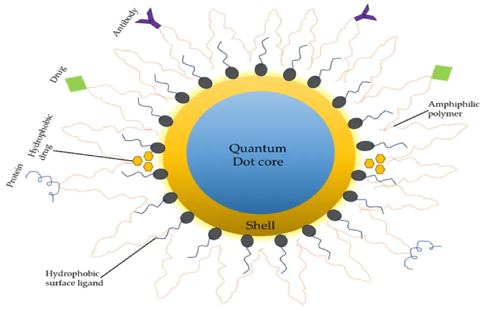
Fig.1: Basic Structure of QDs With Shell, Core & Ligand.
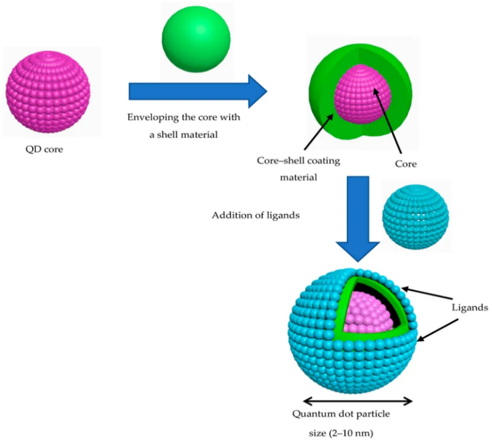
Fig.2: Formation of Quantum Dot Particle.

Synthesis of QDs:
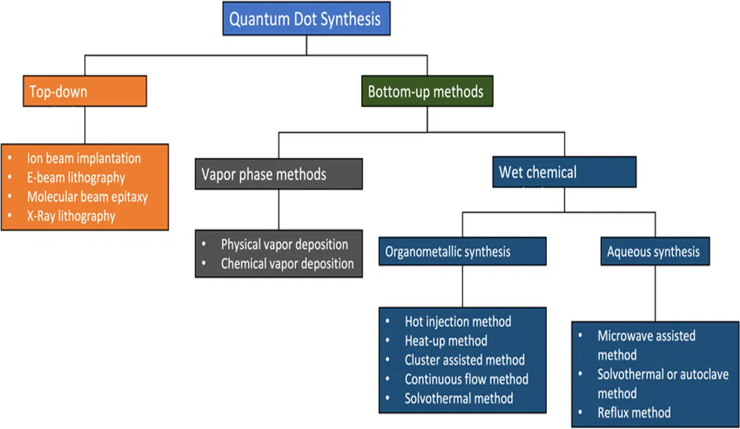
Fig.3: Quantum Dots Synthesis Flow Chart.

QDs Synthesis:

1. Top-down approach

The top-down method involves creating QDs by ablating bulk semiconductor materials. This covers techniques like reactive ion etching, electron beam lithography, and focused ion beam. These procedures create QDs with a diameter of about 30 nm. But these methods have drawbacks, such as the incorporation of pollutants during synthesis [2].

2. Bottom-Up Methods for GQDs Synthesis

Using advanced bottom-up methods that begin with tiny aromatic precursors or other carbon sources, which are also produced by the breakdown of bulk materials, GQDs with regulated morphology and well-distributed sizes have been synthesized.

3. Other Synthesis

Hydrothermal synthesis can also be used to create QDs. This is a method of single-pot synthesis whereby inorganic salts are crystallized from aqueous solution by controlling temperature and pressure. Due to the pressure confinement inside the autoclave, the temperature may be increased to very high levels in this method. This causes partial chemical breakdown and encourages molecular interactions, which leads to the synthesis of QD [2].

The usage of quantum dots (QDs) in the treatment of cancer.

The use of nanotechnology for cancer therapy, known as Nano-Oncology, has seen significant development during the previous ten years. The engineering of nanomaterials for cancer therapy and diagnosis has demonstrated the possibility of transforming the cancer treatment by creating novel theranostic nanoparticles for simultaneous imaging and therapy [13]. QDs can be used in a variety of cancer therapy-related studies, such as identifying molecular targets, sentinel lymph node mapping, drug delivery and tracking, surgical oncology, fluorescence resonance energy transfer and photodynamic therapy, personalized and predictive medicine, and multifunctional design and development [9].

*Imagining:

In identifying the right cancer treatment, imaging is a crucial clinical method [15]. Molecular imaging of cancer using nanotechnology is a potential platform. Due to their distinct optical and electronic properties, quantum dots (QDs) are being extensively investigated as a novel probe for biomedical imaging, both in vitro and in vivo. The physicochemical properties of QDs, including their size, shape, composition, and surface characteristics, have been thoroughly studied in an effort to address the challenges that QDs present for biomedical imaging [12]. Within a photodynamic method for therapy, CNTs have shown a significant potential for imaging in both in vivo (utilizing mice) and in vitro [14]. QDs, a novel type of inorganic nano-fluorescent probe, have demonstrated remarkable benefits in multi-color fluorescence imaging and detection over extended periods of time. The advancement of QD labeling facilitates the study of nano-drugs at the cellular and live animal level. The creation of therapeutic multifunctional nanomedicines and fluorescence imaging technology of quantum dots is anticipated to be used in the diagnosis and treatment of cancer [6].

(a) In vitro tumor imaging:

Tumors In Vitro with QDs are used in imaging, and recent studies have demonstrated that they can create fluorescent in vitro images of tumor cells. QDs are superior to traditional fluorescence organic dyes due to their distinct characteristics [2]. The in-vitro approach allows for the regulated and unnatural handling of organs, tissues, cells, and biomolecules [10]. Using quantum dots, many research produced in vitro fluorescent pictures of human cancerous cells taken from melanoma, ovarian, breast, pancreatic, glioblastoma, ovarian epidermoid, lung, hepatocellular, and adenocarcinoma cancers. [7] Tissue staining, cell imaging, and biomolecular tracking in cells are the main categories indicated by in vitro fluorescent imaging [10]. The toxicity of cadmium-containing QDs remained a significant problem when using QDs for biological imaging and cell research. However, the potential toxicity of cadmium is no longer an issue for in vitro and in vivo imaging experiments thanks to recent advancements in the surface modification of QDs and cadmium-free QDs [11]. In their study, Zhang et al. demonstrated that anti-type-1 insulin-like growth factor receptor (IGF1R) quantum dots are a viable substitute for targeting and imaging breast cancer cells. In that work, it was essential to find that MCF-7 breast cancer cells had an upregulated IGF1R using Quantum dots-hostile to IGFR1 form [7].

(b) tumor imaging in vivo:

Because of their superb fluorescence signals and multiplexing capabilities, QDs are a promising tool for cancer bioimaging, particularly in vivo. Researchers have published numerous instances of employing QDs to image malignancies in vivo [2]. In vivo investigations refer to the depiction and study of biomolecules and biological systems in the whole organism, and they often involve conducting experiments in the animal's vast system. As opposed to in vitro imaging, in vivo QD imaging faces a number of difficulties due to the rise in complexity brought about by multicellular organisms and increasing size. There are four primary categories of QDs in vivo imaging applications: QDs biodistribution, vascular imaging, QDs tracking, and tumor imaging [10]. Fluorescent imaging has been widely used to detect tumors in vivo using aggressive tumor targeting methods and passive enhanced permeation and retention (EPR) effect. For example, Hong et al. created six-armed PEG modified Ag2S (6PEG-Ag2S) QDs as a biocompatible NIR-II imaging contrast agent with high SBR that is free of heavy metals and may be used for passive tumor targeting for organ registration, tumor identification, and tumor angiography [16].
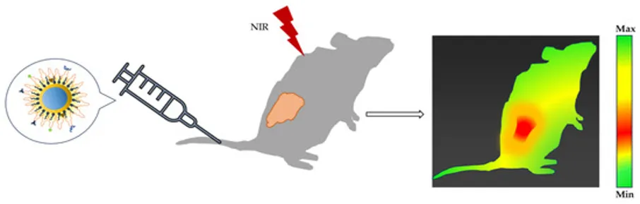
Figure 4: A schematic representation of QD administered into a tumor-bearing mouse for in vivo tumor research is shown.

For targeted imaging and cancer treatment, Zhu et al. created, for example, near-infrared (NIR) fluorescent silver selenide (Ag2Se) QDs labeled with Cetuximab. In contrast to using Ag2Se QDs alone, the multifunctional nanoprobe was said to show fluorescent contrast at the tumor site, and the fluorescence was still easily seen there 24 hours after injection. This nanoprobe, according to their findings, considerably slowed tumor development, and the survival rate of bare mice with orthotopic tongue cancer increased from 0% to 57.1%. According to reports, this platform was able to effectively target orthotopic tongue cancer [2].

Imaging of the blood vessels:

QDs have proven to have excellent micron-level spatial resolution and high penetration depth, making them useful for imaging tumor blood vessels in vivo. In the meantime, the outstanding optical characteristics of NIR-II (Near Infrared-II) QDs can be used to perform real-time, high-resolution angiography in order to investigate the mechanism of angiogenesis and provide hemodynamic data. This is essential in the real-time diagnosis and intervention therapy of cancer, as well as in the non-invasive, accurate identification of benign and malignant tumors in situ and the early diagnosis of malignant solid tumors [16].

  • QDs for Drug Delivery:

Many teams have concentrated on integrating therapeutic and diagnostic skills into a single, nanoparticle-based agent. Because they may serve as the primary nanocarrier or as the fluorescent markers in a more complicated design [5], QDs are excellent candidates for theranostic platforms. QDs are only one instance of the many nanoparticles that have been extensively studied for use in medication delivery. Reports indicate that the antitumor efficacy is improved while the systemic adverse effect is decreased, which is due to the effective nanoparticle entrapment of anti-cancer drugs and the regulated distribution in tissues and cells [2]. Important characteristics for effective targeted delivery are demonstrated by nanoparticle drug carriers, including a sufficiently long blood circulation, protection of the cargo from degradation, a high drug loading capacity, a regulated drug release profile, and the integration of several targeting ligands on their surface. In addition, fluorescence can be used with QD probes to track drug delivery in vivo and monitor the biodistribution of carriers and intracellular uptake [10].

Table 1. Using QDs for targeted drug delivery in vitro and in vivo [2].

QDs Used In Vitro

Drug

Cell Line

Iron oxide carbon QDs encapsulated in chitosan

Curcumin

MCF-7cells

Transferrin (TF) – conjugated Carbon QDs

Doxorubicin

MCF-7cells

Graphene oxide QDs conjugated with glucosamine and boric acid

Doxorubicin

MCF-7cells

Magnesium nitride (Mg/N) doped carbon QDs (CQDs)

Epirubicin (EPI)

4T1 and MCF-7cells

Nitrogen-doped Graphene QDs (N-GQDs)

Methotrexate (MTX)

MCF-7 human breast cancer cells

PEGylated molybdenum disulfide QDs

Doxorubicin

U251cells

Zinc oxide adipic dihydrazide heparin

Paclitaxel

A549cells

Cadmium-sulfide-modified chitosan

Sesamol

MCF-7cells

PEGylated Silver graphene QDs (Ag-GQDs)

Doxorubicin

HeLa and DU145 cells

QDs used in vivo


Graphene QDs

Doxorubicin

MCF-7cells

Silver sulfide (Ag2S) QDs conjugated with chitosan

Doxorubicin

HeLa cells

Manganese doped zinc sulfide (Mn-ZnS) QDs conjugated with folic acid (FA)

5-fluorouracil (5-FU)

4T1 breast cancer cells

PEGylated silver sulfide Ag2S QDs

Doxorubicin

MDA-MB-231 human breast tumour cells

Graphene QD (GQD)-modified magnetic chitosan

Doxorubicin

Hepatocellular carcinoma

Red-emissive carbon QDs

Doxorubicin

HeLa cells

Black phosphorus QDs (BPQDs) encapsulated in platelet-osteosarcoma hybrid membrane

Doxorubicin

Osteosarcoma

Nitrogen-doped carbon QDs conjugated with folic acid

Doxorubicin

4T1 and MCF-7 cells

PEGylated molybdenum disulfide (MoS2) QDs conjugated with arginylglycylaspartic acid (RGD)peptide

Doxorubicin

HepG2 cells

Table 2. The main results of the QDs used for drug delivery in different malignancies [4].

Sr. no.

Types of QDs

Drug

Targeted Diseases

Inference

  1.  

CDs

Epirubicin and

Temo zolomide

Brain Tumour

• Triple conjugated system showed profound cytotoxicity in tumour cells with minimal effects on normal cells.

• Conjugating with transferrin improved the cytotoxicity and cellular uptake of conjugated QDs.

  1.  

GQDs

Imatinib

Blood Cancer

• Higher IC50 values were observed for drug-loaded GQDs.

• Drug-loaded GQDs induced mild apoptosis.

  1.  

CQDs

5-fluorouracil

Breast Cancer

• The nanoconjugate exhibited higher cytotoxicity than the free drug.

• Better release profile in acidic environments.

  1.  

CQDs

Curcumin

Breast Cancer

• Viable cancerous cell count was lowered, and late apoptosis was boosted in MCF-7 breast cancer cell lines upon treatment.

• Controlled drug release by curcumin CQDs.

• The nano composite showed pH-dependent drug release owing to chitosan’s pH sensitivity.

  1.  

CQDs

Camptothecin

Breast Cancer

• Sustained drug release was observed for upto100h, followed by a rapid release phase.

• Dose-dependent cytotoxicity observed.

• pH-responsive drug release observed.

  1.  

CQDs

Doxorubicin

Breast Cancer

• Elevated drug release was observed at acidic pH.

• Higher apoptotic effect of transferrin (Tf) DOX CQDs than free DOX.

•DOX Tf CQDs better internalised than free drugs and DOX CQDs

• Real-time drug release tracking through CQDs.

  1.  

CQDs

Gemcitabine

Breast Cancer

• Elevated drug release in acidic pH compared to physiological pH.

• IC50 value of drug-loaded QDs is 2 times less than the free drug.

• Drug-loaded QDs were localised in the tumour region, sparing the healthy normal cells.

  1.  

Ag QDs

Methotrexate

Cervical Cancer

• IC50 values of drug-loaded targeted QDs were found to be comparable to those of free drug in cancer cell inhibition.

• No significant cytotoxicity was observed after 24 h.

  1.  

CDs

Doxorubicin

Cervical and kidney cancer

• The β-cyclodextrin decorated CDs showed acidic pH-dependent drug release.

• Selective targeting of cancer cells achieved upon decoration with β-cyclodextrin.

  1.  

CDs

Doxorubicin

Cervical cancer and liver cancer

• Showed3timeshigherfluorescence intensity of DOX CDs cubosomes at 24h.

• Significantly lower cell viability by DOX CDs cubosomes.

• In vivo DOX CDs cubosomes exhibited the lowest average tumour volume.

• Reduced cardiotoxicity and hepatotoxicity of DOX CDs cubosomes.

  1.  

Ag QDs

Doxorubicin

Lung Cancer

• FA conjugation augmented the cytotoxic action of the DOX-MUA-QDs.

• FA-MUA-DOX QDs obstructed the colony-forming ability of A549 effectively at concentrations near their cytotoxic thresholds.

• DNA damage in the cells treated by FA-MUA-DOX QDs was genotoxic to the A549 cells.

  1.  

CDs

Doxorubicin

Liver cancer

• Increased drug release at pH 5.2.

• Sustained release from CDs (72 h), whereas free DOX released completely in 10 h.

• DOX-PEI CDs had stronger fluorescence in tumours and improved drug delivery compared to free DOX.

• The DOX-PEI CDs animal group had better survival than the free DOX group, indicating a superior safety profile for the nanoconjugate.

  1.  

BPQDs

Doxorubicin

Osteosarcoma

• Prominent aggregation of DOX in the tumour tissue from the BPQDs.

• The stability of BPQDs is augmented through OPM encapsulation.

• NIR light irradiation triggered DOX release.

• In vivo studies showed notable tumour reduction.

  1.  

GQDs

Doxorubicin

Colorectal cancer

• EpCAM aptamer conjugated nanoparticles encapsulated DOX, ZIF-8, and GQDs facilitated triple delivery for the treatment of colorectal cancer.

• Drug release from the nano conjugate was favoured at acidic pH 5.4 with a burst release prior to gradual release.

  1.  

GQDs

Gemcitabine

Pancreatic cancer

• Conjugation of GQDs with human serum albumin nanoparticles demonstrated sustained drug release and improved stability.

• HA targeted CD44 receptors for enhanced delivery to resistant cells.

  1.  

Ag QDs

Etoposide+ methotrexate

Skin Cancer

• Ferromagnetic QD nanocomposites showed sustained release for 30h.

• Reduced cell viability in a concentration-dependent manner.

  1.  

CdSe QDs

Methotrexate

Throat cancer

• Higher cytotoxicity by drug conjugate than free MTX with 4 times more efficacy.

  1.  

GQDs

Doxorubicin/ Curcumin

Colorectal and breast cancer

• Enhanced tumour penetration due to MiRGD peptide

• Better release profile in an acidic environment, indicating its selective targeting.

• Higher drug accumulation at the tumour site than that of free drugs.

  1.  

CDs

Doxorubicin

Adenoid Cystic Carcinoma

• DOX-CDs biocompatible and non-toxic to normal cells.

• DOX-CDs-induced apoptosis inhibited tumour growth and prolonged the survival of the mice.

  1.  

Ag-In-Zn QDs

Compound C-2028

Lung and prostate cancer

• Strong cytotoxic action against cancer cells with low IC80 values.

• Cytotoxicity reduced upon conjugation withβ-cyclodextrin but vice versa with folic acid conjugation.

CHALLENGES

The toxicity of QDs remains the biggest impediment to clinical translation, despite the possible immense social advantages they provide for human illness. The biochemical processes underlying cytotoxicity are now being understood. The toxicity of QDs has been linked to their charge, size, concentration, and stability, as well as the bioactivity of their outer coating [9].

Despite the enormous potential of QDs in biomedical imaging and detection, toxicological and pharmacological problems, primarily related to heavy metals and colloidal instability, impede progress toward cancer diagnosis and treatment as well as other illnesses. Nanotoxicology The development of these applications in vitro may not be hampered by these issues, but they present significant obstacles to their use in vivo for human cancer imaging [12].

(Cd Se) ZnS is the most prevalent QD makeup. It has a core of cadmium and selenium, both of which are well known to be harmful [9].

Although CdSe and CdTe semiconductor materials have been widely utilized as the core of QDs, they have been shown to exhibit cytotoxicity at low concentrations. As a result, silicon or AgInS2 core-based novel QDs that exclude Cd, Se, and Te elements have already been created, and these QDs have been shown to have minimal toxicity [11].

FUTURE PROSPECTS

Scientists in the domains of chemistry, biology, medical engineering, and pharmaceutical sciences have come together in a shared effort to establish QDs as technological advancements with features that could significantly advance in vivo and in vitro imaging as well as achieve notable advances in the field of nanomedicine research [10].

The flexibility of QDs allows for novel approaches to treating cancer. Recent research has shown that black phosphorus material functions as a "nanoknife," allowing for the exact tumor ablation described in the article. BPQDs are being investigated for use in chemotherapeutic medication delivery and may potentially be used as nanoknives as well as nanocarriers [4].

A variety of efforts should be made to create unique QDs based on their composition, sizes, surface coatings, and valences in order to reduce toxicity and optimize detection performance. Consider, however, challenges such as shell coating degradation brought about by QD [7].

In vivo theranostics [11], an approach that likely allows for the simultaneous diagnosis and treatment of stem cells in vivo, is made possible by the development of hybrid materials between QDs and other functional molecules during the evolution of QDs.

Immunomodulatory capabilities may be included in future QD-based treatments. QDs that are functionalized with immune checkpoint inhibitors or stimulatory molecules may help the immune system identify and destroy cancer cells. For a full cancer treatment, such approaches might combine with chemotherapy and radiation [4].

CONCLUSION

The use of nanoparticles in the battle against cancer has been the subject of considerable study. Because nanoparticles have a number of traits necessary to address the drawbacks of standard cancer therapy methods, they may be used as a basis for early diagnosis and treatment. The most recent nanoparticles to show distinctive characteristics that might revolutionize cancer diagnosis and treatment are quantum dots. The characteristics of these features include their tiny tunable size, consistent photoluminescence, high surface-to-volume ratio, and possibility for biocompatibility. QDs have been widely used for tumor imaging in vitro and in vivo, and they have also been combined with therapeutic medicines for targeted drug delivery in vivo. Early tumor identification and diagnosis, bioimaging, targeted gene-drug delivery, phototherapy, and medication delivery are the main applications of QDs in cancer therapy. Due to their excellent biocompatibility, low cytotoxicity, and high capacity for cell uptake, QDs are also the preferred method of drug administration. There is no doubt that QDs have tremendous promise for use in bio-imaging, sensors, drug delivery, and other fields. To realize the true potential of QDs in clinical practice, a number of challenges must yet be overcome, including overall toxicity, body clearance, the scalability of the synthesis procedure, environmental effects, manufacturing costs, and other factors. Theranostic platforms are always being created by combining QDs with additional kinds of nanoparticles and/or biologically active compounds.

REFERENCES

  1. National Cancer Institute. (2015). What is cancer? U.S. Department of Health and Human Services, National Institutes of Health. (https://www.cancer.gov/about-cancer/understanding/what-is-cancer).
  2. Hamidu, A., Pitt, W. G., & Husseini, G. A. (2023). Recent Breakthroughs in Using Quantum Dots for Cancer Imaging and Drug Delivery Purposes. Nanomaterials (Basel, Switzerland), 13(18), 2566. https://doi.org/10.3390/nano13182566
  3. Tada, H., Higuchi, H., Wanatabe, T. M., & Ohuchi, N. (2007). In vivo real-time tracking of single quantum dots conjugated with monoclonal anti-HER2 antibody in tumors of mice. Cancer research, 67(3), 1138–1144. https://doi.org/10.1158/0008-5472.CAN-06-1185
  4. Pareek, A., Kumar, D., Pareek, A., & Gupta, M. M. (2025). Advancing Cancer Therapy with Quantum Dots and Other Nanostructures: A Review of Drug Delivery Innovations, Applications, and Challenges. Cancers, 17(5), 878. https://doi.org/10.3390/cancers17050878
  5. Matea, C. T., Mocan, T., Tabaran, F., Pop, T., Mosteanu, O., Puia, C., Iancu, C., & Mocan, L. (2017). Quantum dots in imaging, drug delivery and sensor applications. International journal of nanomedicine, 12, 5421–5431. https://doi.org/10.2147/IJN.S138624
  6. Zhao, M. X., & Zhu, B. J. (2016). The Research and Applications of Quantum Dots as Nano-Carriers for Targeted Drug Delivery and Cancer Therapy. Nanoscale research letters, 11(1), 207. https://doi.org/10.1186/s11671-016-1394-9
  7. Devi, S., Kumar, M., Tiwari, A., Tiwari, V., Kaushik, D., Verma, R., Bhatt, S., Sahoo, B. M., Bhattacharya, T., Alshehri, S., Ghoneim, M. M., Babalghith, A. O., & Batiha, G. E.-S. (2022). Quantum dots: An emerging approach for cancer therapy. Frontiers in Materials, 8, 798440. https://doi.org/10.3389/fmats.2021.798440
  8. Iannazzo, D., Celesti, C., & Espro, C. (2021). Recent Advances on Graphene Quantum Dots as Multifunctional Nanoplatforms for Cancer Treatment. Biotechnology journal, 16(2), e1900422. https://doi.org/10.1002/biot.201900422
  9. Luo, G., Long, J., Zhang, B., Liu, C., Ji, S., Xu, J., Yu, X., & Ni, Q. (2012). Quantum dots in cancer therapy. Expert opinion on drug delivery, 9(1), 47–58. https://doi.org/10.1517/17425247.2012.638624
  10. Cheki, M., Moslehi, M., & Assadi, M. (2013). Marvelous applications of quantum dots. European review for medical and pharmacological sciences, 17(9), 1141–1148.
  11. Onoshima, D., Yukawa, H., & Baba, Y. (2015). Multifunctional quantum dots-based cancer diagnostics and stem cell therapeutics for regenerative medicine. Advanced drug delivery reviews, 95, 2–14. https://doi.org/10.1016/j.addr.2015.08.004
  12. Fang, M., Peng, C. W., Pang, D. W., & Li, Y. (2012). Quantum dots for cancer research: current status, remaining issues, and future perspectives. Cancer biology & medicine, 9(3), 151–163. https://doi.org/10.7497/j.issn.2095-3941.2012.03.001
  13. Iannazzo, D., Pistone, A., Celesti, C., Triolo, C., Patané, S., Giofré, S. V., Romeo, R., Ziccarelli, I., Mancuso, R., Gabriele, B., Visalli, G., Facciolà, A., & Di Pietro, A. (2019). A Smart Nanovector for Cancer Targeted Drug Delivery Based on Graphene Quantum Dots. Nanomaterials (Basel, Switzerland), 9(2), 282. https://doi.org/10.3390/nano9020282
  14. Pardo, J., Peng, Z., & Leblanc, R. M. (2018). Cancer Targeting and Drug Delivery Using Carbon-Based Quantum Dots and Nanotubes. Molecules (Basel, Switzerland), 23(2), 378. https://doi.org/10.3390/molecules23020378
  15. Zhang, H., Yee, D., & Wang, C. (2008). Quantum dots for cancer diagnosis and therapy: biological and clinical perspectives. Nanomedicine (London, England), 3(1), 83–91. https://doi.org/10.2217/17435889.3.1.83
  16. Chen, L. L., Zhao, L., Wang, Z. G., Liu, S. L., & Pang, D. W. (2022). Near-Infrared-II Quantum Dots for In Vivo Imaging and Cancer Therapy. Small (Weinheim an der Bergstrasse, Germany), 18(8), e2104567. https://doi.org/10.1002/smll.202104567
  17. Gao, X., Cui, Y., Levenson, R. M., Chung, L. W., & Nie, S. (2004). In vivo cancer targeting and imaging with semiconductor quantum dots. Nature biotechnology, 22(8), 969–976. https://doi.org/10.1038/nbt994
  18. Misra, R. D. (2008). Quantum Dots for Tumor-Targeted Drug Delivery and Cell Imaging. Nanomedicine, 3(3), 271–274. https://doi.org/10.2217/17435889.3.3.271
  19. Ding, H., Zhang, F., Zhao, C., Lv, Y., Ma, G., Wei, W., & Tian, Z. (2017). Beyond a Carrier: Graphene Quantum Dots as a Probe for Programmatically Monitoring Anti-Cancer Drug Delivery, Release, and Response. ACS applied materials & interfaces, 9(33), 27396–27401. https://doi.org/10.1021/acsami.7b08824
  20. Zeng, Q., Shao, D., He, X., Ren, Z., Ji, W., Shan, C., Qu, S., Li, J., Chen, L., & Li, Q. (2016). Carbon dots as a trackable drug delivery carrier for localized cancer therapy in vivo. Journal of Materials Chemistry B, 4(30), 5119-5126. https://doi.org/10.1039/C6TB01259K
  21. Thakor, A. S., & Gambhir, S. S. (2013). Nanooncology: the future of cancer diagnosis and therapy. CA: a cancer journal for clinicians, 63(6), 395–418. https://doi.org/10.3322/caac.21199
  22. Luque-Michel, E., Imbuluzqueta, E., Sebastián, V., & Blanco-Prieto, M. J. (2017). Clinical advances of nanocarrier-based cancer therapy and diagnostics. Expert opinion on drug delivery, 14(1), 75–92. https://doi.org/10.1080/17425247.2016.1205585
  23. Li, J., Liu, F., Gupta, S., & Li, C. (2016). Interventional Nanotheranostics of Pancreatic Ductal Adenocarcinoma. Theranostics, 6(9), 1393–1402. https://doi.org/10.7150/thno.15122
  24. Din, F. U., Aman, W., Ullah, I., Qureshi, O. S., Mustapha, O., Shafique, S., & Zeb, A. (2017). Effective use of nanocarriers as drug delivery systems for the treatment of selected tumors. International journal of nanomedicine, 12, 7291–7309. https://doi.org/10.2147/IJN.S146315
  25. Cheng, C., Li, S., Thomas, A., Kotov, N. A., & Haag, R. (2017). Functional Graphene Nanomaterials Based Architectures: Bio interactions, Fabrications, and Emerging Biological Applications. Chemical reviews, 117(3), 1826–1914. https://doi.org/10.1021/acs.chemrev.6b00520
  26. Gabriele, B., Mancuso, R., Salerno, G., & Veltri, L. (2005). Sequential homobimetallic catalysis: an unprecedented tandem Pd (0)-catalysed deprotection – Pd (II)-catalysed heterocyclisation reaction leading to benzofurans. Chemical communications (Cambridge, England), (2), 271–273. https://doi.org/10.1039/b413240h
  27. Zaharie-Butucel, D., Potara, M., Craciun, A. M., Boukherroub, R., Szunerits, S., & Astilean, S. (2017). Revealing the structure and functionality of graphene oxide and reduced graphene oxide/pyrene carboxylic acid interfaces by correlative spectral and imaging analysis. Physical chemistry chemical physics: PCCP, 19(24), 16038–16046. https://doi.org/10.1039/c7cp02443f
  28. Kano, M. R., Bae, Y., Iwata, C., Morishita, Y., Yashiro, M., Oka, M., Fujii, T., Komuro, A., Kiyono, K., Kaminishi, M., Hirakawa, K., Ouchi, Y., Nishiyama, N., Kataoka, K., & Miyazono, K. (2007). Improvement of cancer-targeting therapy, using nanocarriers for intractable solid tumors by inhibition of TGF-beta signalling. Proceedings of the National Academy of Sciences of the United States of America, 104(9), 3460–3465. https://doi.org/10.1073/pnas.0611660104
  29. Dadwal, A., Baldi, A., & Kumar Narang, R. (2018). Nanoparticles as carriers for drug delivery in cancer. Artificial cells, nanomedicine, and biotechnology, 46(sup2), 295–305. https://doi.org/10.1080/21691401.2018.1457039
  30. Bentolila, L. A., Ebenstein, Y., & Weiss, S. (2009). Quantum dots for in vivo small-animal imaging. Journal of nuclear medicine : official publication, Society of Nuclear Medicine, 50(4), 493–496. https://doi.org/10.2967/jnumed.108.053561
  31. Liang, Z., Khawar, M. B., Liang, J., & Sun, H. (2021). Bio-Conjugated Quantum Dots for Cancer Research: Detection and Imaging. Frontiers in oncology, 11, 749970. https://doi.org/10.3389/fonc.2021.749970
  32. Ferlay, J., Colombet, M., Soerjomataram, I., Parkin, D. M., Piñeros, M., Znaor, A., & Bray, F. (2021). Cancer statistics for the year 2020: An overview. International journal of cancer, 10.1002/ijc.33588. Advance online publication. https://doi.org/10.1002/ijc.33588
  33. Wu, S., Zhu, W., Thompson, P., & Hannun, Y. A. (2018). Evaluating intrinsic and non-intrinsic cancer risk factors. Nature communications, 9(1), 3490. https://doi.org/10.1038/s41467-018-05467-z
  34. Ferrari M. (2005). Cancer nanotechnology: opportunities and challenges. Nature reviews. Cancer, 5(3), 161–171. https://doi.org/10.1038/nrc1566
  35. Duncan R. (2006). Polymer conjugates as anticancer nanomedicines. Nature reviews. Cancer, 6(9), 688–701. https://doi.org/10.1038/nrc1958
  36. Burz, C., Pop, V. V., Buiga, R., Daniel, S., Samasca, G., Aldea, C., & Lupan, I. (2018). Circulating tumor cells in clinical research and monitoring patients with colorectal cancer. Oncotarget, 9(36), 24561–24571. https://doi.org/10.18632/oncotarget.25337
  37. Shamsi, J., Dang, Z., Bianchini, P., Canale, C., Stasio, F. D., Brescia, R., Prato, M., & Manna, L. (2016). Colloidal Synthesis of Quantum Confined Single Crystal CsPbBr3 Nanosheets with Lateral Size Control up to the Micrometer Range. Journal of the American Chemical Society, 138(23), 7240–7243. https://doi.org/10.1021/jacs.6b03166
  38. Karanikolos, G. N., Alexandridis, P., Itskos, G., Petrou, A., & Mountziaris, T. J. (2004). Synthesis and size control of luminescent ZnSe nanocrystals by a microemulsion-gas contacting technique. Langmuir : the ACS journal of surfaces and colloids, 20(3), 550–553. https://doi.org/10.1021/la035397+
  39. K Tiwari, P., Sahu, M., Kumar, G., & Ashourian, M. (2021). Pivotal Role of Quantum Dots in the Advancement of Healthcare Research. Computational intelligence and neuroscience, 2021, 2096208. https://doi.org/10.1155/2021/2096208
  40. Lees, E. E., Nguyen, T. L., Clayton, A. H., Muir, B. W., & Mulvaney, P. (2009). The preparation of colloidally stable, water-soluble, biocompatible, semiconductor nanocrystals with a small hydrodynamic diameter. ACS nano, 3(5), 1121–1128. https://doi.org/10.1021/nn900144n
  41. Heyne, B., Arlt, K., Geßner, A., Richter, A. F., Döblinger, M., Feldmann, J., Taubert, A., & Wedel, A. (2020). Mixed Mercaptocarboxylic Acid Shells Provide Stable Dispersions of InPZnS/ZnSe/ZnS Multishell Quantum Dots in Aqueous Media. Nanomaterials (Basel, Switzerland), 10(9), 1858. https://doi.org/10.3390/nano10091858
  42. Speranskaya, E. S., Beloglazova, N. V., Lenain, P., De Saeger, S., Wang, Z., Zhang, S., Hens, Z., Knopp, D., Niessner, R., Potapkin, D. V., & Goryacheva, I. Y. (2014). Polymer-coated fluorescent CdSe-based quantum dots for application in immunoassay. Biosensors & bioelectronics, 53, 225–231. https://doi.org/10.1016/j.bios.2013.09.045
  43. Carrillo-Carrion, C., & Parak, W. J. (2016). Design of pyridyl-modified amphiphilic polymeric ligands: Towards better passivation of water-soluble colloidal quantum dots for improved optical performance. Journal of colloid and interface science, 478, 88–96. https://doi.org/10.1016/j.jcis.2016.05.058
  44. Khan, M. M. R., Mitra, T., & Sahoo, D. (2020). Metal oxide QD based ultrasensitive microsphere fluorescent sensor for copper, chromium and iron ions in water. RSC advances, 10(16), 9512–9524. https://doi.org/10.1039/c9ra09985a
  45. Smith, A. M., Duan, H., Mohs, A. M., & Nie, S. (2008). Bioconjugated quantum dots for in vivo molecular and cellular imaging. Advanced drug delivery reviews, 60(11), 1226–1240. https://doi.org/10.1016/j.addr.2008.03.015

Reference

  1. National Cancer Institute. (2015). What is cancer? U.S. Department of Health and Human Services, National Institutes of Health. (https://www.cancer.gov/about-cancer/understanding/what-is-cancer).
  2. Hamidu, A., Pitt, W. G., & Husseini, G. A. (2023). Recent Breakthroughs in Using Quantum Dots for Cancer Imaging and Drug Delivery Purposes. Nanomaterials (Basel, Switzerland), 13(18), 2566. https://doi.org/10.3390/nano13182566
  3. Tada, H., Higuchi, H., Wanatabe, T. M., & Ohuchi, N. (2007). In vivo real-time tracking of single quantum dots conjugated with monoclonal anti-HER2 antibody in tumors of mice. Cancer research, 67(3), 1138–1144. https://doi.org/10.1158/0008-5472.CAN-06-1185
  4. Pareek, A., Kumar, D., Pareek, A., & Gupta, M. M. (2025). Advancing Cancer Therapy with Quantum Dots and Other Nanostructures: A Review of Drug Delivery Innovations, Applications, and Challenges. Cancers, 17(5), 878. https://doi.org/10.3390/cancers17050878
  5. Matea, C. T., Mocan, T., Tabaran, F., Pop, T., Mosteanu, O., Puia, C., Iancu, C., & Mocan, L. (2017). Quantum dots in imaging, drug delivery and sensor applications. International journal of nanomedicine, 12, 5421–5431. https://doi.org/10.2147/IJN.S138624
  6. Zhao, M. X., & Zhu, B. J. (2016). The Research and Applications of Quantum Dots as Nano-Carriers for Targeted Drug Delivery and Cancer Therapy. Nanoscale research letters, 11(1), 207. https://doi.org/10.1186/s11671-016-1394-9
  7. Devi, S., Kumar, M., Tiwari, A., Tiwari, V., Kaushik, D., Verma, R., Bhatt, S., Sahoo, B. M., Bhattacharya, T., Alshehri, S., Ghoneim, M. M., Babalghith, A. O., & Batiha, G. E.-S. (2022). Quantum dots: An emerging approach for cancer therapy. Frontiers in Materials, 8, 798440. https://doi.org/10.3389/fmats.2021.798440
  8. Iannazzo, D., Celesti, C., & Espro, C. (2021). Recent Advances on Graphene Quantum Dots as Multifunctional Nanoplatforms for Cancer Treatment. Biotechnology journal, 16(2), e1900422. https://doi.org/10.1002/biot.201900422
  9. Luo, G., Long, J., Zhang, B., Liu, C., Ji, S., Xu, J., Yu, X., & Ni, Q. (2012). Quantum dots in cancer therapy. Expert opinion on drug delivery, 9(1), 47–58. https://doi.org/10.1517/17425247.2012.638624
  10. Cheki, M., Moslehi, M., & Assadi, M. (2013). Marvelous applications of quantum dots. European review for medical and pharmacological sciences, 17(9), 1141–1148.
  11. Onoshima, D., Yukawa, H., & Baba, Y. (2015). Multifunctional quantum dots-based cancer diagnostics and stem cell therapeutics for regenerative medicine. Advanced drug delivery reviews, 95, 2–14. https://doi.org/10.1016/j.addr.2015.08.004
  12. Fang, M., Peng, C. W., Pang, D. W., & Li, Y. (2012). Quantum dots for cancer research: current status, remaining issues, and future perspectives. Cancer biology & medicine, 9(3), 151–163. https://doi.org/10.7497/j.issn.2095-3941.2012.03.001
  13. Iannazzo, D., Pistone, A., Celesti, C., Triolo, C., Patané, S., Giofré, S. V., Romeo, R., Ziccarelli, I., Mancuso, R., Gabriele, B., Visalli, G., Facciolà, A., & Di Pietro, A. (2019). A Smart Nanovector for Cancer Targeted Drug Delivery Based on Graphene Quantum Dots. Nanomaterials (Basel, Switzerland), 9(2), 282. https://doi.org/10.3390/nano9020282
  14. Pardo, J., Peng, Z., & Leblanc, R. M. (2018). Cancer Targeting and Drug Delivery Using Carbon-Based Quantum Dots and Nanotubes. Molecules (Basel, Switzerland), 23(2), 378. https://doi.org/10.3390/molecules23020378
  15. Zhang, H., Yee, D., & Wang, C. (2008). Quantum dots for cancer diagnosis and therapy: biological and clinical perspectives. Nanomedicine (London, England), 3(1), 83–91. https://doi.org/10.2217/17435889.3.1.83
  16. Chen, L. L., Zhao, L., Wang, Z. G., Liu, S. L., & Pang, D. W. (2022). Near-Infrared-II Quantum Dots for In Vivo Imaging and Cancer Therapy. Small (Weinheim an der Bergstrasse, Germany), 18(8), e2104567. https://doi.org/10.1002/smll.202104567
  17. Gao, X., Cui, Y., Levenson, R. M., Chung, L. W., & Nie, S. (2004). In vivo cancer targeting and imaging with semiconductor quantum dots. Nature biotechnology, 22(8), 969–976. https://doi.org/10.1038/nbt994
  18. Misra, R. D. (2008). Quantum Dots for Tumor-Targeted Drug Delivery and Cell Imaging. Nanomedicine, 3(3), 271–274. https://doi.org/10.2217/17435889.3.3.271
  19. Ding, H., Zhang, F., Zhao, C., Lv, Y., Ma, G., Wei, W., & Tian, Z. (2017). Beyond a Carrier: Graphene Quantum Dots as a Probe for Programmatically Monitoring Anti-Cancer Drug Delivery, Release, and Response. ACS applied materials & interfaces, 9(33), 27396–27401. https://doi.org/10.1021/acsami.7b08824
  20. Zeng, Q., Shao, D., He, X., Ren, Z., Ji, W., Shan, C., Qu, S., Li, J., Chen, L., & Li, Q. (2016). Carbon dots as a trackable drug delivery carrier for localized cancer therapy in vivo. Journal of Materials Chemistry B, 4(30), 5119-5126. https://doi.org/10.1039/C6TB01259K
  21. Thakor, A. S., & Gambhir, S. S. (2013). Nanooncology: the future of cancer diagnosis and therapy. CA: a cancer journal for clinicians, 63(6), 395–418. https://doi.org/10.3322/caac.21199
  22. Luque-Michel, E., Imbuluzqueta, E., Sebastián, V., & Blanco-Prieto, M. J. (2017). Clinical advances of nanocarrier-based cancer therapy and diagnostics. Expert opinion on drug delivery, 14(1), 75–92. https://doi.org/10.1080/17425247.2016.1205585
  23. Li, J., Liu, F., Gupta, S., & Li, C. (2016). Interventional Nanotheranostics of Pancreatic Ductal Adenocarcinoma. Theranostics, 6(9), 1393–1402. https://doi.org/10.7150/thno.15122
  24. Din, F. U., Aman, W., Ullah, I., Qureshi, O. S., Mustapha, O., Shafique, S., & Zeb, A. (2017). Effective use of nanocarriers as drug delivery systems for the treatment of selected tumors. International journal of nanomedicine, 12, 7291–7309. https://doi.org/10.2147/IJN.S146315
  25. Cheng, C., Li, S., Thomas, A., Kotov, N. A., & Haag, R. (2017). Functional Graphene Nanomaterials Based Architectures: Bio interactions, Fabrications, and Emerging Biological Applications. Chemical reviews, 117(3), 1826–1914. https://doi.org/10.1021/acs.chemrev.6b00520
  26. Gabriele, B., Mancuso, R., Salerno, G., & Veltri, L. (2005). Sequential homobimetallic catalysis: an unprecedented tandem Pd (0)-catalysed deprotection – Pd (II)-catalysed heterocyclisation reaction leading to benzofurans. Chemical communications (Cambridge, England), (2), 271–273. https://doi.org/10.1039/b413240h
  27. Zaharie-Butucel, D., Potara, M., Craciun, A. M., Boukherroub, R., Szunerits, S., & Astilean, S. (2017). Revealing the structure and functionality of graphene oxide and reduced graphene oxide/pyrene carboxylic acid interfaces by correlative spectral and imaging analysis. Physical chemistry chemical physics: PCCP, 19(24), 16038–16046. https://doi.org/10.1039/c7cp02443f
  28. Kano, M. R., Bae, Y., Iwata, C., Morishita, Y., Yashiro, M., Oka, M., Fujii, T., Komuro, A., Kiyono, K., Kaminishi, M., Hirakawa, K., Ouchi, Y., Nishiyama, N., Kataoka, K., & Miyazono, K. (2007). Improvement of cancer-targeting therapy, using nanocarriers for intractable solid tumors by inhibition of TGF-beta signalling. Proceedings of the National Academy of Sciences of the United States of America, 104(9), 3460–3465. https://doi.org/10.1073/pnas.0611660104
  29. Dadwal, A., Baldi, A., & Kumar Narang, R. (2018). Nanoparticles as carriers for drug delivery in cancer. Artificial cells, nanomedicine, and biotechnology, 46(sup2), 295–305. https://doi.org/10.1080/21691401.2018.1457039
  30. Bentolila, L. A., Ebenstein, Y., & Weiss, S. (2009). Quantum dots for in vivo small-animal imaging. Journal of nuclear medicine : official publication, Society of Nuclear Medicine, 50(4), 493–496. https://doi.org/10.2967/jnumed.108.053561
  31. Liang, Z., Khawar, M. B., Liang, J., & Sun, H. (2021). Bio-Conjugated Quantum Dots for Cancer Research: Detection and Imaging. Frontiers in oncology, 11, 749970. https://doi.org/10.3389/fonc.2021.749970
  32. Ferlay, J., Colombet, M., Soerjomataram, I., Parkin, D. M., Piñeros, M., Znaor, A., & Bray, F. (2021). Cancer statistics for the year 2020: An overview. International journal of cancer, 10.1002/ijc.33588. Advance online publication. https://doi.org/10.1002/ijc.33588
  33. Wu, S., Zhu, W., Thompson, P., & Hannun, Y. A. (2018). Evaluating intrinsic and non-intrinsic cancer risk factors. Nature communications, 9(1), 3490. https://doi.org/10.1038/s41467-018-05467-z
  34. Ferrari M. (2005). Cancer nanotechnology: opportunities and challenges. Nature reviews. Cancer, 5(3), 161–171. https://doi.org/10.1038/nrc1566
  35. Duncan R. (2006). Polymer conjugates as anticancer nanomedicines. Nature reviews. Cancer, 6(9), 688–701. https://doi.org/10.1038/nrc1958
  36. Burz, C., Pop, V. V., Buiga, R., Daniel, S., Samasca, G., Aldea, C., & Lupan, I. (2018). Circulating tumor cells in clinical research and monitoring patients with colorectal cancer. Oncotarget, 9(36), 24561–24571. https://doi.org/10.18632/oncotarget.25337
  37. Shamsi, J., Dang, Z., Bianchini, P., Canale, C., Stasio, F. D., Brescia, R., Prato, M., & Manna, L. (2016). Colloidal Synthesis of Quantum Confined Single Crystal CsPbBr3 Nanosheets with Lateral Size Control up to the Micrometer Range. Journal of the American Chemical Society, 138(23), 7240–7243. https://doi.org/10.1021/jacs.6b03166
  38. Karanikolos, G. N., Alexandridis, P., Itskos, G., Petrou, A., & Mountziaris, T. J. (2004). Synthesis and size control of luminescent ZnSe nanocrystals by a microemulsion-gas contacting technique. Langmuir : the ACS journal of surfaces and colloids, 20(3), 550–553. https://doi.org/10.1021/la035397+
  39. K Tiwari, P., Sahu, M., Kumar, G., & Ashourian, M. (2021). Pivotal Role of Quantum Dots in the Advancement of Healthcare Research. Computational intelligence and neuroscience, 2021, 2096208. https://doi.org/10.1155/2021/2096208
  40. Lees, E. E., Nguyen, T. L., Clayton, A. H., Muir, B. W., & Mulvaney, P. (2009). The preparation of colloidally stable, water-soluble, biocompatible, semiconductor nanocrystals with a small hydrodynamic diameter. ACS nano, 3(5), 1121–1128. https://doi.org/10.1021/nn900144n
  41. Heyne, B., Arlt, K., Geßner, A., Richter, A. F., Döblinger, M., Feldmann, J., Taubert, A., & Wedel, A. (2020). Mixed Mercaptocarboxylic Acid Shells Provide Stable Dispersions of InPZnS/ZnSe/ZnS Multishell Quantum Dots in Aqueous Media. Nanomaterials (Basel, Switzerland), 10(9), 1858. https://doi.org/10.3390/nano10091858
  42. Speranskaya, E. S., Beloglazova, N. V., Lenain, P., De Saeger, S., Wang, Z., Zhang, S., Hens, Z., Knopp, D., Niessner, R., Potapkin, D. V., & Goryacheva, I. Y. (2014). Polymer-coated fluorescent CdSe-based quantum dots for application in immunoassay. Biosensors & bioelectronics, 53, 225–231. https://doi.org/10.1016/j.bios.2013.09.045
  43. Carrillo-Carrion, C., & Parak, W. J. (2016). Design of pyridyl-modified amphiphilic polymeric ligands: Towards better passivation of water-soluble colloidal quantum dots for improved optical performance. Journal of colloid and interface science, 478, 88–96. https://doi.org/10.1016/j.jcis.2016.05.058
  44. Khan, M. M. R., Mitra, T., & Sahoo, D. (2020). Metal oxide QD based ultrasensitive microsphere fluorescent sensor for copper, chromium and iron ions in water. RSC advances, 10(16), 9512–9524. https://doi.org/10.1039/c9ra09985a
  45. Smith, A. M., Duan, H., Mohs, A. M., & Nie, S. (2008). Bioconjugated quantum dots for in vivo molecular and cellular imaging. Advanced drug delivery reviews, 60(11), 1226–1240. https://doi.org/10.1016/j.addr.2008.03.015

Photo
Anas J. Shaikh
Corresponding author

Department of Pharmaceutical Chemistry, Matoshri College of Pharmacy, Eklahre, Nashik.

Photo
Aman J. Shaikh
Co-author

Department of Pharmaceutical Chemistry, Matoshri College of Pharmacy, Eklahre, Nashik.

Anas J. Shaikh, Aman J. Shaikh, Quantum Dots for Imaging and Drug Delivery in Cancer Therapy, Int. J. of Pharm. Sci., 2026, Vol 4, Issue 8, 4985-4997. https://doi.org/10.5281/zenodo.22163531

More related articles
An Overview of Green Silver Nanoparticle Synthesis...
Jay Kumar Chandra, Aman Panda, Dusmanta Kumar Pradhan, Vibhor Kum...
A Review on Mucoadhesive Drug Delivery Systems: Fo...
Bhupendra Saratkar, Nimita Manocha, Nayany Sharma, Nadeem Farooqu...
A Review- Molecular Mechanisms of Azithromycin Res...
Dr. Ria Memoria, Vankar Varsha, Uttam Kakra, Surbhi Pathak, Soumy...
Adverse Drug Reactions Associated With Sodium-Glucose Cotransporter-2 Inhibitors...
Dr. Ajo John, Abinesh R., Dr. Kenneth N., Divya D., Blessed Mebi K., Adrine Rexice A., Shijin P....
Related Articles
Evaluation of NSAID Prescription Patterns in a Tertiary Care Teaching Hospital u...
Ruban Jose, J. S. Venkatesh, Dr. Santhosh Uttangi, Yuvaraja H, Sameer Khan N...
Development and Optimization of Polyethylene Glycol-Based Solid Dispersion of Pi...
Anjana Thakur, Pankaj Bhateja, Dr. Mona Piplani, Nishant Sharma...
Identification of Hub Genes associated with Ribose-5-Phosphate Isomerase A (RPIA...
Dr. Anita Chauhan, Luvina Dogra, Aarti Lagwal, Tanvi Sharma...
An Overview of Green Silver Nanoparticle Synthesis, Characterization and Applica...
Jay Kumar Chandra, Aman Panda, Dusmanta Kumar Pradhan, Vibhor Kumar Jain...
More related articles
An Overview of Green Silver Nanoparticle Synthesis, Characterization and Applica...
Jay Kumar Chandra, Aman Panda, Dusmanta Kumar Pradhan, Vibhor Kumar Jain...
A Review on Mucoadhesive Drug Delivery Systems: Formulation Strategies and Evalu...
Bhupendra Saratkar, Nimita Manocha, Nayany Sharma, Nadeem Farooqui...
A Review- Molecular Mechanisms of Azithromycin Resistance in Neisseria gonorrhoe...
Dr. Ria Memoria, Vankar Varsha, Uttam Kakra, Surbhi Pathak, Soumya Tiwari...
An Overview of Green Silver Nanoparticle Synthesis, Characterization and Applica...
Jay Kumar Chandra, Aman Panda, Dusmanta Kumar Pradhan, Vibhor Kumar Jain...
A Review on Mucoadhesive Drug Delivery Systems: Formulation Strategies and Evalu...
Bhupendra Saratkar, Nimita Manocha, Nayany Sharma, Nadeem Farooqui...
A Review- Molecular Mechanisms of Azithromycin Resistance in Neisseria gonorrhoe...
Dr. Ria Memoria, Vankar Varsha, Uttam Kakra, Surbhi Pathak, Soumya Tiwari...