
We use cookies to ensure our website works properly and to personalise your experience. Cookies policy
Srinath college of pharmacy
Microneedles (MN) technology represents a transformative advancement in transdermal drug delivery, offering a minimally invasive, painless alternative to hypodermic needles while overcoming the limitations of traditional transdermal patches. These micro-scale devices can deliver a broad range of drugs across the skin barrier, including vaccines, biologics, and small molecules. In recent years, significant progress has been made in microneedle materials, design, fabrication techniques, and functional integration, enhancing precision, biocompatibility, and therapeutic efficacy. Innovations such as hydrogel-forming microneedles, stimuli-responsive materials, and integration with biosensors have opened new frontiers in personalized medicine and smart drug delivery systems.Microneedles are being explored for applications in chronic disease management (e.g., diabetes), vaccine administration (e.g., influenza, COVID-19), wound healing, dermatology, and diagnostics. Despite these advances, challenges remain in optimizing drug loading, ensuring consistent skin penetration across populations, and navigating regulatory pathways. This review provides a detailed overview of MN types, materials, fabrication techniques, functional enhancements, safety profiles, and clinical applications, concluding with a discussion on current challenges and future perspectives.
projections designed to penetrate the stratum corneum, the skin's outermost barrier, allowing for efficient transdermal drug delivery. These systems include various types—solid, hollow, coated, dissolving, and hydrogel-forming microneedles—each offering unique advantages depending on therapeutic needs1–3 .
The transdermal route offers several benefits: it bypasses the gastrointestinal tract and hepatic first-pass metabolism, improves patient compliance due to its pain-free nature, and allows for sustained drug release1,2,4. Historically, transdermal delivery was limited by the skin’s barrier properties, especially the stratum corneum. Only a few small, lipophilic molecules could passively diffuse across the skin.
Microneedle technology was developed to address these limitations, providing enhanced precision, safety, and compatibility with a wide range of therapeutic molecules1,2,5 . The field has evolved into multifunctional platforms capable of combining diagnosis and therapy (theranostics), with increased focus on controlled release and personalized medicine 6–8.
The transdermal route is an appealing, non-invasive way to deliver drugs. It offers high bioavailability by bypassing the gastrointestinal tract and avoiding first-pass metabolism by the liver. When drugs are delivered through the skin, they often accumulate in the skin layers before entering the bloodstream, which helps provide a sustained release over time.
Despite its advantages, transdermal drug delivery is currently limited to only a few drugs that meet specific requirements. Ideally, a drug suitable for this route should have a molecular weight under 500 Daltons and a log P value between 2 and 3, which reflects a good balance between water and fat solubility. The main barrier to effective drug absorption through the skin is the outermost layer, called the stratum corneum.
To overcome this barrier and improve drug absorption, several enhancement technologies have been developed—such as iontophoresis, sonophoresis, magnetophoresis, electroporation, and laser microporation. However, these techniques can be expensive and may not be suitable for widespread use
Traditional methods like intradermal injections are still commonly used to tackle the limitations of transdermal delivery. But injections come with their own challenges, including the risk of needle injuries, fear of needles, the need for trained healthcare personnel, and higher overall costs1.
2.1 Mechanism of Action : Transdermal Drug Delivery

Figure 1:Transdermal Drug Delivery
2.1.1 Application of the Drug Formulation
The process begins when a transdermal system (such as a patch, cream, gel, or microneedle array) is placed on the skin.
The system contains the active drug along with excipients (like permeation enhancers or stabilizers) that help in controlled drug release.
The drug must be in a form that allows it to dissolve and diffuse through the layers of the skin.
2.1.2 Contact with the Stratum Corneum (Main Barrier)
Stratum corneum is the outermost layer of the epidermis and acts as the primary barrier to penetration.
It is made up of dead, keratinized cells (corneocytes) embedded in a lipid matrix often described as a “brick and mortar” structure.
Because of this dense and water-resistant structure, only small, lipophilic (fat-soluble) and non-ionized molecules can naturally pass through easily.
Therefore, this step determines how much drug can actually begin to move into the body.
2.1.3 Penetration Pathways Across the Skin
Once the drug encounters the stratum corneum, it can enter through three main routes:
1. Transcellular (through the cells):
The drug passes directly through the corneocytes and lipid layers. Requires both water and lipid solubility for efficient transport.
2. Intracellular (between the cells):
The drug diffuses through the lipid channels between cells. This is the most common and important route for most drugs.
3. Appendageal (through skin appendages):
The drug enters through hair follicles, sweat glands, and sebaceous glands.
Through these cover a small area, they are useful for delivering larger molecules or rapid onset drugs.
2.1.4 Diffusion Through the Viable Epidermis
Once past the stratum corneum, the drug reaches the viable epidermis, which is composed of living cells but lacks blood vessels.
Here, the drug continues to diffuse passively through cell layers, driven by the concentration gradient (from high concentration at the skin surface to low concentration inside).
The epidermis acts as a rate-controlling layer in drug transport.
2.1.5 Passage into the Dermis
After the viable epidermis, the drug reaches the dermis, a thicker layer (~2 mm) rich in connective tissue, nerves, lymphatics, and capillaries.
The dermis does not significantly hinder drug movement.Once the drug diffuses into the capillary network, it can enter systemic circulation or act locally in the skin.
2.1.6 Absorption into the Bloodstream
From the dermal blood vessels, the drug enters the systemic circulation, distributing throughout the body to reach its target tissues.
The absorption rate depends on factors such as:
°Drug concentration and formulation
°Skin thickness and hydration
° Body site of application (thinner skin = faster absorption)
° Duration of application
2.1.7 Controlled and Sustained Release
Most transdermal systems are designed to release the drug slowly and steadily over hours or even days. This provides consistent plasma drug levels, avoiding the peaks (toxicity) and troughs (inefficacy) often seen with oral or injectable routes. Controlled release also improves patient compliance, as fewer doses are needed.
2.1.8 Avoidance of First-Pass Metabolism
Drugs delivered through the skin bypass the gastrointestinal tract and liver, meaning they avoid first-pass metabolism. This increases bioavailability and allows for lower doses to achieve the same effect.It also benefits patients with swallowing difficulties or gastrointestinal side effects.
2.1.9 Role of Permeation Enhancement Techniques
To overcome the strong barrier of the stratum corneum, enhancement strategies are often used:
Chemical Enhancers: Alcohols, fatty acids, surfactants, and terpenes temporarily disrupt lipid structure to increase permeability.
Physical Methods:
Microneedles: Create microscopic pores in the skin to allow larger molecules to pass Iontophoresis: Uses a mild electric current to push charged drug molecules through the skin. Sonophoresis: Uses ultrasound waves to enhance penetration.
Thermal methods: Slightly heat the skin to increase permeability
2.1.10 Pharmacological Action:
Once in the bloodstream, the drug reaches its site of action and exerts its therapeutic effect.
Alternatively, for local delivery systems (like pain patches), the drug may act directly on tissues beneath the application site.
2.1.11 Final Elimination:
After performing its function, the drug is metabolized and excreted via normal physiological pathways (liver and kidneys), just like drugs delivered by other routes.2

3. Classification & Types of Microneedle Systems
Microneedles are classified based on their structure and mechanism of drug delivery:
• Solid Microneedles: Used to create microchannels followed by application of a drug patch (“poke and patch”)2,3
Solid microneedles can be used to prepare the skin for drug delivery. They are briefly inserted and then removed, creating tiny, microscopic pores on the skin’s surface — a process known
as the “poke and patch” approach. These microchannels allow drugs to pass more easily through the skin and reach the deeper layers, improving absorption.
Research on rat skin has shown that when the treated area is kept covered (for example, with an occlusive tape), the tiny pores can remain open for up to 72 hours after microneedle application. However, if the skin is left uncovered, the pores close much more quickly. Importantly, these microchannels heal rapidly — typically within about two hours — which helps minimize the risk of infection.9
• Coated Microneedles: Drugs are coated onto the microneedle surface and delivered upon insertion (“coat and poke”)2,4
• Hollow Microneedles: Serve as microinjectors, delivering liquid formulations through their lumen (“poke and flow”)10,11.
• Dissolving/Degradable Microneedles: Made from biodegradable polymers that dissolve in the skin, releasing the drug (“shove and release”)12–14 .
• Hydrogel-Forming Microneedles: Absorb interstitial fluid, swelling to form a drug- conductive pathway; can also extract fluid for diagnostics12,15 .
Each type offers unique advantages and challenges in terms of fabrication, drug loading, and patient outcomes.
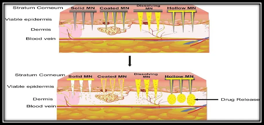
Figure 2: Mechanism of Action: microneedle technology for transdermal drug delivery
Table 1: A detailed list of advantages, disadvantages and method of delivery of various MNs
|
MN Classification |
Advantages |
Disadvantages |
Method of Drug Delivery |
|
Solid |
Can be made from range of material |
Microneedle fracture under skin limited surface area for drug absorption |
Createmicro conduitsin skin to which drug is applied |
|
Hollow |
High drug load can be injected |
Must be fabricated with strong material withstand flow pressure |
Pressure driven flow through needle |
|
Dissolving |
Easy manufacturing |
Only biodegradable material can be used |
Dissolve under skin to release drug payload |
|
Coated |
Used for potential drugs requiring low doses |
Associated with drug loss while manufacturing , temperature limitations |
Coating drug release |
4. Materials & Fabrication Techniques
4.1 Materials Used
Polymers: Both natural (e.g., gelatin, chitosan) and synthetic (e.g., PVP, PLA, PLGA) polymers are widely used due to their biocompatibility and biodegradability12–14 .
Metals: Stainless steel and titanium offer excellent mechanical strength but lack biodegradability3,16.
Ceramics and Composites: Offer high mechanical strength but are more brittle and complex to fabricate11 .
4.2 Fabrication Methods
° Micromolding: Most common, scalable, and cost-effective method for polymer MNs17.
°Photolithography and Laser Etching: High precision, ideal for silicon and metal MNs 18.
°3D Printing: Innovations such as vat polymerization and two-photon polymerization allow customized MN arrays with complex geometries 18,19..
Recent advances focus on improving reproducibility, reducing production costs, and enabling the use of novel, stimuli-responsive materials6,7,19 .
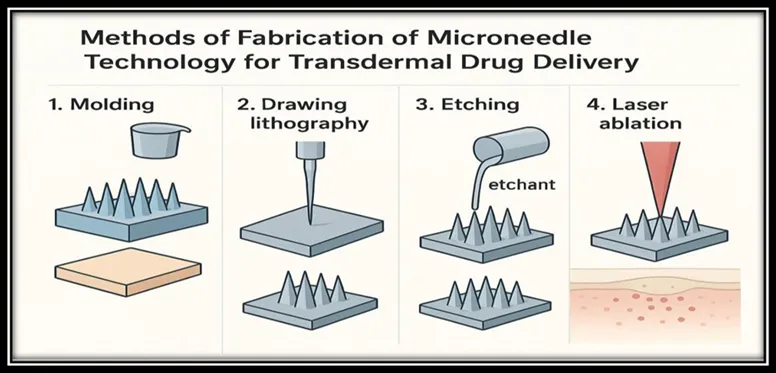
Figure 3: Methods of Fabrication of Microneedle technology for transdermal drug delivery
5. Functional Enhancements & Smart Features
Modern microneedle systems often integrate advanced functionalities:
Controlled/Sustained Release: Layered or encapsulated designs allow for time-controlled delivery of drugs3,13.
Stimuli-Responsive Systems: Respond to pH, temperature, or light for targeted release7,8.
Smart Systems: Integration with biosensors allows real-time monitoring and closed-loop drug delivery (e.g., insulin pumps for diabetes)7,19.
Enhanced Mechanical Properties: Advanced materials and geometric designs ensure reliable skin penetration and adhesion18,20.
These enhancements make microneedles suitable for precision medicine and theranostic applications.
6. Safety, Biocompatibility, and Skin Response
Insertion Mechanics: MNs are generally pain-free due to shallow skin penetration. Needle geometry and force directly influence pain and efficacy 3,6,20.Biocompatibility: Most polymeric microneedles are safe and non-immunogenic. Biodegradable MNs reduce infection risk by leaving no residual sharp waste4,12.
Long-Term Effects: Studies suggest minimal skin irritation and no significant long-term adverse effects when properly fabricated and sterilized 4,13.
Nonetheless, safety must be assessed for each new material and formulation to ensure clinical acceptability.
7. Characterization and Testing Methods
Mechanical Testing: Includes fracture force, insertion force, and penetration depth. These are tested in synthetic skin models and ex vivo tissues17,20.
Drug Release & Permeation:
In Vitro: Franz diffusion cells evaluate drug permeation across skin samples13. In Vivo: Microdialysis techniques measure drug levels in interstitial fluid4,15.
Such rigorous testing ensures consistent performance, safety, and therapeutic efficacy.
8. Applications
1. Vaccines: Microneedles offer dose sparing, thermostability, and improved compliance for vaccines like influenza, anthrax, and COVID-196,8,9.
2. Biologics Delivery: Effective for insulin, GLP-1 analogs, and other peptides or proteins16,19 .
3. Chronic Disease Management: Long-acting delivery systems for diseases like diabetes and hypertension6,19.
4. Wound Healing: MNs with embedded nanomaterials enhance tissue regeneration, angiogenesis, and antimicrobial activity8.
5. Local Therapies: Targeted treatment of skin diseases and superficial tumors2,12.
6. Diagnostics: Hydrogel MNs can sample interstitial fluid for real-time biomarker analysis 8,15.
7. Cosmetics: Used for skin rejuvenation and anti-aging therapies, often in combination with hyaluronic acid12 .
9. Case Studies / Examples of Novel Systems
Iron Nanoparticle Patch: A microneedle patch for anemia offering sustained iron release over several days8 .
3D Printed Dissolving MNs: Customizable for drug loading and shape, showing promise for public health applications18,19 .
Wound Healing MNs: Integration of nanomaterials like silver or zinc oxide to accelerate healing and reduce infection 8.
These case studies highlight the translational potential of MN systems for real-world healthcare challenges.
10. Challenges & Limitations
1. Mechanical Strength vs. Penetration: Balancing strength with skin compatibility remains a challenge 18,20.
2. Drug Loading: Limited space on or within MNs for large molecules like antibodies11,16.
3. Skin Variability: Differences in skin type, hydration, or disease status affect performance 3,20
4. Stability: Sensitive drugs may degrade during storage or under heat/moisture 8,9
5. Biocompatibility Concerns: Material selection is critical to avoid immune responses
12,13.
6. Manufacturing Scalability: Cost-effective, reproducible mass production is still being optimized17 .
7. Regulatory Hurdles: Unclear pathways delay clinical translation and approval 6.
11. Future Perspectives / Directions
1. Standardization: Need for uniform testing protocols to compare across devices .
2. Smart MN Systems: Development of real-time responsive and closed-loop devices .
3. Hybrid MNs: Combining therapy and diagnostics for personalized healthcare .
4. Improved Materials: High-loading, stable materials to enhance delivery of biologics.
5. Customization: Patient-specific patches for better therapeutic targeting .
6. Regulatory Clarity: Streamlined approval processes can accelerate clinical adoption .
7. The future of microneedle technology lies in intelligent systems, hybrid designs, and clinical personalization.6,7.
Table 2: Drugs Used In Microneedle Technology
|
Drug |
Polymer |
Microneedle Type /Method |
Uses |
Reference |
|
Doxorubicin |
Polyvinyl Alcohol |
Polymeric micromolding |
Chemotherapeutic delivery |
Nquven et al .[50] |
|
Insulin |
Hollow metal microneedle |
Hallow metal microneedle |
Diabetes Management |
Davis et al.[29] |
|
Hyaluronic acid microneedle |
Hyaluronic acid |
Dissolving microneedle |
Enhanced skin Permeation model for macromolecule |
Liu et al [31] |
|
Lidocaine & Rhodamine B |
Surface plasma modified stainless steel microneedles( porous polymer coated) |
Solid microneedles |
Local anesthesia + dye delivery |
Ullah et al. [38] |
|
Lidocaine |
PEG – Matrix coated on solid microneedles |
Coated microneedles |
Local anesthesia |
Ma et al .[36] |
|
Calcein |
Titanium porous microneedles |
Metal microneedles array |
Transdermal drug delivery/drug permeation enhancement |
Li et al.[37] |
|
Vaccines ( various antigens) |
Carboxymethylcellulose/
Hyaluronanceramic ( Nano - porous) |
Dissolving microneedles & porous ceramic microneedles |
Vaccines & immune delivery |
Boks et al .[71] |
|
Insulin |
( other studies) |
Dissolving polymer |
Dissolving microneedles |
Blood glucose lowering diabetic mice |
|
Human IgG ( |
model monoclonal antibody) |
Maltose microneedles |
Dissolving sugar microneedles |
Protein permeability enhancement |
|
Various cosmetic/ dermatology drug |
Stainless steel/ Titanium |
Solid microneedles rollers |
( derma roller) |
Skin pore creation |
CONCLUSION
Microneedle (MN) technology has emerged as a revolutionary advancement in transdermal drug delivery, offering a minimally invasive, patient-friendly, and versatile platform for delivering a wide range of therapeutics. The field has evolved from simple solid needles to highly sophisticated, smart microneedle systems capable of controlled release, biosensing, and even closed-loop therapeutic management.
Recent innovations in materials science, fabrication techniques, and functional design have significantly enhanced the performance, safety, and applicability of microneedles. Clinical and preclinical studies have demonstrated their efficacy in diverse areas, including vaccine delivery, chronic disease management, wound healing, diagnostics, and cosmetic applications.
However, several challenges remain, including limitations in drug loading (particularly for biologics), variability in skin penetration across different patient populations, long-term safety concerns, and regulatory uncertainties. These gaps must be addressed to translate laboratory innovations into scalable, commercially viable, and clinically accepted solutions.
Looking ahead, future research should focus on the development of personalized and responsive microneedle systems, better materials for high-capacity drug delivery, standardization of evaluation protocols, and clearer regulatory frameworks. As these hurdles are progressively overcome, microneedle technology is poised to become a cornerstone of next-generation drug delivery and diagnostics, especially in resource-limited and remote healthcare settings.
REFERENCES


Vaishnavi Aher, Shreya Adhave, Shrikant Avhale, Ranaveer Babar, Dr. Santosh Shelke, Monika Madibone, Recent Advances in Microneedle Technology for Transdermal Drug Delivery, Int. J. of Pharm. Sci., 2026, Vol 4, Issue 6, 2607-2616, https://doi.org/10.5281/zenodo.20620969
 10.5281/zenodo.20620969