View Article

  • Research paper on development and evaluation of hydrocortisone nanosponges loaded hydrogel for the topical treatment of psoriasis

  • Baddi university of emerging sciences and technology.

Abstract

Psoriasis is a chronic, immune-mediated inflammatory skin disorder characterized by hyperproliferation of keratinocytes. The present study aims to develop and evaluate a novel hydrocortisone nanosponges loaded hydrogel for enhanced topical delivery in the management of psoriasis.Hydrocortisone nanosponges were prepared using the emulsion solvent diffusion method employing ethyl cellulose as a polymer and polyvinyl alcohol as a stabilizer. The prepared nanosponges were optimized based on particle size, entrapment efficiency, and in-vitro drug release. The optimized formulation was incorporated into a Carbopol 934-based hydrogel system to enhance patient compliance and drug retention at the site of action.The nanosponge formulations were characterized for particle size, drug content, and entrapment efficiency. The hydrogel formulations were evaluated for pH, viscosity, spreadability, drug content, and in-vitro drug release using Franz diffusion cell and stability studies were performed as per ICH guidelines.The optimized nanosponge hydrogel showed controlled and sustained drug release, improved skin permeation, and better retention compared to conventional formulations. The study concludes that hydrocortisone nanosponges loaded hydrogel is a promising approach for effective topical delivery in psoriasis with enhanced therapeutic efficacy and reduced side effects.

Keywords

Psoriasis, Hydrocortisone, Nanosponges, Hydrogel, Topical drug delivery, Controlled release

Introduction

× Popup Image

Psoriasis

Psoriasis is a chronic, immune-mediated inflammatory skin disorder characterized by accelerated epidermal turnover, resulting in the formation of distinct erythematous plaques covered with silvery scales [1, 2]. This condition affects approximately 2-5% of the global population, with prevalence varying across geographical regions and ethnicities [3, 4]. The disease manifests in several clinical forms, with psoriasis vulgaris (chronic plaque psoriasis) being the most prevalent, accounting for nearly 90% of cases [5]. Other variants include guttate psoriasis, inverse psoriasis, pustular psoriasis, and erythrodermic psoriasis, the latter representing a medical emergency requiring immediate intervention [5, 6].

The pathophysiology of psoriasis involves a complex interplay between genetic predisposition and environmental triggers. Genome-wide association studies have identified susceptibility loci, including the HLA-Cw6 gene and psoriasis susceptibility 1 (PSORS1) gene, which significantly increase disease risk [7, 8]. Environmental factors such as stress, infections (particularly streptococcal upper respiratory infections), trauma (Koebner phenomenon), medications (lithium, beta-blockers, antimalarials), and lifestyle factors including smoking and alcohol consumption can precipitate or exacerbate the condition [8, 9].

At the molecular level, psoriasis is driven by dysregulation of the immune system, particularly involving T-helper 17 (Th17), Th22, and Th1 cell subsets [10]. These activated T cells infiltrate the dermis and epidermis, releasing pro-inflammatory cytokines including interleukin (IL)-17, IL-22, IL-23, tumor necrosis factor-alpha (TNF-α), and interferon-gamma (IFN-γ) [10, 11]. This cytokine cascade activates keratinocytes, promoting their hyperproliferation and abnormal differentiation. While normal keratinocyte maturation and shedding occur over approximately 30 days, in psoriatic skin this process is accelerated to merely 6-8 days [3, 12]. The rapidly proliferating keratinocytes accumulate without adequate desquamation, forming the characteristic plaques and scales observed clinically [3, 12].

The disease burden extends beyond physical manifestations, significantly impairing patients' quality of life through psychosocial distress, stigmatization, and association with multiple comorbidities [4, 13]. These comorbidities include psoriatic arthritis affecting up to 30% of patients, cardiovascular disease, metabolic syndrome, obesity, depression, and inflammatory bowel disease [8, 13].

Nanosponge-Loaded Hydrogels: A Synergistic Approach

The integration of nanosponges within hydrogel matrices represents an innovative convergence of two advanced drug delivery technologies, creating composite systems with synergistic advantages [23, 32, 33]. Nanosponge-loaded hydrogels combine the high drug loading capacity, controlled release, and skin reservoir formation of nanosponges with the excellent spreading properties, patient compliance, and moisturizing effects of hydrogels [23, 32, 33].

From a formulation perspective, this approach addresses limitations inherent to each individual system. Nanosponges alone, while excellent drug carriers, may exhibit poor retention on the skin surface and require incorporation into a suitable vehicle for practical application [23]. Conversely, hydrogels alone demonstrate limited capacity for hydrophobic drug encapsulation and uncontrolled drug release kinetics for many therapeutic agents [3, 33]. The composite system overcomes these drawbacks: nanosponges provide efficient encapsulation and sustained release of hydrophobic drugs like hydrocortisone, while the hydrogel matrix ensures uniform application, prolonged skin contact, and enhanced patient acceptability [23, 33].

The drug release mechanism from nanosponge-loaded hydrogels involves multiple kinetic processes: diffusion of drug from nanosponge nanocavities, partitioning between nanosponge and hydrogel phases, and subsequent diffusion through the hydrogel matrix to the skin surface [23, 32]. This complex release profile typically follows sustained kinetics, reducing application frequency and minimizing peak concentration-related side effects [23, 33].

Recent investigations have demonstrated the efficacy of this approach for various therapeutic agents in dermatological disorders. Kumar and colleagues developed clobetasol propionate-loaded cyclodextrin nanosponge hydrogel for psoriasis treatment, reporting significantly enhanced drug solubility (45-fold increase), controlled in-vitro release (86.25% over 24 hours), and superior in -vivo antipsoriatic activity in the mouse tail model compared to conventional hydrogel [41]. The nanosponge formulation demonstrated remarkable increase in orthokeratosis degree and reduction in epidermal thickness, with improved biochemical parameters including oxidative stress biomarkers [41]. Similarly, studies on nanosponge hydrogels containing anti-inflammatory agents have confirmed improved therapeutic outcomes with reduced local and systemic toxicity [32, 33].

 AIM AND OBJECTIVES

The present work entitles “Development and Evaluation of Hydrocortisone Nanosponges Loaded Hydrogel for the Topical Treatment of Psoriasis

Aim

The primary aim of this research is to develop and evaluate hydrocortisone-loaded nanosponges incorporated into a hydrogel system as a novel topical drug delivery approach for the effective treatment of psoriasis. The study focuses on enhancing drug retention, controlled release, skin permeation, stability, and therapeutic efficacy of hydrocortisone through nanosponge-based hydrogel formulation, thereby improving patient compliance and minimizing adverse effects associated with conventional topical therapies.

Objectives

  • To enhance the topical delivery and therapeutic efficacy of hydrocortisone by encapsulating the drug into nanosponge carriers.
  • To formulate hydrocortisone-loaded nanosponges using suitable polymers and stabilizers for sustained and controlled drug release.
  • To incorporate the optimized hydrocortisone nanosponges into a suitable hydrogel base for effective topical application in psoriasis management.
  • To use suitable polymers (e.g., ethyl cellulose, polyvinyl alcohol, carbopol) for the preparation of stable and porous nanosponge structures compatible with hydrocortisone.

Drug and Excipients profile

Drug Profile

Hydrocortisone is a corticosteroid widely used in the management of inflammatory and autoimmune skin disorders such as psoriasis, eczema, dermatitis, and allergic skin conditions. It exerts anti-inflammatory, antipruritic, and vasoconstrictive effects by inhibiting the release of inflammatory mediators including prostaglandins and leukotrienes. Hydrocortisone suppresses the migration of polymorphonuclear leukocytes and reverses capillary permeability, thereby reducing erythema, edema, and itching associated with psoriatic lesions [65-67].

Despite its therapeutic effectiveness, the conventional topical administration of hydrocortisone is associated with limitations such as poor skin penetration, rapid drug removal from the skin surface, low retention time, and systemic side effects upon prolonged use. The hyperkeratotic nature of psoriatic skin further reduces drug permeation [68].

To overcome these limitations, hydrocortisone was formulated into nanosponges. Nanosponges are porous polymeric nanocarriers capable of entrapping drugs and providing controlled drug release with enhanced skin penetration and retention [67].

Hydrocortisone (API)


Table: Drug profile of Hydrocortisone

Parameter

Description

Synonyms

Cortisol, Hydrocortisone acetate

IUPAC Name

11β,17α,21-Trihydroxypregn-4-ene-3,20-dione

Molecular Formula

C21H30O5

Molecular Weight

362.46 g/mol

Solubility

Slightly soluble in water; freely soluble in ethanol and methanol

Melting Point

215–220°C [65]

Pharmacodynamics

Anti-inflammatory corticosteroid used for suppression of inflammatory responses

MOA

Inhibits phospholipase A2 activity and inflammatory mediator release

Half Life

Approximately 1.5–2 hours

Absorption

Absorbed through skin after topical application

Distribution

Widely distributed and highly protein bound

Metabolism

Metabolized primarily in liver

Excretion

Excreted through urine [70]


Excipients Profile

In the formulation of hydrocortisone nanosponges loaded hydrogel, excipients such as ethyl cellulose, PVA, dichloromethane, DMSO, Carbopol 934, and triethanolamine play important roles in nanosponge formation and hydrogel preparation.

Ethyl cellulose acts as a polymer forming the nanosponge matrix and provides sustained release characteristics. PVA acts as a stabilizer and emulsifying agent. DCM serves as an organic solvent during nanosponge preparation. DMSO enhances drug solubility and permeation. Carbopol 934 is used as a gelling agent to prepare hydrogel, while triethanolamine neutralizes Carbopol and adjusts pH [71].

Ethyl Cellulose


Table: Excipient profile of Ethyl Cellulose

Parameter

Description

Chemical Name

Ethyl cellulose

State

White odorless powder

Solubility

Insoluble in water; soluble in organic solvents

Application

Sustained release polymer and nanosponge former [72]


Polyvinyl Alcohol (PVA)


Table: Excipient profile of PVA

Parameter

Description

Chemical Name

Polyvinyl alcohol

State

White powder

Solubility

Soluble in water

Application

Stabilizer and emulsifying agent [73]


Dichloromethane (DCM)


Table: Excipient profile of Dichloromethane

Parameter

Description

Chemical Name

Dichloromethane

State

Colorless volatile liquid

Solubility

Miscible with organic solvents

Application

Organic solvent in solvent evaporation technique [74]


Carbopol 934


Table: Excipient profile of Carbopol 934

Parameter

Description

Chemical Name

Carbopol 934

State

White fluffy powder

Solubility

Swells in water

Application

Gelling agent for hydrogel preparation [75]


Formulation of Hydrocortisone Nanosponges

(Solvent Emulsion Evaporation Method)

Hydrocortisone-loaded nanosponges were prepared using solvent emulsion evaporation method employing ethyl cellulose as polymer and PVA as stabilizer [64,85].

Materials Used


Table-Materials used for formulation

Sr. No.

Material

Use

Quantity

1.

Hydrocortisone

API

50 mg

2.

Ethyl Cellulose

Polymer

100 mg

3.

PVA (0.5% w/v)

Stabilizer

100 mL

4.

Dichloromethane

Solvent

10 mL

5.

DMSO

Co-solvent

0.5 mL


Procedure

Preparation of Organic Phase

Ethyl cellulose was dissolved in dichloromethane under magnetic stirring. Hydrocortisone was dissolved separately in DMSO and added into polymeric solution.


Preparation of Aqueous Phase

PVA solution (0.5% w/v) was prepared in distilled water under heating and stirring.

Emulsification

Organic phase was added dropwise into aqueous phase under continuous stirring at 1000–1500 rpm for 2 hours for evaporation of DCM and formation of nanosponges.

Collection and Drying

Nanosponges were collected by centrifugation at 10,000 rpm for 20 minutes and washed with distilled water. The product was dried in vacuum oven at 40°C [76,80,85,86].

Preparation of Hydrocortisone Nanosponge Loaded Hydrogel

Hydrogel containing hydrocortisone-loaded nanosponges was prepared using Carbopol 934 as gelling agent.

Procedure:

  1. Carbopol 934 (1% w/w) was dispersed in distilled water with continuous stirring.
  2. The dispersion was allowed to hydrate overnight.
  3. Prepared nanosponges were incorporated into hydrated Carbopol gel.
  4. Propylene glycol was added as humectant and penetration enhancer.
  5. Triethanolamine was added dropwise to adjust pH and obtain clear hydrogel.
  6. Final hydrogel was packed in airtight containers [71,76,80,87].

Evaluation of Nanosponges and Hydrogel

Particle Size and Polydispersity Index

Particle size and PDI were determined using Dynamic Light Scattering (DLS).


Table No: Polydispersity index [80,88,89]

Polydispersity Index

Nature of Dispersion

0–0.05

Monodisperse

0.05–0.08

Nearly monodisperse

0.08–0.7

Mid-range polydispersity

>0.7

Very polydisperse


Surface Morphology

Surface morphology of nanosponges was studied using Scanning Electron Microscopy (SEM) [80,90].

Zeta Potential

Zeta potential was determined using zeta sizer to evaluate stability of nanosponge dispersion [88,91,92].

Entrapment Efficiency

Entrapment efficiency was determined by dissolving nanosponges in methanol and analyzing drug content [80,86].

Entrapment Efficiency (%)=Actual drug contentTheoretical drug content×100



Production Yield [80,85]

Production Yield (%)=Practical mass of nanospongesTheoretical mass of drug + polymer×100


pH Determination

The pH of hydrogel was determined using digital pH meter [79].

Viscosity

Viscosity of hydrogel formulation was measured using Brookfield viscometer [93].

Spreadability

Spreadability was determined by measuring spreading diameter between two glass slides [94].

Drug Content

Drug content was analyzed by dissolving hydrogel in suitable solvent followed by UV or HPLC analysis [78,79].

In-Vitro Drug Release Study

Drug release study was carried out using Franz diffusion cell with phosphate buffer pH 7.4 as receptor medium.

Samples were withdrawn at predetermined intervals and analyzed spectrophotometrically [79,95,96].

Stability Studies

Stability studies were conducted according to ICH guidelines at different storage conditions to evaluate physical appearance, pH, viscosity, and drug content [79,97,98,99].

RESULTS AND DISCUSSION

Preformulation Studies

Organoleptic Properties

The organoleptic properties of Hydrocortisone were evaluated visually to confirm its identity and purity before formulation development.


Table: Organoleptic properties of Hydrocortisone

Property

Hydrocortisone

Appearance

White to off-white crystalline powder

Odor

Odorless

Taste

Slightly bitter

Solubility

Slightly soluble in water, freely soluble in methanol and DMSO


Discussion

The observed organoleptic properties of hydrocortisone matched standard pharmacopeial specifications. The drug appeared as a white crystalline powder without any characteristic odor, confirming its acceptable quality and purity for formulation purposes.

Solubility Studies

Solubility studies were carried out in various solvents to identify suitable solvents for nanosponge preparation and analytical studies.


Table No 5.2: Solubility studies of Hydrocortisone

Solvent

Solubility of Hydrocortisone (mg/mL)

Distilled Water

0.28

Dichloromethane (DCM)

2.5

DMSO

5.4

Ethanol

3.1

Methanol

4.7

Phosphate Buffer pH 7.4

0.65


Discussion

Hydrocortisone exhibited poor aqueous solubility and comparatively higher solubility in organic solvents such as DMSO, methanol, and dichloromethane. These findings supported the use of DCM and DMSO in nanosponge preparation by solvent evaporation method.

Conclusion

The low aqueous solubility of hydrocortisone justified the need for nanosponge formulation to enhance drug solubility and topical delivery.

Melting Point Determination

Melting point determination was performed using capillary melting point apparatus to confirm identity and purity of hydrocortisone.


Table: Melting point determination of Hydrocortisone

Drug

Reported MP (°C)

Observed MP (°C)

Hydrocortisone

215–220

218


Discussion

The observed melting point was found within the official range reported in literature, indicating purity of the drug sample and absence of impurities.

High Performance Liquid Chromatography (HPLC)

Objective

To develop a reliable and reproducible HPLC method for quantitative estimation of hydrocortisone during formulation and evaluation studies.

Chromatographic Conditions

  • Instrument: Shimadzu HPLC with UV detector
  • Column: C18 Column (250 mm × 4.6 mm, 5 µm)
  • Mobile Phase: Methanol: Water (70:30 v/v)
  • Flow Rate: 1.0 mL/min
  • Detection Wavelength: 242 nm
  • Injection Volume: 20 µL
  • Run Time: 10 minutes
  • Column Temperature: Ambient


Table: Observations from chromatogram of Hydrocortisone

Parameter

Value

Retention Time (Rt)

9.25 min

Peak Shape

Sharp and symmetrical

Peak Height

~0.30 AU

Baseline Interference

None observed

Additional Peaks

None detected

Total Run Time

10 minutes


Discussion

A sharp and symmetrical peak was observed at retention time 9.25 min without any baseline interference. The absence of additional peaks confirmed purity and specificity of the method.

Conclusion

The developed HPLC method was found suitable for estimation of hydrocortisone in nanosponge formulations and drug release studies.

pKa Determination


Table: pKa determination

Drug

pKa Value

Implication

Hydrocortisone

12.5

Weakly acidic steroid compound


Discussion

The pKa value suggested that hydrocortisone remains predominantly unionized at physiological pH, favoring skin permeation and topical absorption.

Post-formulation Studies

Visual Inspection and Surface Morphology

Method

Prepared nanosponge suspensions were visually examined for color, uniformity, and aggregation. SEM analysis was performed to evaluate morphology.

Result


Table: Visual inspection and surface morphology

Observation

Result

Color

White to off-white

Appearance

Homogeneous

Sedimentation

Absent

Morphology (SEM)

Spherical and porous


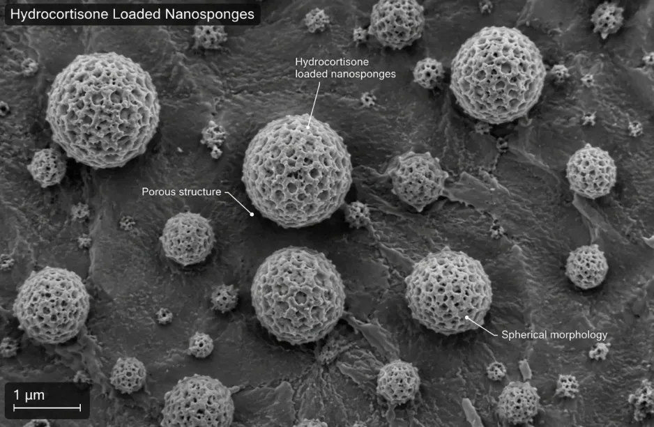
The prepared formulation appeared homogeneous without phase separation or aggregation. SEM analysis showed spherical porous nanosponges, confirming successful nanosponge formation.

Particle Size and Polydispersity Index (PDI)

Particle size and PDI were measured using Dynamic Light Scattering (DLS).


Table: Particle size and PDI

Formulation

Average Particle Size (nm)

PDI

F1

178.3 ± 4.4

0.216

F2

196.5 ± 5.2

0.231

F3

221.7 ± 5.9

0.286


Discussion

The particle size ranged between 178–222 nm, which is suitable for topical drug delivery. PDI values below 0.3 indicated uniform particle distribution and good formulation homogeneity.

Visual Inspection and Surface Morphology Method

Prepared nanosponge suspensions were visually examined for uniformity, color, turbidity, and sedimentation. Surface morphology was observed under Scanning Electron Microscopy (SEM). Result:

 • The formulation appeared as a milky white, homogeneous suspension without any phase separation or aggregation.

SEM images revealed spherical, porous structures with rough surfaces, characteristic of nanosponges.


Table: Visual Inspection and Surface Morphology

Observation

Result

Color

White to off-white

Appearance

Homogeneous, smooth

Sedimentation

Absent

Morphology (SEM)

Spherical, porous


Figure: SEM of Hydrocortisone loaded Nanosponges


Drug Content and Entrapment Efficiency



Entrapment efficiency was determined using UV spectrophotometric analysis.


Table: Entrapment efficiency and drug content

Formulation

Entrapment Efficiency (%)

Drug Content (%)

F1

83.8 ± 2.1

88.6 ± 1.5

F2

87.5 ± 1.8

91.3 ± 1.4

F3

81.9 ± 2.3

86.9 ± 1.7


Discussion

The high entrapment efficiency confirmed successful encapsulation of hydrocortisone within nanosponge matrix. F2 formulation showed highest entrapment efficiency due to optimized polymer concentration.

Evaluation of Hydrogel

pH of Hydrogel


Table: pH of hydrogel

Formulation

pH

F1

6.4

F2

6.8

F3

6.6


Discussion

The pH values were found within acceptable skin pH range, indicating suitability for topical application without causing irritation.

Viscosity


Table: Viscosity of hydrogel

Formulation

Viscosity (cps)

F1

4280

F2

4520

F3

4395


Discussion

The viscosity values indicated good consistency and spreadability of hydrogel formulations suitable for topical administration.

Spreadability


Table: Spreadability of hydrogel

Formulation

Spreadability (g·cm/sec)

F1

18.6

F2

20.4

F3

19.1


DISCUSSION

Hydrogel formulations exhibited good spreadability, ensuring easy application over affected psoriatic skin.

Stability Studies

The optimized formulation was subjected to accelerated stability studies at 40°C ± 2°C / 75% RH for one month.


Table: Stability studies

Parameter

Initial

After 1 Month

Appearance

White, homogeneous

No significant change

Particle Size (nm)

178.3 ± 4.4

186.5 ± 5.0

Entrapment Efficiency (%)

87.5 ± 1.8

84.9 ± 2.0

Drug Content (%)

91.3 ± 1.4

88.6 ± 1.5


Discussion

No significant changes were observed in physical appearance, particle size, drug content, or entrapment efficiency after stability studies, indicating acceptable stability of the optimized hydrocortisone nanosponge hydrogel formulation.


CONCLUSION

The present study was undertaken with the objective of developing and evaluating a hydrocortisone nanosponges loaded hydrogel for topical delivery in the management of psoriasis. Psoriasis is a chronic inflammatory skin disorder that requires long-term therapy, and conventional topical formulations often suffer from limitations such as poor skin penetration, low bioavailability, frequent dosing, and associated side effects. Therefore, the development of an advanced drug delivery system capable of overcoming these limitations was considered essential.

In this research, hydrocortisone was selected as the model drug due to its well-established anti-inflammatory and immunosuppressive activity. The formulation strategy involved the preparation of nanosponges using the emulsion solvent diffusion method, followed by their incorporation into a Carbopol-based hydrogel. This dual delivery system was designed to achieve controlled drug release, enhanced skin retention, and improved therapeutic efficacy.

The nanosponges were successfully formulated with desirable physicochemical characteristics. The optimized formulation exhibited nanoscale particle size, uniform distribution, and porous morphology, which are essential for effective drug encapsulation and release. The entrapment efficiency was found to be satisfactory, indicating efficient incorporation of hydrocortisone within the nanosponge structure. These results confirmed that the selected method was suitable for the preparation of stable and efficient nanocarriers.

The incorporation of nanosponges into the hydrogel base was achieved without any compatibility issues. The prepared hydrogel exhibited acceptable physical properties, including smooth texture, good homogeneity, appropriate viscosity, and excellent spreadability. The pH of the formulation was found to be within the acceptable range for topical application, ensuring minimal risk of skin irritation. These characteristics indicate that the formulation is patient-friendly and suitable for dermal use.

In-vitro drug release studies demonstrated that the nanosponge formulation provided a controlled release profile, which was further prolonged upon incorporation into the hydrogel. The release pattern showed sustained drug delivery over an extended period, with no significant burst release observed. This behavior can be attributed to the porous structure of nanosponges, which act as reservoirs for the drug, and the hydrogel matrix, which provides an additional barrier to drug diffusion. Such a sustained release profile is highly beneficial in reducing the frequency of application and improving patient compliance.

The drug release kinetics analysis indicated that the release mechanism followed diffusion-controlled behavior, best described by the Higuchi and Korsmeyer–Peppas models. This suggests that the drug release is governed by a combination of diffusion and matrix-controlled mechanisms, which is ideal for achieving sustained therapeutic action.

Stability studies conducted under accelerated conditions demonstrated that the formulation remained stable with minimal changes in drug content and physical properties over time. This confirms the robustness and suitability of the formulation for long-term storage and practical use.

Overall, the findings of the study clearly indicate that the hydrocortisone nanosponges loaded hydrogel is a promising and effective topical drug delivery system. The formulation successfully addresses the limitations of conventional therapies by providing controlled drug release, enhanced skin permeation, improved stability, and better patient compliance. The integration of nanosponges with a hydrogel base offers a synergistic effect, resulting in a dual-controlled release system that enhances therapeutic performance.

In conclusion, the developed formulation represents a significant advancement in topical drug delivery for psoriasis management. It has the potential to improve treatment outcomes, reduce side effects, and enhance patient adherence. The study provides a strong foundation for further research and development in this area and supports the application of nanotechnology-based systems in dermatological therapy.

REFERENCES

  1. Shetty K, Sherje AP. Nano intervention in topical delivery of corticosteroid for psoriasis and atopic dermatitis-a systematic review. J Mater Sci Mater Med. 2021;32(8):88.
  2. Mhetre V, Chaudhari PM. Hydrogel-driven psoriasis therapies: Mechanisms, applications, and future directions. Indian J Physiol Allied Sci. 2025;77(4):4-13.
  3. Xiao L, Kuang X, Zhang T, Tang Y, Wu J, Zheng Y. Research progress of biomaterial-based hydrogels in the treatment of psoriasis. Int J Pharm. 2025;677:125190.
  4. Parisi R, Iskandar IYK, Kontopantelis E, Augustin M, Griffiths CEM, Ashcroft DM. National, regional, and worldwide epidemiology of psoriasis: systematic analysis and modelling study. BMJ. 2020;369:m1590.
  5. Rendon A, Schäkel K. Psoriasis pathogenesis and treatment. Int J Mol Sci. 2019;20(6):1475.
  6. Griffiths CEM, Armstrong AW, Gudjonsson JE, Barker JNWN. Psoriasis. Lancet. 2021;397(10281):1301-1315.
  7. Nair RP, Duffin KC, Helms C, et al. Genome-wide scan reveals association of psoriasis with IL-23 and NF-κB pathways. Nat Genet. 2009;41(2):199-204.
  8. Kamiya K, Kishimoto M, Sugai J, Komine M, Ohtsuki M. Risk factors for the development of psoriasis. Int J Mol Sci. 2019;20(18):4347.
  9. Zeng J, Luo S, Huang Y, Lu Q. Critical role of environmental factors in the pathogenesis of psoriasis. J Dermatol. 2017;44(8):863-872.
  10. Hawkes JE, Yan BY, Chan TC, Krueger JG. Discovery of the IL-23/IL-17 signaling pathway and the treatment of psoriasis. J Immunol. 2018;201(6):1605-1613.
  11. Lowes MA, Suárez-Fariñas M, Krueger JG. Immunology of psoriasis. Annu Rev Immunol. 2014;32:227-255.
  12. Zhou X, Chen Y, Cui L, Shi Y, Guo C. Advances in the pathogenesis of psoriasis: from keratinocyte perspective. Cell Death Dis. 2022;13(1):81.
  13. Takeshita J, Grewal S, Langan SM, et al. Psoriasis and comorbid diseases: Epidemiology. J Am Acad Dermatol. 2017;76(3):377-390.
  14. Menter A, Gelfand JM, Connor C, et al. Joint American Academy of Dermatology-National Psoriasis Foundation guidelines of care for the management of psoriasis with systemic nonbiologic therapies. J Am Acad Dermatol. 2020;82(6):1445-1486.
  15. Elmets CA, Korman NJ, Prater EF, et al. Joint AAD-NPF guidelines of care for the management and treatment of psoriasis with topical therapy and alternative medicine modalities for psoriasis severity measures. J Am Acad Dermatol. 2021;84(2):432-470.
  16. Shetty K, Sherje AP. Nano intervention in topical delivery of corticosteroid for psoriasis and atopic dermatitis-a systematic review. J Mater Sci Mater Med. 2021;32(8):88.
  17. Ference JD, Last AR. Choosing topical corticosteroids. Am Fam Physician. 2009;79(2):135-140.
  18. Alkilani AZ, McCrudden MTC, Donnelly RF. Transdermal drug delivery: innovative pharmaceutical developments based on disruption of the barrier properties of the stratum corneum. Pharmaceutics. 2015;7(4):438-470.
  19. Hengge UR, Ruzicka T, Schwartz RA, Cork MJ. Adverse effects of topical glucocorticosteroids. J Am Acad Dermatol. 2006;54(1):1-15.
  20. Gupta R, Dwadasi BS, Rai B, Mitragotri S. Effect of chemical permeation enhancers on skin permeability: In silico screening using molecular dynamics simulations. Sci Rep. 2019;9(1):1456.
  21. Prow TW, Grice JE, Lin LL, et al. Nanoparticles and microparticles for skin drug delivery. Adv Drug Deliv Rev. 2011;63(6):470-491.
  22. Sala M, Diab R, Elaissari A, Fessi H. Lipid nanocarriers as skin drug delivery systems: Properties, mechanisms of skin interactions and medical applications. Int J Pharm. 2018;535(1-2):1-17.
  23. N N A, Sharma A, Chauhan N, Singh U, Sikarwar GS, Madhuria N. Nanosponges as a revolutionary drug delivery system for skin disorders: advancements, challenges, and future perspectives. J Neonatal Surg. 2025;14(15S):544-555.
  24. Attri M, Raghav A, Rao K, Pandey P, Minocha N. A review on nanosponges: an idiosyncratic approach for delivery of proactive molecules. Int J Pharm Sci Nanotechnol. 2024;9(3):193-208.
  25. Trotta F, Zanetti M, Cavalli R. Cyclodextrin-based nanosponges as drug carriers. Beilstein J Org Chem. 2012;8:2091-2099.
  26. Swaminathan S, Pastero L, Serpe L, et al. Cyclodextrin-based nanosponges encapsulating camptothecin: physicochemical characterization, stability and cytotoxicity. Eur J Pharm Biopharm. 2010;74(2):193-201.
  27. Cavalli R, Trotta F, Tumiatti W. Cyclodextrin-based nanosponges for drug delivery. J Incl Phenom Macrocycl Chem. 2006;56(1-2):209-213.
  28. Tejashri G, Amrita B, Darshana J. Cyclodextrin-based nanosponges for pharmaceutical use: A review. Acta Pharm. 2013;63(3):335-358.
  29. Ansari KA, Torne SJ, Vavia PR, Trotta F, Cavalli R. Cyclodextrin-based nanosponges for delivery of resveratrol: In-vitro characterisation. J Incl Phenom Macrocycl Chem. 2011;69(1-2):139-147.
  30. Bhowmik H, Venkatesh DN, Kuila A, Kumar KH. Nanosponges: A review. Int J Appl Pharm. 2018;10(4):1-5.
  31. Lopedota AA, Denora N, Iacobellis NS, et al. Cyclodextrin-based nanosponges for topical delivery of poorly soluble drugs. Int J Pharm. 2021;606:120879.
  32. Date RB, Mulla JAS. Nanosponges loaded hydrogels for anti-inflammatory drug delivery: characterization evaluation and applications. Asian J Pharm Res. 2026;16(1):27-32.
  33. Farsana P, Sivakumar R, Haribabu Y. Hydrogel-based nanosponges drug delivery for topical applications: An updated review. Res J Pharm Technol. 2021;14(1):527-530.
  34. Peppas NA, Hoffman AS. Hydrogels for biomedical applications. Adv Drug Deliv Rev. 2021;168:1-3.
  35. Bashir S, Hina M, Iqbal J, et al. Fundamental concepts of hydrogels: Synthesis, properties, and their applications. Polymers (Basel). 2020;12(11):2702.
  36. Ho TC, Chang CC, Chan HP, et al. Hydrogels: Properties and applications in biomedicine. Molecules. 2022;27(9):2902.
  37. Jacob S, Nair AB, Shah J, Sreeharsha N, Gupta S, Shinu P. Emerging role of hydrogels in drug delivery systems for treatment of skin diseases. Gels. 2021;7(4):253.
  38. Ullah F, Othman MBH, Javed F, Ahmad Z, Akil HM. Classification, processing and application of hydrogels: A review. Mater Sci Eng C Mater Biol Appl. 2015;57:414-433.
  39. Mulla JAS, Karande BS. Microemulsion based hydrogel formulation for topical drug delivery - A concise review. Indian J Novel Drug Deliv. 2021;13(2):63-69.
  40. Sala M, Elaissari A, Fessi H. Advances in psoriasis physiopathology and treatments: Up to date of mechanistic insights and perspectives of novel therapies based on innovative skin drug delivery systems (ISDDS). J Control Release. 2016;239:182-202.
  41. Kumar S, Prasad M, Rao R. Topical delivery of clobetasol propionate loaded nanosponge hydrogel for effective treatment of psoriasis: Formulation, physicochemical characterization, antipsoriatic potential and biochemical estimation. Mater Sci Eng C Mater Biol Appl.
  42. Gün, E., Sevgi, S., Demirçi, B. et al. A review of current therapeutics for psoriasis: Systemic and topical use of non-biological drugs. Eur J Clin Pharmacol 82, 53 (2026). https://doi.org/10.1007/s00228-025-03986-5
  43. Armstrong, A.W., Blauvelt, A., Callis Duffin, K. et al. Psoriasis. Nat Rev Dis Primers 11, 45 (2025). https://doi.org/10.1038/s41572-025-00630-5
  44. Aneja, P., Guleria, R., & Dahiya, D. P. (2025). Targeted drug delivery with nanosponges and nanocrystals: Innovations, formulation strategies, and applications. ASSAY and Drug Development Technologies, 23(7), 337–366. https://doi.org/10.1089/adt.2025.026
  45. Gupta, M., Reddy, V. R., Alam, M. S., Rajput, S. S., Mohammed, J. S., Pawara, P., Taru, P. P., Sahu, B., Singh, A., & Mandal, S. (2025). Development of sulfasalazine-loaded nanosponges in hydrogel for enhanced topical psoriasis management. Journal of Neonatal Surgery, 14, Article 2571. https://doi.org/10.52783/jns.v14.2571
  46. Ez-zoubi, A., El Fadili, M., Fadil, M., Alami, A., & Farah, A. (2025). Synthesis of carbonate beta-cyclodextrin nanosponges utilizing choline chloride-ZnCl2 deep eutectic solvent. Journal of the Indian Chemical Society, 102(6), 101722. https://doi.org/10.1016/j.jics.2025.101722
  47. Kumar, S., Nair, A. B., Kadian, V., Dalal, P., Jangir, B. L., Aldhubiab, B., Almuqbil, R. M., Alnaim, A. S., Alwadei, N., & Rao, R. (2025). Development and evaluation of hydrogel-based sulfasalazine-loaded nanosponges for enhanced topical psoriasis therapy. Pharmaceuticals, 18(3), 391. https://doi.org/10.3390/ph18030391
  48. Xiao, L., Kuang, X., Zhang, T., Tang, Y., Wu, J., & Zheng, Y. (2025). Research progress of biomaterial-based hydrogels in the treatment of psoriasis. International Journal of Pharmaceutics, 686, 126353. https://doi.org/10.1016/j.ijpharm.2025.12635
  49. Parihar, A., Paliwal, H., Kaur, J., Pal, S., Prajapati, B., & Sharma, R. (2025). Advancing psoriasis care: Efficacy of nanosuspensions in targeted skin therapies. Biomedical Materials & Devices. https://doi.org/10.1007/s44174-025-00590-0
  50. Kumar, S., Nair, A. B., Kadian, V., Dalal, P., Jangir, B. L., Aldhubiab, B., Almuqbil, R. M., Alnaim, A. S., Alwadei, N., & Rao, R. (2025). Development and evaluation of hydrogel-based sulfasalazine-loaded nanosponges for enhanced topical psoriasis therapy. Pharmaceuticals, 18(3), 391. https://doi.org/10.3390/ph18030391
  51. Alam, M. R., Ansari, V. A., Singh, A., Ansari, T. M., & Ahsan, F. (2025). An overview of potential nanotechnological methods for psoriasis treatment. Journal of Chemical Health Risks, 15(1), 380–390. https://jchr.org/index.php/JCHR/article/view/7423
  52. Bodnár, K., Fehér, P., Ujhelyi, Z., Bácskay, I., & Józsa, L. (2024). Recent approaches for the topical treatment of psoriasis using nanoparticles. Pharmaceutics, 16(4), 449. https://doi.org/10.3390/pharmaceutics16040449
  53. Cao, H., Wang, Y., Li, X., Zhang, L., & Chen, J. (2024). Hydrogel-based topical drug delivery systems for psoriasis therapy: Recent advances and future perspectives. International Journal of Biological Macromolecules, 276, 133845. https://doi.org/10.1016/j.ijbiomac.2024.133845
  54. Sundar, V. D., Dhanaraju, M. D., Vadaga, A., & Madhulatha, S. N. S. V. (2024). Design and evaluation of telmisartan-loaded nanosponges for hypertension treatment. Advances in Bioresearch, 15(4), 1–13. https://doi.org/10.15515/abr.0976-4585.15.4.113
  55. Utzeri, G., Matias, P. M. C., Murtinho, D., & Valente, A. J. M. (2022). Cyclodextrin-based nanosponges: Overview and opportunities. Frontiers in Chemistry, 10, 859406. https://doi.org/10.3389/fchem.2022.859406
  56. Pawar, A. Y., Jadhav, K. R., Rathod, S. P., Sanap, A. S., & Umekar, M. J. (2023). Formulation and evaluation of nanosponges loaded hydrogel of metformin hydrochloride. Indian Journal of Pharmaceutical Education and Research, 57(1), 53–61. https://doi.org/10.5530/001954641980
  57. Vij, M., Dand, N., Kumar, L., Wadhwa, P., Wani, S. U. D., Mahdi, W. A., Alshehri, S., Alam, P., & Shakeel, F. (2023). Optimisation of a greener-approach for the synthesis of cyclodextrin-based nanosponges for the solubility enhancement of domperidone, a BCS class II drug. Pharmaceuticals, 16(4), 567. https://doi.org/10.3390/ph16040567
  58. Kumar, S., Jangir, B. L., & Rao, R. (2022). A new perspective for psoriasis: Dithranol nanosponge loaded hydrogels. Applied Surface Science Advances, 12, 100347. https://doi.org/10.1016/j.apsadv.2022.100347
  59. Rana, K., Sharma, R., & Sharma, A. K. (2022). Current understanding of psoriasis pathogenesis and therapeutic approaches targeting inflammatory pathways. International Immunopharmacology, 113, 109322. https://doi.org/10.1016/j.intimp.2022.109322
  60. awar, A. Y., Jadhav, K. R., Rao, J. B., Tapkir, A. D., Malpure, P. S., & Bachhav, R. S. (2022). Development and characterization of griseofulvin nanosponges to enhance bioavailability. International Journal of Life Science and Pharma Research, 12(5), P99–P111. https://doi.org/10.22376/ijpbs/lpr.2022.12.5.P99-111
  61. Rodrigues, D., Barbosa, A. I., Costa Lima, S. A., & Reis, S. (2022). Smart topical delivery systems based on nanocarriers for anti-inflammatory therapy. Pharmaceutics, 14(6), 1167. https://doi.org/10.3390/pharmaceutics14061167
  62. Almutairy, B. K., Alharbi, W. S., Alruwaili, N. K., Alshahrani, S. M., Alanazi, F. K., & Alshehri, S. (2021). Development and evaluation of olmesartan medoxomil-loaded nanosponges for enhancement of solubility and antihypertensive activity. Gels, 7(4), 190. https://doi.org/10.3390/gels7040190
  63. El-Assal, M. I., El-Sayed, A. M., & El-Houssieny, B. M. (2019). Development and evaluation of nifedipine loaded nanosponges for controlled drug delivery. Bulletin of Faculty of Pharmacy, Cairo University, 57(2), 251–261. https://doi.org/10.1016/j.bfopcu.2019.06.001
  64. Rao, M. R. P., Borate, S. G., Thanki, K. C., & Ranpise, A. A. (2018). Preparation and characterization of β-cyclodextrin based nanosponges for solubility enhancement of rilpivirine. Journal of Drug Delivery Science and Technology, 45, 255–264. https://doi.org/10.1016/j.jddst.2018.03.021
  65. Brayfield, A. (Ed.). (2024). Martindale: The complete drug reference (42nd ed.). Pharmaceutical Press.
  66. Katzung, B. G., & Vanderah, T. W. (2021). Basic and clinical pharmacology (15th ed.). McGraw-Hill Education.
  67. Griffiths, C. E. M., Armstrong, A. W., Gudjonsson, J. E., & Barker, J. N. W. N. (2021). Psoriasis. The Lancet, 397(10281), 1301–1315. https://doi.org/10.1016/S0140-6736(20)32549-6
  68. Date, R. B., & Mulla, J. A. S. (2026). A review of current therapeutics for psoriasis: Systemic and topical use of non-biological drugs. European Journal of Clinical Pharmacology, 82(2), 53. https://doi.org/10.1007/s00228-025-03986-5
  69. Kumar, S., Jangir, B. L., & Rao, R. (2022). A new perspective for psoriasis: Dithranol nanosponge loaded hydrogels. Applied Surface Science Advances, 12, 100347. https://doi.org/10.1016/j.apsadv.2022.100347
  70. Katzung, B. G., & Vanderah, T. W. (2021). Basic and clinical pharmacology (15th ed.). McGraw-Hill Education.
  71. Rowe, R. C., Sheskey, P. J., & Quinn, M. E. (Eds.). (2020). Handbook of pharmaceutical excipients (8th ed.). Pharmaceutical Press.
  72. https://pubchem.ncbi.nlm.nih.gov/compound/Ethyl-cellulose
  73. https://pubchem.ncbi.nlm.nih.gov/compound/Polyvinyl-alcohol
  74. https://pubchem.ncbi.nlm.nih.gov/compound/Dichloromethane
  75. https://pubchem.ncbi.nlm.nih.gov/compound/Carbomer
  76. Sinko, P. J. (2017). Martin’s physical pharmacy and pharmaceutical sciences (7th ed.). Wolters Kluwer.
  77. Beckett, A. H., & Stenlake, J. B. (2018). Practical pharmaceutical chemistry (4th ed.). CBS Publishers & Distributors.
  78. Indian Pharmacopoeia Commission. (2022). Indian Pharmacopoeia (Vol. II). Ministry of Health and Family Welfare, Government of India.
  79. United States Pharmacopeial Convention. (2023). United States Pharmacopeia and National Formulary (USP–NF). USP Convention.
  80. Aulton, M. E., & Taylor, K. (2018). Aulton’s pharmaceutics: The design and manufacture of medicines (5th ed.). Elsevier.
  81. Chatwal, G. R., & Anand, S. K. (2019). Instrumental methods of chemical analysis (6th ed.). Himalaya Publishing House.
  82. Snyder, L. R., Kirkland, J. J., & Dolan, J. W. (2020). Introduction to modern liquid chromatography (4th ed.). Wiley.
  83. Stuart, B. (2015). Infrared spectroscopy: Fundamentals and applications. Wiley.
  84. Dong, M. W. (2019). Modern HPLC for practicing scientists. Wiley.
  85. Sharma, P., Dinesh, K., & Singh, R. (2021). Nanosponges: A novel approach for drug delivery system. Drug Development and Industrial Pharmacy, 47(6), 903–915.
  86. Trotta, F. (2013). Cyclodextrin nanosponges and their applications. Journal of Inclusion Phenomena and Macrocyclic Chemistry, 80, 17–25.
  87. Allen, L. V., Popovich, N. G., & Ansel, H. C. (2014). Ansel’s pharmaceutical dosage forms and drug delivery systems (10th ed.). Wolters Kluwer.
  88. Danaei, M., Dehghankhold, M., Ataei, S., Hasanzadeh Davarani, F., Javanmard, R., Dokhani, A., Khorasani, S., & Mozafari, M. R. (2018). Impact of particle size and polydispersity index on the clinical applications of lipidic nanocarrier systems. Pharmaceutics, 10(2), 57. https://doi.org/10.3390/pharmaceutics10020057
  89. International Organization for Standardization. (2017). ISO 22412: Particle size analysis — Dynamic light scattering (DLS). ISO.
  90. Goldstein, J., Newbury, D. E., Michael, J. R., Ritchie, N. W. M., Scott, J. H. J., & Joy, D. C. (2018). Scanning electron microscopy and X-ray microanalysis (4th ed.). Springer.
  91. Bhattacharjee, S. (2016). DLS and zeta potential – What they are and what they are not? Journal of Controlled Release, 235, 337–351. https://doi.org/10.1016/j.jconrel.2016.06.017
  92. ISO. (2016). ISO 13099-2: Colloidal systems — Methods for zeta-potential determination. International Organization for Standardization.
  93. Brookfield Engineering Laboratories. (2020). Brookfield viscometers: Operating manual and viscosity measurement principles. AMETEK Brookfield.
  94. Lachman, L., Lieberman, H. A., & Kanig, J. L. (2014). The theory and practice of industrial pharmacy (4th ed.). CBS Publishers & Distributors.
  95. Franz, T. J. (1975). Percutaneous absorption. On the relevance of in-vitro data. Journal of Investigative Dermatology, 64(3), 190–195. https://doi.org/10.1111/1523-1747.ep12533356
  96. Hadgraft, J., & Lane, M. E. (2010). Skin permeation: The years of enlightenment. International Journal of Pharmaceutics, 415(1–2), 2–5. https://doi.org/10.1016/j.ijpharm.2011.02.039
  97. International Council for Harmonisation (ICH). (2003). ICH Q1A(R2): Stability testing of new drug substances and products. ICH Harmonised Tripartite Guideline.
  98. World Health Organization. (2018). WHO guidelines on stability testing of pharmaceutical products. WHO Technical Report Series.

International Council for Harmonisation. (2003). ICH Q1B: Photostability testing of new drug substances and products. ICH Harmonised Tripartite


Reference

  1. Shetty K, Sherje AP. Nano intervention in topical delivery of corticosteroid for psoriasis and atopic dermatitis-a systematic review. J Mater Sci Mater Med. 2021;32(8):88.
  2. Mhetre V, Chaudhari PM. Hydrogel-driven psoriasis therapies: Mechanisms, applications, and future directions. Indian J Physiol Allied Sci. 2025;77(4):4-13.
  3. Xiao L, Kuang X, Zhang T, Tang Y, Wu J, Zheng Y. Research progress of biomaterial-based hydrogels in the treatment of psoriasis. Int J Pharm. 2025;677:125190.
  4. Parisi R, Iskandar IYK, Kontopantelis E, Augustin M, Griffiths CEM, Ashcroft DM. National, regional, and worldwide epidemiology of psoriasis: systematic analysis and modelling study. BMJ. 2020;369:m1590.
  5. Rendon A, Schäkel K. Psoriasis pathogenesis and treatment. Int J Mol Sci. 2019;20(6):1475.
  6. Griffiths CEM, Armstrong AW, Gudjonsson JE, Barker JNWN. Psoriasis. Lancet. 2021;397(10281):1301-1315.
  7. Nair RP, Duffin KC, Helms C, et al. Genome-wide scan reveals association of psoriasis with IL-23 and NF-κB pathways. Nat Genet. 2009;41(2):199-204.
  8. Kamiya K, Kishimoto M, Sugai J, Komine M, Ohtsuki M. Risk factors for the development of psoriasis. Int J Mol Sci. 2019;20(18):4347.
  9. Zeng J, Luo S, Huang Y, Lu Q. Critical role of environmental factors in the pathogenesis of psoriasis. J Dermatol. 2017;44(8):863-872.
  10. Hawkes JE, Yan BY, Chan TC, Krueger JG. Discovery of the IL-23/IL-17 signaling pathway and the treatment of psoriasis. J Immunol. 2018;201(6):1605-1613.
  11. Lowes MA, Suárez-Fariñas M, Krueger JG. Immunology of psoriasis. Annu Rev Immunol. 2014;32:227-255.
  12. Zhou X, Chen Y, Cui L, Shi Y, Guo C. Advances in the pathogenesis of psoriasis: from keratinocyte perspective. Cell Death Dis. 2022;13(1):81.
  13. Takeshita J, Grewal S, Langan SM, et al. Psoriasis and comorbid diseases: Epidemiology. J Am Acad Dermatol. 2017;76(3):377-390.
  14. Menter A, Gelfand JM, Connor C, et al. Joint American Academy of Dermatology-National Psoriasis Foundation guidelines of care for the management of psoriasis with systemic nonbiologic therapies. J Am Acad Dermatol. 2020;82(6):1445-1486.
  15. Elmets CA, Korman NJ, Prater EF, et al. Joint AAD-NPF guidelines of care for the management and treatment of psoriasis with topical therapy and alternative medicine modalities for psoriasis severity measures. J Am Acad Dermatol. 2021;84(2):432-470.
  16. Shetty K, Sherje AP. Nano intervention in topical delivery of corticosteroid for psoriasis and atopic dermatitis-a systematic review. J Mater Sci Mater Med. 2021;32(8):88.
  17. Ference JD, Last AR. Choosing topical corticosteroids. Am Fam Physician. 2009;79(2):135-140.
  18. Alkilani AZ, McCrudden MTC, Donnelly RF. Transdermal drug delivery: innovative pharmaceutical developments based on disruption of the barrier properties of the stratum corneum. Pharmaceutics. 2015;7(4):438-470.
  19. Hengge UR, Ruzicka T, Schwartz RA, Cork MJ. Adverse effects of topical glucocorticosteroids. J Am Acad Dermatol. 2006;54(1):1-15.
  20. Gupta R, Dwadasi BS, Rai B, Mitragotri S. Effect of chemical permeation enhancers on skin permeability: In silico screening using molecular dynamics simulations. Sci Rep. 2019;9(1):1456.
  21. Prow TW, Grice JE, Lin LL, et al. Nanoparticles and microparticles for skin drug delivery. Adv Drug Deliv Rev. 2011;63(6):470-491.
  22. Sala M, Diab R, Elaissari A, Fessi H. Lipid nanocarriers as skin drug delivery systems: Properties, mechanisms of skin interactions and medical applications. Int J Pharm. 2018;535(1-2):1-17.
  23. N N A, Sharma A, Chauhan N, Singh U, Sikarwar GS, Madhuria N. Nanosponges as a revolutionary drug delivery system for skin disorders: advancements, challenges, and future perspectives. J Neonatal Surg. 2025;14(15S):544-555.
  24. Attri M, Raghav A, Rao K, Pandey P, Minocha N. A review on nanosponges: an idiosyncratic approach for delivery of proactive molecules. Int J Pharm Sci Nanotechnol. 2024;9(3):193-208.
  25. Trotta F, Zanetti M, Cavalli R. Cyclodextrin-based nanosponges as drug carriers. Beilstein J Org Chem. 2012;8:2091-2099.
  26. Swaminathan S, Pastero L, Serpe L, et al. Cyclodextrin-based nanosponges encapsulating camptothecin: physicochemical characterization, stability and cytotoxicity. Eur J Pharm Biopharm. 2010;74(2):193-201.
  27. Cavalli R, Trotta F, Tumiatti W. Cyclodextrin-based nanosponges for drug delivery. J Incl Phenom Macrocycl Chem. 2006;56(1-2):209-213.
  28. Tejashri G, Amrita B, Darshana J. Cyclodextrin-based nanosponges for pharmaceutical use: A review. Acta Pharm. 2013;63(3):335-358.
  29. Ansari KA, Torne SJ, Vavia PR, Trotta F, Cavalli R. Cyclodextrin-based nanosponges for delivery of resveratrol: In-vitro characterisation. J Incl Phenom Macrocycl Chem. 2011;69(1-2):139-147.
  30. Bhowmik H, Venkatesh DN, Kuila A, Kumar KH. Nanosponges: A review. Int J Appl Pharm. 2018;10(4):1-5.
  31. Lopedota AA, Denora N, Iacobellis NS, et al. Cyclodextrin-based nanosponges for topical delivery of poorly soluble drugs. Int J Pharm. 2021;606:120879.
  32. Date RB, Mulla JAS. Nanosponges loaded hydrogels for anti-inflammatory drug delivery: characterization evaluation and applications. Asian J Pharm Res. 2026;16(1):27-32.
  33. Farsana P, Sivakumar R, Haribabu Y. Hydrogel-based nanosponges drug delivery for topical applications: An updated review. Res J Pharm Technol. 2021;14(1):527-530.
  34. Peppas NA, Hoffman AS. Hydrogels for biomedical applications. Adv Drug Deliv Rev. 2021;168:1-3.
  35. Bashir S, Hina M, Iqbal J, et al. Fundamental concepts of hydrogels: Synthesis, properties, and their applications. Polymers (Basel). 2020;12(11):2702.
  36. Ho TC, Chang CC, Chan HP, et al. Hydrogels: Properties and applications in biomedicine. Molecules. 2022;27(9):2902.
  37. Jacob S, Nair AB, Shah J, Sreeharsha N, Gupta S, Shinu P. Emerging role of hydrogels in drug delivery systems for treatment of skin diseases. Gels. 2021;7(4):253.
  38. Ullah F, Othman MBH, Javed F, Ahmad Z, Akil HM. Classification, processing and application of hydrogels: A review. Mater Sci Eng C Mater Biol Appl. 2015;57:414-433.
  39. Mulla JAS, Karande BS. Microemulsion based hydrogel formulation for topical drug delivery - A concise review. Indian J Novel Drug Deliv. 2021;13(2):63-69.
  40. Sala M, Elaissari A, Fessi H. Advances in psoriasis physiopathology and treatments: Up to date of mechanistic insights and perspectives of novel therapies based on innovative skin drug delivery systems (ISDDS). J Control Release. 2016;239:182-202.
  41. Kumar S, Prasad M, Rao R. Topical delivery of clobetasol propionate loaded nanosponge hydrogel for effective treatment of psoriasis: Formulation, physicochemical characterization, antipsoriatic potential and biochemical estimation. Mater Sci Eng C Mater Biol Appl.
  42. Gün, E., Sevgi, S., Demirçi, B. et al. A review of current therapeutics for psoriasis: Systemic and topical use of non-biological drugs. Eur J Clin Pharmacol 82, 53 (2026). https://doi.org/10.1007/s00228-025-03986-5
  43. Armstrong, A.W., Blauvelt, A., Callis Duffin, K. et al. Psoriasis. Nat Rev Dis Primers 11, 45 (2025). https://doi.org/10.1038/s41572-025-00630-5
  44. Aneja, P., Guleria, R., & Dahiya, D. P. (2025). Targeted drug delivery with nanosponges and nanocrystals: Innovations, formulation strategies, and applications. ASSAY and Drug Development Technologies, 23(7), 337–366. https://doi.org/10.1089/adt.2025.026
  45. Gupta, M., Reddy, V. R., Alam, M. S., Rajput, S. S., Mohammed, J. S., Pawara, P., Taru, P. P., Sahu, B., Singh, A., & Mandal, S. (2025). Development of sulfasalazine-loaded nanosponges in hydrogel for enhanced topical psoriasis management. Journal of Neonatal Surgery, 14, Article 2571. https://doi.org/10.52783/jns.v14.2571
  46. Ez-zoubi, A., El Fadili, M., Fadil, M., Alami, A., & Farah, A. (2025). Synthesis of carbonate beta-cyclodextrin nanosponges utilizing choline chloride-ZnCl2 deep eutectic solvent. Journal of the Indian Chemical Society, 102(6), 101722. https://doi.org/10.1016/j.jics.2025.101722
  47. Kumar, S., Nair, A. B., Kadian, V., Dalal, P., Jangir, B. L., Aldhubiab, B., Almuqbil, R. M., Alnaim, A. S., Alwadei, N., & Rao, R. (2025). Development and evaluation of hydrogel-based sulfasalazine-loaded nanosponges for enhanced topical psoriasis therapy. Pharmaceuticals, 18(3), 391. https://doi.org/10.3390/ph18030391
  48. Xiao, L., Kuang, X., Zhang, T., Tang, Y., Wu, J., & Zheng, Y. (2025). Research progress of biomaterial-based hydrogels in the treatment of psoriasis. International Journal of Pharmaceutics, 686, 126353. https://doi.org/10.1016/j.ijpharm.2025.12635
  49. Parihar, A., Paliwal, H., Kaur, J., Pal, S., Prajapati, B., & Sharma, R. (2025). Advancing psoriasis care: Efficacy of nanosuspensions in targeted skin therapies. Biomedical Materials & Devices. https://doi.org/10.1007/s44174-025-00590-0
  50. Kumar, S., Nair, A. B., Kadian, V., Dalal, P., Jangir, B. L., Aldhubiab, B., Almuqbil, R. M., Alnaim, A. S., Alwadei, N., & Rao, R. (2025). Development and evaluation of hydrogel-based sulfasalazine-loaded nanosponges for enhanced topical psoriasis therapy. Pharmaceuticals, 18(3), 391. https://doi.org/10.3390/ph18030391
  51. Alam, M. R., Ansari, V. A., Singh, A., Ansari, T. M., & Ahsan, F. (2025). An overview of potential nanotechnological methods for psoriasis treatment. Journal of Chemical Health Risks, 15(1), 380–390. https://jchr.org/index.php/JCHR/article/view/7423
  52. Bodnár, K., Fehér, P., Ujhelyi, Z., Bácskay, I., & Józsa, L. (2024). Recent approaches for the topical treatment of psoriasis using nanoparticles. Pharmaceutics, 16(4), 449. https://doi.org/10.3390/pharmaceutics16040449
  53. Cao, H., Wang, Y., Li, X., Zhang, L., & Chen, J. (2024). Hydrogel-based topical drug delivery systems for psoriasis therapy: Recent advances and future perspectives. International Journal of Biological Macromolecules, 276, 133845. https://doi.org/10.1016/j.ijbiomac.2024.133845
  54. Sundar, V. D., Dhanaraju, M. D., Vadaga, A., & Madhulatha, S. N. S. V. (2024). Design and evaluation of telmisartan-loaded nanosponges for hypertension treatment. Advances in Bioresearch, 15(4), 1–13. https://doi.org/10.15515/abr.0976-4585.15.4.113
  55. Utzeri, G., Matias, P. M. C., Murtinho, D., & Valente, A. J. M. (2022). Cyclodextrin-based nanosponges: Overview and opportunities. Frontiers in Chemistry, 10, 859406. https://doi.org/10.3389/fchem.2022.859406
  56. Pawar, A. Y., Jadhav, K. R., Rathod, S. P., Sanap, A. S., & Umekar, M. J. (2023). Formulation and evaluation of nanosponges loaded hydrogel of metformin hydrochloride. Indian Journal of Pharmaceutical Education and Research, 57(1), 53–61. https://doi.org/10.5530/001954641980
  57. Vij, M., Dand, N., Kumar, L., Wadhwa, P., Wani, S. U. D., Mahdi, W. A., Alshehri, S., Alam, P., & Shakeel, F. (2023). Optimisation of a greener-approach for the synthesis of cyclodextrin-based nanosponges for the solubility enhancement of domperidone, a BCS class II drug. Pharmaceuticals, 16(4), 567. https://doi.org/10.3390/ph16040567
  58. Kumar, S., Jangir, B. L., & Rao, R. (2022). A new perspective for psoriasis: Dithranol nanosponge loaded hydrogels. Applied Surface Science Advances, 12, 100347. https://doi.org/10.1016/j.apsadv.2022.100347
  59. Rana, K., Sharma, R., & Sharma, A. K. (2022). Current understanding of psoriasis pathogenesis and therapeutic approaches targeting inflammatory pathways. International Immunopharmacology, 113, 109322. https://doi.org/10.1016/j.intimp.2022.109322
  60. awar, A. Y., Jadhav, K. R., Rao, J. B., Tapkir, A. D., Malpure, P. S., & Bachhav, R. S. (2022). Development and characterization of griseofulvin nanosponges to enhance bioavailability. International Journal of Life Science and Pharma Research, 12(5), P99–P111. https://doi.org/10.22376/ijpbs/lpr.2022.12.5.P99-111
  61. Rodrigues, D., Barbosa, A. I., Costa Lima, S. A., & Reis, S. (2022). Smart topical delivery systems based on nanocarriers for anti-inflammatory therapy. Pharmaceutics, 14(6), 1167. https://doi.org/10.3390/pharmaceutics14061167
  62. Almutairy, B. K., Alharbi, W. S., Alruwaili, N. K., Alshahrani, S. M., Alanazi, F. K., & Alshehri, S. (2021). Development and evaluation of olmesartan medoxomil-loaded nanosponges for enhancement of solubility and antihypertensive activity. Gels, 7(4), 190. https://doi.org/10.3390/gels7040190
  63. El-Assal, M. I., El-Sayed, A. M., & El-Houssieny, B. M. (2019). Development and evaluation of nifedipine loaded nanosponges for controlled drug delivery. Bulletin of Faculty of Pharmacy, Cairo University, 57(2), 251–261. https://doi.org/10.1016/j.bfopcu.2019.06.001
  64. Rao, M. R. P., Borate, S. G., Thanki, K. C., & Ranpise, A. A. (2018). Preparation and characterization of β-cyclodextrin based nanosponges for solubility enhancement of rilpivirine. Journal of Drug Delivery Science and Technology, 45, 255–264. https://doi.org/10.1016/j.jddst.2018.03.021
  65. Brayfield, A. (Ed.). (2024). Martindale: The complete drug reference (42nd ed.). Pharmaceutical Press.
  66. Katzung, B. G., & Vanderah, T. W. (2021). Basic and clinical pharmacology (15th ed.). McGraw-Hill Education.
  67. Griffiths, C. E. M., Armstrong, A. W., Gudjonsson, J. E., & Barker, J. N. W. N. (2021). Psoriasis. The Lancet, 397(10281), 1301–1315. https://doi.org/10.1016/S0140-6736(20)32549-6
  68. Date, R. B., & Mulla, J. A. S. (2026). A review of current therapeutics for psoriasis: Systemic and topical use of non-biological drugs. European Journal of Clinical Pharmacology, 82(2), 53. https://doi.org/10.1007/s00228-025-03986-5
  69. Kumar, S., Jangir, B. L., & Rao, R. (2022). A new perspective for psoriasis: Dithranol nanosponge loaded hydrogels. Applied Surface Science Advances, 12, 100347. https://doi.org/10.1016/j.apsadv.2022.100347
  70. Katzung, B. G., & Vanderah, T. W. (2021). Basic and clinical pharmacology (15th ed.). McGraw-Hill Education.
  71. Rowe, R. C., Sheskey, P. J., & Quinn, M. E. (Eds.). (2020). Handbook of pharmaceutical excipients (8th ed.). Pharmaceutical Press.
  72. https://pubchem.ncbi.nlm.nih.gov/compound/Ethyl-cellulose
  73. https://pubchem.ncbi.nlm.nih.gov/compound/Polyvinyl-alcohol
  74. https://pubchem.ncbi.nlm.nih.gov/compound/Dichloromethane
  75. https://pubchem.ncbi.nlm.nih.gov/compound/Carbomer
  76. Sinko, P. J. (2017). Martin’s physical pharmacy and pharmaceutical sciences (7th ed.). Wolters Kluwer.
  77. Beckett, A. H., & Stenlake, J. B. (2018). Practical pharmaceutical chemistry (4th ed.). CBS Publishers & Distributors.
  78. Indian Pharmacopoeia Commission. (2022). Indian Pharmacopoeia (Vol. II). Ministry of Health and Family Welfare, Government of India.
  79. United States Pharmacopeial Convention. (2023). United States Pharmacopeia and National Formulary (USP–NF). USP Convention.
  80. Aulton, M. E., & Taylor, K. (2018). Aulton’s pharmaceutics: The design and manufacture of medicines (5th ed.). Elsevier.
  81. Chatwal, G. R., & Anand, S. K. (2019). Instrumental methods of chemical analysis (6th ed.). Himalaya Publishing House.
  82. Snyder, L. R., Kirkland, J. J., & Dolan, J. W. (2020). Introduction to modern liquid chromatography (4th ed.). Wiley.
  83. Stuart, B. (2015). Infrared spectroscopy: Fundamentals and applications. Wiley.
  84. Dong, M. W. (2019). Modern HPLC for practicing scientists. Wiley.
  85. Sharma, P., Dinesh, K., & Singh, R. (2021). Nanosponges: A novel approach for drug delivery system. Drug Development and Industrial Pharmacy, 47(6), 903–915.
  86. Trotta, F. (2013). Cyclodextrin nanosponges and their applications. Journal of Inclusion Phenomena and Macrocyclic Chemistry, 80, 17–25.
  87. Allen, L. V., Popovich, N. G., & Ansel, H. C. (2014). Ansel’s pharmaceutical dosage forms and drug delivery systems (10th ed.). Wolters Kluwer.
  88. Danaei, M., Dehghankhold, M., Ataei, S., Hasanzadeh Davarani, F., Javanmard, R., Dokhani, A., Khorasani, S., & Mozafari, M. R. (2018). Impact of particle size and polydispersity index on the clinical applications of lipidic nanocarrier systems. Pharmaceutics, 10(2), 57. https://doi.org/10.3390/pharmaceutics10020057
  89. International Organization for Standardization. (2017). ISO 22412: Particle size analysis — Dynamic light scattering (DLS). ISO.
  90. Goldstein, J., Newbury, D. E., Michael, J. R., Ritchie, N. W. M., Scott, J. H. J., & Joy, D. C. (2018). Scanning electron microscopy and X-ray microanalysis (4th ed.). Springer.
  91. Bhattacharjee, S. (2016). DLS and zeta potential – What they are and what they are not? Journal of Controlled Release, 235, 337–351. https://doi.org/10.1016/j.jconrel.2016.06.017
  92. ISO. (2016). ISO 13099-2: Colloidal systems — Methods for zeta-potential determination. International Organization for Standardization.
  93. Brookfield Engineering Laboratories. (2020). Brookfield viscometers: Operating manual and viscosity measurement principles. AMETEK Brookfield.
  94. Lachman, L., Lieberman, H. A., & Kanig, J. L. (2014). The theory and practice of industrial pharmacy (4th ed.). CBS Publishers & Distributors.
  95. Franz, T. J. (1975). Percutaneous absorption. On the relevance of in-vitro data. Journal of Investigative Dermatology, 64(3), 190–195. https://doi.org/10.1111/1523-1747.ep12533356
  96. Hadgraft, J., & Lane, M. E. (2010). Skin permeation: The years of enlightenment. International Journal of Pharmaceutics, 415(1–2), 2–5. https://doi.org/10.1016/j.ijpharm.2011.02.039
  97. International Council for Harmonisation (ICH). (2003). ICH Q1A(R2): Stability testing of new drug substances and products. ICH Harmonised Tripartite Guideline.
  98. World Health Organization. (2018). WHO guidelines on stability testing of pharmaceutical products. WHO Technical Report Series.

International Council for Harmonisation. (2003). ICH Q1B: Photostability testing of new drug substances and products. ICH Harmonised Tripartite

Photo
Ritika Rana
Corresponding author

Baddi university of emerging sciences and technology

Ritika Rana, Development and Evaluation of Hydrocortisone Nanosponges loaded Hydrogel for the Topical Treatment of Psoriasis, Int. J. of Pharm. Sci., 2026, Vol 4, Issue 7, 4081-4098, https://doi.org/10.5281/zenodo.21470715

More related articles
A Review on Dual Drug Niosomal Transdermal Patches...
Naveen V, Muhammed Sinan A K, Kannan V, Jeeva S, Asifullah R, Aru...
Green Synthesis, Characterization, And Antimycobac...
Sneha M.V. , Divya Easwaran, Ramesan C.K.V. , Prajina P., Ayana N...
Benzimidazole Derivatives: A Comprehensive Review ...
Tejal Patil, Arpita Kharche, Shailesh Malkar, Vitthal Chopade...
A Review on Curcumin-Piperine Synergy in Alopecia: Phytochemical Profiling, Mole...
Sanjana Mulay, Dr. Sachin Aglawe, Dr. Vijay Jadhav, Dr. Nitin Jain, Karveer Aghade...
Role of Medicinal Plant-Derived Homeopathic Mother Tinctures in Menstrual and Ho...
Riya Ghate, Prajakta Shukla, Sakshi Suraskar, Lipika Walde, Sejal Gajbhiye, Mahima Turkar, Kavita Ma...
Formulation And Evaluation of Herbal Dosage Forms Utilizing Brahmi...
Ayisha Bint Ashraf, Vyshnavy Devy D K, Fathimath Shijana M C, Fathimath Rizwana K, Anusree Pavithran...
Related Articles
A Review of Hot-Melt Extrusion : Process Technology to Pharmaceutical Products ...
Nikesh Kawana, Manisha Kitukale, Rushikesh Wanole, Harshal Yadav, Aditya kale...
Formulation And Evaluation of Antifungal Gel by Using Natural Polymer...
Jadhav Ankita, Kolhe Shruti, Kendre poonam, Hange Sanket...
A Review on Dual Drug Niosomal Transdermal Patches for Anti- Inflammatory Therap...
Naveen V, Muhammed Sinan A K, Kannan V, Jeeva S, Asifullah R, Arun Pandiyan R, Alagar Raja M, Christ...
More related articles
A Review on Dual Drug Niosomal Transdermal Patches for Anti- Inflammatory Therap...
Naveen V, Muhammed Sinan A K, Kannan V, Jeeva S, Asifullah R, Arun Pandiyan R, Alagar Raja M, Christ...
Green Synthesis, Characterization, And Antimycobacterial Activity Of Silver Nano...
Sneha M.V. , Divya Easwaran, Ramesan C.K.V. , Prajina P., Ayana N....
Benzimidazole Derivatives: A Comprehensive Review Of Synthesis, Biological Evalu...
Tejal Patil, Arpita Kharche, Shailesh Malkar, Vitthal Chopade...
A Review on Dual Drug Niosomal Transdermal Patches for Anti- Inflammatory Therap...
Naveen V, Muhammed Sinan A K, Kannan V, Jeeva S, Asifullah R, Arun Pandiyan R, Alagar Raja M, Christ...
Green Synthesis, Characterization, And Antimycobacterial Activity Of Silver Nano...
Sneha M.V. , Divya Easwaran, Ramesan C.K.V. , Prajina P., Ayana N....
Benzimidazole Derivatives: A Comprehensive Review Of Synthesis, Biological Evalu...
Tejal Patil, Arpita Kharche, Shailesh Malkar, Vitthal Chopade...