

We use cookies to ensure our website works properly and to personalise your experience. Cookies policy
IVM'S Krishnarao Bhegade Institute of Pharmaceutical Education and Research, Talegaon Dabhade, Pune.
Aptamers are short, single-stranded DNA or RNA oligonucleotides selected in vitro via SELEX (Systematic Evolution of Ligands by Exponential Enrichment) that fold into defined 3-D structures and bind cognate targets with antibody-like affinity yet superior versatility. Thirty-five years after their simultaneous disclosure by Ellington & Szostak and Tuerk & Gold, the technology has matured from proof-of-principle to clinical reality (pegaptanib, 2004) and, more recently, to precision tools for COVID-19, cancer and environmental surveillance. Continuous innovation cell-SELEX, capillary-electrophoresis SELEX, sol-gel multiplexing, magnetic-bead automation, microfluidic integration and AI-guided design has compressed selection times from weeks to hours and expanded target space from purified proteins to whole cells, small molecules and in vivo antigens. Consequently, aptamers now serve as diagnostic reagents in electrochemical and colorimetric biosensors, as therapeutics that block ligand-receptor signalling, as chaperones that ferry siRNAs, CRISPR RNPs or chemotherapy payloads to HER2-, PSMA- or CD19-expressing cells, and as imaging agents that deliver radionuclides or fluorophores with high signal-to-noise ratios. Their low cost, negligible batch-to-batch variability, thermal stability and ease of chemical conjugation position them as next-generation surrogates to antibodies in personalised medicine, point-of-care devices and field-deployable sensors. Ongoing efforts focus on nuclease resistance, renal-clearance modulation and large-scale GMP manufacture to broaden the clinical pipeline beyond ocular neovascularisation into oncology, thrombosis, neurodegeneration and regenerative medicine.
Since their initial description in 1990, aptamers have developed into highly advanced instruments in the domains of molecular biology, nanotechnology, and medicine. They exhibit remarkable selectivity and affinity for a variety of target molecules, including proteins, peptides, metal ions, tiny molecules, and even entire cells. By creating stable three-dimensional structures, single stranded oligonucleotides known as aptamers can attach to a variety of molecular targets with high affinity and selectivity [1]. Aptamers attach to protein targets and change the activity of proteins, just like antibodies do. On closer examination, it becomes evident that high affinity and high specificity interactions between oligonucleotides and proteins are a natural part of many biological processes, including transcription, translation, RNA interference, and others. This may seem counterintuitive to someone who is unfamiliar with aptamers [2]. Once the concept of high affinity oligonucleotide protein contact and its functional consequence is recognised, one might imagine that this potential can be leveraged pharmacologically to identify non-naturally occurring oligonucleotides that bind to target proteins of interest and affect protein function [3]. This provides the theoretical basis for therapeutic aptamers. The therapeutic principle has progressed to the point where many aptamers are presently undergoing clinical development, and one product the antivascular endothelial growth factor (VEGF) aptamer MacugenTM, is presently available for purchase. In recent years, aptamers have become important tools for improving point of care diagnostics, especially for infectious disorders. Because of their versatility and stability, they are used in lateral flow assays, electrochemical biosensors, and fluorescence based systems for the rapid detection of illnesses such as SARS-CoV-2, HIV, and bacterial toxins. Aptamers are highly useful for reacting to emergency epidemics because, unlike antibodies, they are cheap and simple to produce. Aptamers can also be designed to bind a target, impede its function, or act as carriers for targeted drug delivery by attaching themselves to therapeutic cargo or nanoparticles [4]. A potential use for this adaptability in personalised medicine is the ability to customise aptamers to bind certain biomarkers that are unique to a patient's disease profile. Their interoperability with microfluidics and lab on a chip technology further supports their integration into next generation diagnostic instruments. Aptamers have several important advantages over antibodies, including limited batch to batch variation, a longer shelf life, lower production costs, and little to no toxicity and immunogenicity [5]. The selection method typically employed to acquire them is SELEX (systematic evolution of ligands by exponential enrichment: Figure 1).
In the early 1990s, aptamers were initially created as molecular recognition components. An important turning point was the simultaneous publication of the SELEX (Systematic Evolution of Ligands by Exponential enrichment) method by two different research groups in 1990
Following their discovery:
Table No. 1: Historical Overview Table
|
Year |
Event |
|
1990 |
Discovery of RNA aptamers (Ellington & Szostak; Tuerk & Gold) |
|
1992 |
DNA aptamers introduced |
|
1995 |
Variants of SELEX developed [Cell SELEX , Counter SELESX ] |
|
2004 |
FDA approval of Macugen, first aptamer based drug |
|
2010 |
Advances in HTSELEX, SOMAmers, aptamerdrug conjugates |
|
2020 |
AI integration, COVID19 applications, new delivery methods |
Aptamers' Importance: Advantages Compared to Traditional Antibodies. Because they are powerful molecular recognition components with several obvious advantages over traditional antibodies, aptamers are crucial to both clinical and scientific research. Unlike antibodies, which are protein based and created by biological processes in animals, aptamers are short, synthetic nucleic acid sequences (DNA or RNA) selected in vitro, utilising the Systematic Evolution of Ligands by Exponential enrichment (SELEX) approach [10]. There are some noteworthy benefits to this synthetic nature: High purity and repeatability Scalable production and precise batch to batch homogeneity are made possible by the chemical synthesis of aptamers. Additionally, aptamers are safer to utilise in diagnosis and treatment since they are less immunogenic and toxic. They can swiftly enter tissue and attach to particular epitopes that larger antibodies (~150 kDa) are unable to reach due to their small size (~6-30 kDa) [11].
It is simple to chemically alter aptamers to enhance their pharmacokinetics and boost their nuclease resistance. They also have a high degree of thermal stability. Aptamers are incredibly versatile for a variety of applications, including targeted drug administration, biosensing, and molecular imaging. They can also be quickly created to bind a wide range of targets, including proteins, ions, small molecules, and even entire cells. Aptamers are in a good position to either replace or supplement antibodies in next-generation diagnostics and therapeutics because of all these benefits. The accompanying table No. 2 will help you comprehend the unique characteristics and benefits of aptamers over conventional antibodies [12].
Table No. 2: A comprehensive comparison of aptamers and antibodies
|
Feature |
Aptamers |
Antibodies |
|
Nature |
synthetic molecules of RNA or single-stranded DNA |
Immune system produced protein molecules |
|
Size |
Small (630kDa) |
Large (150kDa) |
|
Production Method |
SELEX in vitro chemical synthesis
|
In vivo manufacturing in cell cultures or animals |
|
Batch Consistency |
Highquality, precisely controlled chemical synthesis |
Batch variation may result from variable, biological manufacturing. |
|
Thermal Stability |
High; capable of withstanding a wide |
Moderate; sensitive to heat and storage conditions |
|
Target Diversity |
Wide range: small molecules, ions, proteins, cells |
Mainly proteins and larger antigens |
|
Modification |
Easily chemically modified (e.g., labeling, PEGylation) |
More challenging to chemically modify |
|
Tissue Penetration |
Excellent, due to small size |
Limited by larger size |
|
Production Time |
Rapid (weeks) |
Longer (months) |
|
Cost of Production |
Relatively low and scalable |
Higher, due to animal use and complex purification |
|
Shelf Life |
Long, stable at room temperature |
Often require refrigeration |
|
In Vivo Stability |
Generally lower, requires modification |
higher, naturally Generally stable in biological systems |
|
Clinical Use |
Few approved aptamer medicines, but growing |
Many approved medicines and widespread use |
A specific target such as a protein, small molecule, cell, or virus is chosen. The quality and purity of the target are important for successful aptamer selection [13].
The target is mixed with a large library of random single stranded DNA or RNA sequences. Some sequences in the library bind to the target due to favorable interactions [14].
Bound sequences are separated from unbound ones using methods such as filtration, magnetic beads, affinity chromatography, or centrifugation [15].
The nucleic acid sequences that bind to the target are released by changing conditions such as salt concentration, temperature, or pH [16].
The eluted sequences are amplified using PCR for DNA aptamers or RT-PCR for RNA aptamers to generate enough material for the next round [17].
Steps of incubation, partitioning, elution, and amplification are repeated multiple times. With each round, sequences with higher affinity and specificity are enriched [18].
After sufficient enrichment, the selected sequences are cloned and sequenced. Individual aptamers are then tested for binding affinity, specificity, and functional activity [19].
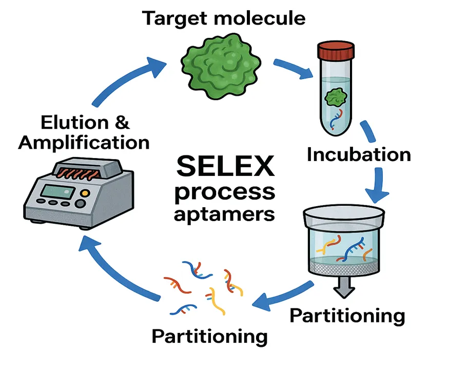
Figure No. 1: SELEX Process
Systematic Evolution of Ligands by Exponential enrichment (SELEX) is a novel in vitro selection technique that finds aptamers, or nucleic acid ligands, with high affinity and specificity for a range of molecular targets [20]. Complex tissues, proteins, cells, viruses, and even minuscule chemical compounds and peptides are among these targets. Since its simultaneous development by Tuerk & Gold (1990) and Ellington & Szostak (1990), SELEX has emerged as a key element of molecular recognition technology in fields such as targeted therapy, biosensing, diagnostics, and nanotechnology [21]. SELEX is essentially an iterative molecular evolution method that mimics natural selection at the molecular level. First, a highly diverse library of synthetic single stranded DNA or RNA molecules is produced, each with a random core region around by consistent sequences that are ready for amplification. Sequences with the highest affinity and specificity for the target are preferentially enriched by repeated cycles of target binding, partitioning, and amplification. The resulting aptamer candidates can match or even outperform antibodies in terms of binding strength, although having clear advantages such reduced size, ease of chemical production, thermal stability, low immunogenicity, and modifiability [22]. Because SELEX is target agnostic, aptamers can be selected even when little is known about the target's molecular structure or epitopes. SELEX also allows the fine tuning of selection pressures to produce aptamers with desirable qualities, such as high affinity in physiological contexts, resistance to nucleases, or the ability to distinguish between extremely similar targets. Numerous specialised variations of SELEX, such as Cell SELEX, Toggle SELEX, CESELEX, and in vivo SELEX, have been developed as a result of the substantial improvements in SELEX during the last thirty years. These developments have increased the method's applicability to more difficult targets and stricter selection criteria, enhancing aptamer discovery's effectiveness and accuracy. SELEX continues to be an essential technique for producing robust and adaptable molecular recognition components as interest in aptamer based technologies grows, particularly in precision medicine and point of care diagnostics [23].
In order to increase the technique's applicability and enhance the effectiveness, specificity, and physiological significance of aptamer selection, several SELEX variants have been created over the last thirty years. The necessity for purified targets, lengthy selection timeframes, and restricted in vivo relevance are only a few of the shortcomings of the conventional SELEX procedure that are addressed by these modifications [24].
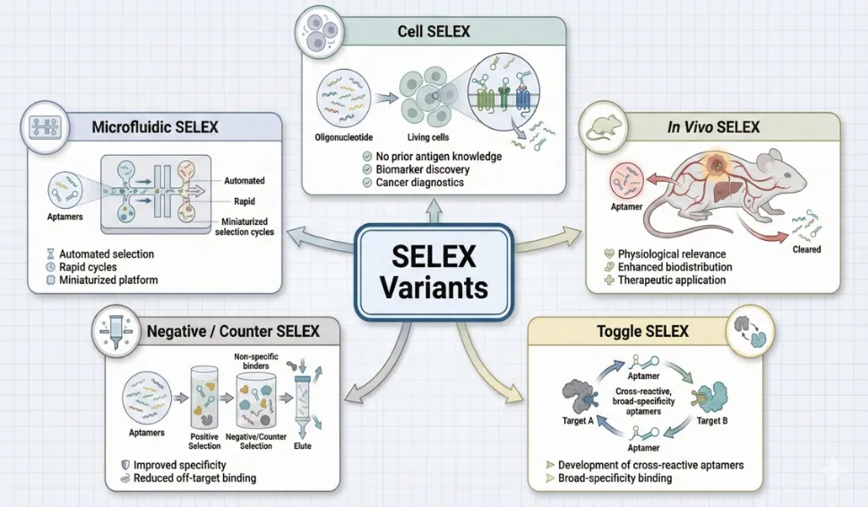
Figure No. 2: SELEX Variants
Aptamer affinity for protein target and selection efficiency have significantly improved when capillary electrophoresis (CE) was recently applied to SELEX. In CESELEX, binding species are chosen according to a mobility shift brought on by complex formation with the target. Increased separation power, decreased nonspecific binding, and the capacity to carry out the selection in free solution are some benefits of CESELEX. These benefits allow for the acquisition of high affinity aptamers in 24 rounds of selection as opposed to the 8-12 rounds that are typical of traditional SELEX. Nevertheless, CESELEX's selection process was limited to smaller targets. [26] Additionally, a non-SELEX aptamer selection procedure that involves many partitioning steps without amplification was shown. They selected aptamers against the hRas protein using nonequilibrium capillary electrophoresis of equilibrium mixture (NECEEM). Here, they discovered that the non-SELEX method's three NECEEM-based partitioning phases were adequate to increase a DNA library's affinity for a target protein. In contrast to the days or weeks needed for a normal SELEX operation by conventional partitioning methods, NECEEM-based non-SELEX selection was remarkably quick just one hour. [27]
Protein and nucleic acid separation has also been thought to benefit from the employment of functionalised magnetic adsorbent particles with the magnetic separation technology. Affinity chromatography is a traditional separation technique used in SELEX, where the targets are typically immobilised on column materials. Nonetheless, the cross reactivity of a nonspecific binder may also be influenced by the presence of linker molecules that are utilised to bind target proteins to columns. Additionally, it is challenging to build and maintain columns and to elute the strongest binders among the nucleic acid sequences. On the other hand, relatively little target is needed when using magnetic beads for target immobilisation, which allows for pleasant handling [28]. This separation technology is also a lot quicker, simpler, and more efficient technique. FluMagSELEX was created by Stoltenburg et al. using magnetic beads as the target molecules' immobilisation matrix. In other words, the magnetic beads coated with streptavidin served as a model target. Using a magnetic separation stand, enhanced aptamers for the target streptavidin were separated during each selection cycle. The benefits of magnetic separation technology and fluorescence tagging of DNA are combined in this SELEX technique. Following the initial Flu Mag SELEX round, the chosen DNA was tagged with fluorescein by PCR using a 5′modified primer. This allowed for direct quantification of the DNA in each SELEX fraction during subsequent selection rounds without the need for reagents or solutions. [29] During the incubation step of the typical SELEX, target proteins are either incubated with a DNA or RNA library in free solution, or are bound to some sort of solid support. However, in the case of binding target proteins to solid supports, this fixation may prevent the conjugation side of the molecule from interacting with the library, hence limiting the evolution of high-affinity aptamers. Yang et al. reported an attractive alternative selection method using bead-bound combinatorial oligonucleoside phosphorothioate (SODNs) and phosphorodithioate (S2ODNs) thioaptamer library. Thioaptamers offer advantages over traditional aptamers in their enhanced affinity and specificity and higher stability, largely due to the properties of the sulfur backbone modifications [30]. Using one-bead one oligonucleotide (SODNs or S2ODNs) combinational library, they screened and identified specific aptamers that bound to NFκB protein using their PCR-based identification tag on the selected bead.[39] In this procedure, each unique member of the library is attached to a separate support bead. By binding the targets to the beads and then using staining and imaging techniques to determine which beads have bound the target, it is possible to choose targets that bind tightly to only a few of the potentially millions of different support beads. For the final sorting of aptamer beads into microcentrifuge tubes for the PCR phase, flow cytometry can readily detect the high fluorescence intensities of individual beads that make up a library.
In addition to this, various bead-based SELEX techniques have shown how adaptable and widely applicable magnetic separation is. For instance, Xie et al. isolated ssDNA aptamers against the herbicide metamitron using magnetic bead SELEX, and after ten rounds of selection, they achieved nanomolar affinity. [31]
One of the most significant developments in aptamer technology is cell based SELEX (cell SELEX), which makes it possible to create molecular probes that can identify intact living cells without the need for prior knowledge of particular surface indicators. Cell SELEX allows the selection of aptamers that bind physiologically relevant epitopes by maintaining the native conformation, spatial orientation, and posttranslational modifications of membrane proteins, in contrast to standard SELEX, which usually needs isolated proteins. In the traditional method, target cells are treated with a randomised ssDNA or RNA library under carefully monitored conditions to enable selective binding. Cell attached oligonucleotides are eluted, amplified, and exposed to counterselection using phenotypically similar but nontarget cells after nonbinding sequences have been washed away.
Cell type specific aptamers are gradually enriched while nonspecific binders are reduced by this cyclic positive-negative enrichment. To track enrichment and identify the ideal selection round endpoint, flow cytometry is commonly employed. Shangguan et al. used two haematopoietic cancer cell lines, Ramos (Burkitt's lymphoma) and HL60, as a model system in one of the fundamental demonstrations of the power of cell SELEX. Their subtraction based approach produced the first well validated DNA aptamers specific to cancer cells and made it possible to distinguish clearly between the molecular signatures of the two cell types. These aptamers were demonstrated to bind distinct surface markers that were difficult to identify using traditional proteomic techniques, and they demonstrated exceptional affinity and selectivity [32]. Oncological applications have provided additional validation for Cell SELEX. Aptamers have been effectively isolated for the specific identification of Burkitt's lymphoma; the chosen sequences demonstrated great discriminatory strength against non-lymphoma controls and a high affinity for Ramos cells. Similar to this, Cerchia et al. used RET-expressing cells as templates to create the first aptamers against the receptor tyrosine kinase RET. The therapeutic potential of these aptamers was highlighted by the fact that they were not only selective binders but also strong antagonists that could block RET-mediated signalling pathways. Cell SELEX has demonstrated efficacy in targeting well characterized tumor associated chemicals in addition to discovering new biomarkers. The aptamer against tenascin-C, an extracellular matrix glycoprotein that is overexpressed during tumour invasion and tissue remodelling, is a well known example. Currently a top contender for targeted drug administration and molecular imaging, the tenascin-C aptamer is effectively internalised by a range of solid tumours [33].
Cell SELEX's adaptability has expanded into therapeutic and diagnostic engineering in addition to biomarker discovery. Tan et al. showed that leukaemia cells from diverse sources could be selectively captured, imaged, and isolated using aptamer functionalized magnetic and fluorescent nanoparticles. Additionally, aptamer nanoparticle conjugates function as targeted delivery vehicles when combined with controlled release polymer carriers. They bind precisely to cancer cells and release chemotherapeutic drugs at the disease site. When combined, cell SELEX offers a potent platform for the development of highly selective targeting ligands and the identification of structure specific chemical fingerprints of living cells. Further developments in precision diagnostics, biomarker identification, and targeted therapeutic delivery are anticipated as a result of its ongoing improvement, which includes microfluidic cell SELEX, hight throughput sequencing analysis, and in vivo cell SELEX [34].
Achieving high specificity for unique protein domains or active areas is essential for aptamer discovery. Target stability presents a significant obstacle because many proteins are susceptible to denaturing during traditional SELEX due to their sensitivity to temperature or solvent conditions. Sol-Gel matrices provide a gentle and encouraging environment for immobilising proteins in order to address this. Instead of chemically tethering proteins, this technique traps them within the gel network, maintaining their original structures. Sol-Gel encapsulated proteins are more stable, exhibit better resistance to chemical and thermal denaturation, and continue to function for longer [35]. Sol-Gel matrices' intrinsically nanoporous structure is particularly appealing because the pores are big enough to let aptamers flow in while holding onto the bigger entrapped target proteins, allowing for efficient binding and partitioning. A microfluidic Sol-Gel device using nanoporous Sol-Gel droplets spotted into chambers over individually addressable microheaters was created by Seung Min Park, Jiyoung Ahn, and associates. Oligonucleotide libraries were incubated with target proteins (such as TATA-binding protein, transcription factor IIB, heat shock factor, etc.) contained in these sol-gel spots. Following washing, localised heat was applied via the microheaters to selectively elute bound aptamers, enabling accurate, multiplexed recovery of aptamers from various compartments. High throughput, parallel aptamer selection is made possible by this design, which allows for the simultaneous immobilisation of many proteins in separate sol-gel spots and the independent heat actuation of each spot for elution. By preventing cross contamination between spots during incubation and elution, pneumatic valves in the microfluidic network enhance specificity [36]. Crucially, the authors demonstrated significantly increased efficiency by producing high affinity aptamers in fewer SELEX cycles than conventional techniques. Additional improvements include "SGSELEX" (Sol-Gel SELEX) on nanoporous silicon substrates, which isolates aptamers without the need for chemical linkers by trapping small molecule targets (such as insoluble compounds) in the Sol-Gel. For instance, this method was used to isolate high affinity ssDNA aptamers against azoxystrobin. More recently, small molecule targets (such as pesticides) have been immobilised for SELEX using sol-gel-coated anodised aluminium oxide (AAO) membranes; these technologies allow for heat elution and have been coupled with next generation sequencing to find high affinity aptamers. All things considered, sol-gel based SELEX offers a strong, mild, and multiplexable platform that is ideal for both protein and small molecule aptamer discovery because it maintains protein structure, permits simultaneous selection against numerous targets, and shortens SELEX cycle durations [37].
The process by which particles in a fluid sink to the bottom due to gravity is known as sedimentation. By using this method, a protein potion can be forced to precipitate by changing the buffer's composition. Centrifugation was previously used to select an RNA aptamer against the disease related prion protein from scrapie-associated fibrils (SAF). The reaction mixture was centrifuged at 25,000 x g for one hour at 10oC in order to separate the complex from free RNA molecules [38]. Bound RNA was successfully isolated after the unbound RNA was extracted from the supernatant. Centrifugation was also used as a separation technique to select an RNA aptamer against African trypanosomes. When combined with additional methods like blotting, flowcytometry, hybridisation, and DNA fingerprint identification, gold conjugations are now acknowledged as crucial instruments for the detection and measurement of proteins, antigens, and nucleic acids. A suspension (or colloid) of sub micro meter-sized gold particles in a fluid is called colloidal gold, sometimes referred to as “nanogold” [39]. To choose high affinity DNA aptamers against KMP11, Moreno et al. developed a selection technique utilising colloidal gold. This technique relied on the target protein attaching to colloidal gold to increase the protein's mass so that it could be further purified by centrifugation. The gold labelled protein was combined with the denatured ssDNA/RNA pool in each SELEX cycle, and bound species were sorted from unbound ones by centrifugation. This method's primary benefit was that the centrifuge procedures could be shortened from several hours to ten to fifteen minutes.Huang et al. also reported a colorimetric technique that used nanoparticle aggregation to detect platelet derived growth factor (PDGFBB) and its isoforms (PDGFAB and PDGFAA) [40].
In order to gradually enrich sequences that bind well to the target, the SELEX technique entails several rounds of alternating selection and amplification phases. Each round involves incubating the nucleic acid library with the target, discarding unbound molecules, and amplifying the binders using PCR; RNA synthesis and reverse transcription are also necessary for RNA SELEX. SELEX is time consuming and difficult because it usually takes 8-20 selection rounds before no more enrichment is observed. This has led to the development of automated, high throughput systems that can execute SELEX in parallel, handle a large number of samples, provide flexibility in buffer and reagent selection, and standardise selection settings. A Beckman Biomek 2000 pipetting robot was utilised in the first automated SELEX protocol, which greatly decreased manual labour and allowed for the concurrent processing of several selection lines [41]. This technique was further modified by Cox and Ellington to select aptamers against biotinylated lysozyme immobilised on beads, showcasing the workflow's capacity to manage the binding, washing, elution, and partitioning stages automatically. Eulberg et al. described a completely automated in vitro SELEX workstation, which introduced more automation. Their device performed full SELEX cycles without direct human interaction by combining a robotic liquid handler (RoboAmp 4200E), ultrafiltration units, fluorescence detection, and semiquantitative PCR [42]. The development of microfluidic based automation in more recent times has significantly decreased cycle times and manual handling by integrating selection, washing, elution, and amplification on a chip. For instance, four rounds of aptamer enrichment for IgE were accomplished in roughly ten hours, utilising a microfluidic SELEX apparatus that used electrokinetic and hydrodynamic control. Additionally, aptamers were chosen against cancer tissue samples using an optimisation chipbased SELEX system, which enhanced buffer conditions, stringency, and throughput [43].
Aptamers are now widely used molecular tools in bioanalytical research, targeted delivery, medicines, environmental monitoring, and diagnostics. They are effective substitutes for conventional antibodies because to their special blend of high affinity, structural flexibility, chemical stability, and simplicity of modification.
In biosensors intended to identify proteins, small chemicals, pathogens, and cancer biomarkers, aptamers are useful recognition components. Their fast binding kinetics and consistent folding make them easy to incorporate into electrochemical, fluorescent, colorimetric, and microfluidic platforms. Additionally, aptamer based assays are compatible with pointofcare diagnostics, allowing for the sensitive identification of disease markers such circulating tumour cells, thrombin, VEGF, and troponin [44].
2. Therapeutic Applications
Aptamers can be used therapeutically to disrupt ligand-receptor interactions or bind active regions of disease related proteins. Pegaptanib, which is approved for age related macular degeneration, is one of the most well known examples. Numerous modern therapeutic aptamers are being studied for viral infections, thrombosis, cancer, and inflammatory conditions. Chemical changes improve stability and lower immunogenicity, while their tiny size permits deep tissue penetration and quick systemic clearance [45].
3. Targeted Drug and Gene Delivery
Aptamers serve as precise delivery agents, directing medications, siRNAs, nanoparticles, and imaging probes to specific cells or tissues. They can be modified to identify markers like HER2 on breast cancer cells or PSMA on prostate cancer cells. This focused strategy reduces off target toxicity and increases therapeutic efficacy. Additionally, regulated release and improved intracellular uptake are made possible by aptamer–nanoparticle conjugates [46].
4. Environmental and Food Safety Monitoring
Aptamers are becoming more and more important in environmental bio-surveillance because of their capacity to bind poisons, antibiotics, pesticides, heavy metals, and pathogens. Tetracycline, mercury ions, ochratoxin, and aflatoxin B1 have all been found in water and food systems using apta-sensors. They are perfect for field based monitoring since they are stable in challenging environments and work with portable devices [47].
5. Biomarker Discovery and Molecular Imaging
Aptamers can selectively attach and enrich molecules to identify patterns linked to disease. Aptamers labelled with fluorophores, radionuclides, or MRI contrast agents enable high resolution imaging of thrombi, tumours, and inflammatory tissues. Strong signal to noise ratios are produced in vivo by their quick binding and clearance characteristics [49].
6. Regenerative Medicine and Cell Biology
Aptamers help guide tissue engineering structures, isolate particular stem cell populations, and modify signalling pathways that are critical for regeneration. Viable cells can be gently captured and released without changing their cellular phenotype thanks to their reversible binding [50].
CONCLUSION
From the first RNA ligands reported in 1990 to today’s AI-optimised, microfluidically selected aptamers, the field has undergone a paradigm shift: what began as a test-tube evolution curiosity is now a mature platform that rivals and often surpasses antibodies in specificity, stability and manufacturability. Convergence of automated SELEX, cell-specific selection, sol–gel multiplexing and nanomaterial integration has enabled rapid, low-cost isolation of reagents against virtually any molecular or cellular target, fueling advances in early disease detection, real-time environmental surveillance and ligand-directed therapy. As chemical biology refines backbone chemistries (Spiegelmers, XNAs) and bioinformatics accelerates hit identification, aptamers are poised to become central building blocks in the emerging toolkit of precision medicine, offering clinicians customisable recognition elements that diagnose, treat and monitor disease with unprecedented accuracy and minimal patient risk.
REFERENCES

Pallavi Desale, Amol Rakte, Dimpal Patil, Jyotiraditya Fiske, SELEX to Clinic: Evolution, Innovations and Applications of Aptamers in Precision Medicine., Int. J. of Pharm. Sci., 2026, Vol 4, Issue 7, 1862-1876. https://doi.org/10.5281/zenodo.21267887
 10.5281/zenodo.21267887