

We use cookies to ensure our website works properly and to personalise your experience. Cookies policy
Department of Biotechnology, St. Peter’s College of Engineering and Technology, College Road, Avadi – 600054, India.
Psoriasis is a chronic immune-mediated inflammatory skin disorder characterized by epidermal hyperproliferation, which leads to increased skin cell turnover. Although several therapeutic agents are available, long-term management is often associated with adverse effects and limited efficacy. Natural products derived from medicinal plants have gained considerable attention because of their potential anti-inflammatory, anticancer, and immunomodulatory activities. This study investigates the antipsoriatic potential of the fruit pulp of Melia dubia against human 5-lipoxygenase (PDB ID: 3V99) using an integrated experimental and computational approach. The ethanolic extract of Melia dubia pulp obtained by Soxhlet extraction was subjected to qualitative phytochemical screening for the presence of secondary metabolites, followed by GC-MS analysis and in silico studies. GC-MS characterization revealed that oleic acid, betulin, lupeol, linoleic acid, and palmitic acid constituted the primary chemical fractions. Betulin was selected as the lead compound for subsequent computational screening. Molecular docking was performed using PyRx and AutoDock Vina and revealed a strong binding affinity of -9.2 kcal/mol, indicating the potential for enzyme inhibition by betulin. ADMET analysis predicted favorable drug-likeness and low toxicity. In conclusion, betulin from Melia dubia pulp may serve as a potential lead for developing novel antipsoriatic therapeutics. To further evaluate this lead, additional molecular dynamics simulations and extensive in vitro and in vivo studies are essential to substantiate its therapeutic efficacy and safety.
Psoriasis is a chronic immune-mediated inflammatory skin disorder in which the immune system becomes overactive, causing skin cells to multiply too quickly. It affects the global population and is influenced by genetic, immunological, and environmental factors. In addition to cutaneous manifestations, psoriasis is associated with systemic comorbidities, including cardiovascular disorders, mental health problems, cancer, diabetes, and metabolic syndrome. Common symptoms of psoriasis include patches of thick, red skin with silvery-white scales, poor sleep quality, and dry, cracked skin that itches or bleeds. Contemporary research emphasizes the therapeutic potential of medicinal plants in treatment of psoriasis due to their anti-inflammatory and immunomodulatory properties (Rajashekar et al., 2016). Medicinal plants represent a rich repository of bioactive compounds with diverse pharmacological activities. Melia dubia is considered one of the largest fast-growing trees belonging to the family Meliaceae. The common name of Melia dubia is Malabar neem. The fruit of Melia dubia is small, yellow, fleshy, and dorsally compressed. It has important industrial applications and is increasingly used as a source of energy. Apart from its industrial applications, Melia dubia fruit is widely used for various medicinal applications because of its reported anticancer, anti-inflammatory, and antimicrobial activities (Halliwell, 2007). The inflammatory cascade of psoriasis is regulated by a complex network of enzymes and signalling molecules, among which Human 5-Lipoxygenase (PDB ID: 3V99) plays a pivotal role in leukotriene biosynthesis and mediator production. Targeting human 5-lipoxygenase offers a strategy to diminish leukotriene-mediated inflammation and mitigate disease progression. Recent advances in computational biology significantly accelerate the selection of effective drugs by reducing the cost and time associated with conventional experimental screening. Molecular docking is a structure-based methodology that helps identify interaction patterns and binding affinities between bioactive compounds and proteins, thereby facilitating the identification of potential therapeutic agents for psoriasis (Senthilkumar et al., 2018). Therefore, in this work, various computational techniques were used to identify potential phytocompounds against the protein associated with psoriasis (Lionta et al., 2014). Furthermore, characterizing pharmacokinetic parameters through predictive ADMET modelling provides information on drug-likeness, oral bioavailability and toxicity profiles, improving the selection of lead molecules. In the present study, the ethanolic extract of Melia dubia fruit was characterised through qualitative screening and GC-MS analysis. Pharmacologically significant phytocompounds were docked against Human 5-Lipoxygenase (PDB ID: 3V99) using PyRx AutoDock Vina. The top-performing lead molecule was subjected to subsequent Swiss ADME. This integrated workflow reveals anti-psoriatic potential of Melia dubia for downstream experimental testing.
2. MATERIALS AND METHODS
Fresh fruits of Melia dubia were collected from Chennai district, Tamil Nadu, India. Fruits were washed with distilled water to remove surface debris. The fruit pulp was isolated manually and then shade-dried at room temperature for seven days (Azwanida, 2015). The dried material was then milled into a fine powder using a mechanical grinder and preserved in a sealed container. To ensure sample integrity handling at every stage of the process was performed under sterile conditions.
For the extraction of bioactive components, 50 g of powdered fruit flesh was macerated in 250 mL of 95% ethanol and subjected to Soxhlet extraction. The process was continued for 6–8 h until complete extraction was achieved. The obtained concentrate was aliquoted into vials and stored at 4ºC for further use.
Phytochemical analysis is a preliminary step in the analysis of medicinal plants. These tests enable the identification of secondary metabolites present in the sample (Harborne, 1998). In the present work, the crude extract of Melia dubia underwent qualitative phytochemical tests to identify various compounds present. The major classes of secondary metabolites are alkaloids, flavonoids, tannins, glycosides, saponins, and steroids. The presence of phytocompounds was confirmed by chemical reactions in the sample, which resulted in colour changes, precipitate formation, and ring formation. Each test was performed under aseptic laboratory conditions (Trease & Evans, 2009).
2.4 GC-MS Analysis:
GC-MS is an analytical technique used to separate and identify volatile compounds. The volatile compounds are separated on the basis of their mass-to-charge ratios, generating mass spectra for identification (Harborne, 1998). The GC-MS analysis for this work was carried out at the Vellore Institute of Technology, Chennai, using an Agilent 7890A gas chromatograph coupled with a 5975C mass spectrometer. The sample was injected at temperature of 250ºC using a 1:10 split ratio. The mass spectra of the compounds were recorded and matched with NIST database to identify them (de Hoffmann & Stroobant, 2007).
Ligand preparation is a crucial, automated process of converting the 2D molecules into accurate, high quality 3D structure for molecular docking. The phytocompounds obtained from the GC-MS analysis were used as ligands for docking studies (Morris et al., 2009). PubChem also helps study the physical and chemical properties of the ligands (Meng et al., 2011). In order to prevent the steric hindrance and to obtain the stable conformation, energy minimization of the ligands was carried out. Converting the ligands from PDB to PDBQT format was essential to guarantee computational compatibility during the simulation process.
The target protein, human 5-lipoxygenase, was retrieved from the Protein Data Bank (PDB) (RCSB PDB, 2025) in its 3D structure format. The protein retrieved from the PDB was subjected to refinement and preparation of protein before the docking process. Protein refinement was performed by removing the water molecules, co-crystallized ligands and other non-essential compounds to ensure proper docking of the protein with the selected ligand. This preparation of protein ensures the reliability of the docking results.
2.7 Molecular Docking:
Molecular docking was performed to identify the the interaction between the selected ligand and target protein. Molecular docking was carried out using AutoDock Vina (Eberhardt et al., 2021) and PyRx (Dallakyan & Olson, 2015). PyRx was used for docking. In this work, the selected phytocompounds from Melia dubia were used as ligands to interact with the target protein associated with psoriasis. The grid box was set up in the active-site region of protein to define the binding interactions of the ligands. The stable binding orientation of ligand and target protein was predicted based on binding-affinity values expressed in Kcal/mol. Lower binding-energy values indicate stronger and more stable interactions, whereas higher binding-energy values indicate weaker and less stable interactions between ligand and target protein (Morris & Lim-Wilby, 2008).
2.8 ADMET Analysis
ADMET analysis (Jiangyu Yan et al., 2021) was performed to identify the the pharmacokinetic and toxicity properties of the selected phytocompounds. ADMET represents the Absorption, Distribution, Metabolism, Excretion and Toxicity which are parameters used to predict the drug-likeness and toxicity of of the phytocompounds. In this work, the phytocompounds from Melia dubia that showed the best docking scores were selected and subjected to ADMET analysis. SwissADME was used for ADMET analysis. In this tool, the chemical structures of the selected phytocompounds were retrieved in SMILES format and uploaded. The compounds were analysed for pharmacokinetic properties to predict their potential as effective drug candidates for psoriasis. The obtained ADMET results were compared and interpreted with standard values to identify the phytocompounds with suitable pharmacokinetic properties for psoriasis (Lipinski et al., 2001).
3. RESULTS
3.1 Phytochemical Analysis
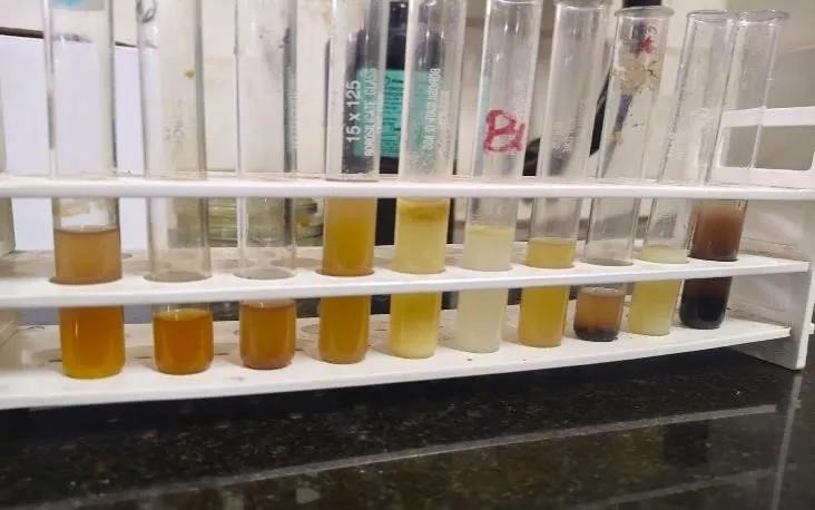
Phytochemical analysis of Melia dubia revealed the presence of flavonoids, phenols, tannins, steroids, polyphenols and saponins. Glycosides and quinones were not detected. The rich content of flavonoids and phenolic compounds have a strong ability to bind and interact with the target protein. The presence of flavonoids, phenols and polyphenols have strong antioxidant properties whereas the presence of saponins enhances the medicinal value of the extract through its effective immunomodulatory, anti-inflammatory and membrane permeabilizing properties. Tannins show antimicrobial and anti-inflammatory properties. These findings suggest that Melia dubia may have potential for further investigation as an antipsoriatic agent.
Table 1: Interpretation of Phytochemical Analysis
|
Phytochemical Compounds |
Result |
|
Flavonoids |
+ |
|
Phenols |
+ |
|
Tannins |
+ |
|
Steroids |
+ |
|
Polyphenols |
+ |
|
Saponins |
+ |
|
Glycoside |
- |
|
Quinones |
- |
|
1 2 3 4 5 6 7 8 9 10 |
3.2 GC-MS Analysis
GC-MS analysis of Melia dubia was performed and it showed the detailed chemical profile of the bioactive compounds present in Melia dubia. The analysis provided a well-defined chromatogram with multiple peaks, representing the different phytocompounds which were separated based on their volatility and interaction with the stationary phase of the GC column. A total of 15 compounds were identified based on their peak areas, percentages, and retention times, which were compared with NST library. From the GC-MS analysis the analysis indicated the predominant presence of Phthalic acid derivatives, triterpenoids and long-chain hydrocarbons in the chromatogram. Based on biological and pharmacological activities, Betulin, Linoleic acid, Palmitic Acid, Oleic acid and Lupeol were selected. Among these five compounds, betulin and lupeol belong to pentacyclic triterpenoids, whereas oleic acid, linoleic acid, and palmitic acid belong to fatty acids. Palmitic acid was detected with a peak area of 3.44% at retention time of 15.521 min. Linoleic acid and oleic acid were detected with peak areas of 26.97% and 10.44% at retention time of 17.333 min and 17.376 min. Betulin and Lupeol were detected with a retention time of 22.48 min and a peak area percentage of 1.53%. Based on this analysis, triterpenoids may have potential therapeutic effects relevant to antipsoriatic activity.
Table 2. GC-MS Results and Interpretation of Melia dubia
|
Peak |
RT (min) |
Compound Identified |
Molecular Formula |
Molecular Weight (g/mol) |
Major Reported Biological Activities |
|
1 |
4.064 |
Glycerin (Glycerol) |
C₃H₈O₃ |
92 |
Humectant, antioxidant, antimicrobial carrier |
|
2 |
11.350 |
Phthalic acid, di-(1-hexen-5-yl) ester |
C₂₀H₂₆O₄ |
330 |
Plasticizer; generally considered a contaminant in GC–MS analyses |
|
3 |
15.521 |
n-Hexadecanoic acid (Palmitic acid) |
C₁₆H₃₂O₂ |
256 |
Antioxidant, antimicrobial, anti-inflammatory |
|
4 |
17.333 |
10(E),12(Z)-Conjugated linoleic acid |
C₁₈H₃₂O₂ |
280 |
Antioxidant, anticancer, anti-inflammatory, immunomodulatory |
|
5 |
17.376 |
9-Octadecenoic acid (E)- (Elaidic acid) |
C₁₈H₃₄O₂ |
282 |
Antimicrobial; fatty acid derivative |
|
6 |
17.489 |
trans,trans-9,12-Octadecadienoic acid, propyl ester |
C₂₁H₃₈O₂ |
322 |
Antioxidant, antimicrobial |
|
7 |
17.548 |
Octadecanoic acid (Stearic acid) |
C₁₈H₃₆O₂ |
284 |
Antimicrobial, anti-inflammatory |
|
8 |
21.254 |
Lanosta-8,24-dien-3-one |
C₃₀H₄₈O |
424 |
Triterpenoid; antioxidant, anti-inflammatory, anticancer potential |
|
9 |
22.481 |
Lupeol, trifluoroacetate |
C₃₂H₄₉F₃O₂ |
522 |
Anti-inflammatory, antioxidant, anticancer (derivatized lupeol) |
|
10 |
23.490 |
1,6,10,14,18,22-Tetracosahexaen-3-ol, 2,6,10,15,19,23-hexamethyl-, (all-E)- (Phytol-related compound) |
C₃₀H₅₀O |
426 |
Antioxidant, antimicrobial, anti-inflammatory |
|
11 |
24.501 |
Tetratetracontane |
C₄₄H₉₀ |
618 |
Long-chain hydrocarbon; reported antimicrobial activity in some natural extracts |
|
12 |
25.963 |
Minor compound (identify from full spectrum) |
— |
— |
Requires confirmation |
|
13 |
27.563 |
Minor compound (identify from full spectrum) |
— |
— |
Requires confirmation |
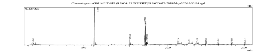
Fig. 2. GC-MS Analysis of Ethanolic Extract of Melia dubia
3.3 Molecular Docking
Molecular docking was carried out between the identified phytocompounds and the target protein. In this work, human 5-lipoxygenase was selected from the PDB as the target protein (Id: 3V99). Based on the GC-MS analysis, five ligands were selected for molecular docking, including Betulin, Linoleic Acid, Palmitic Acid, Oleic Acid and Lupeol. Molecular docking between the selected target protein and ligands was performed using PyRx software. Based on the docking analysis, betulin and linoleic acid showed strong binding affinities of -9.2 kcal/mol and -8.8 kcal/mol respectively. These energy values indicate that these two ligands have strong binding affinities and the ability to form stable interactions. Therefore, betulin and linoleic acid may have potential for further investigation as antipsoriatic candidates.
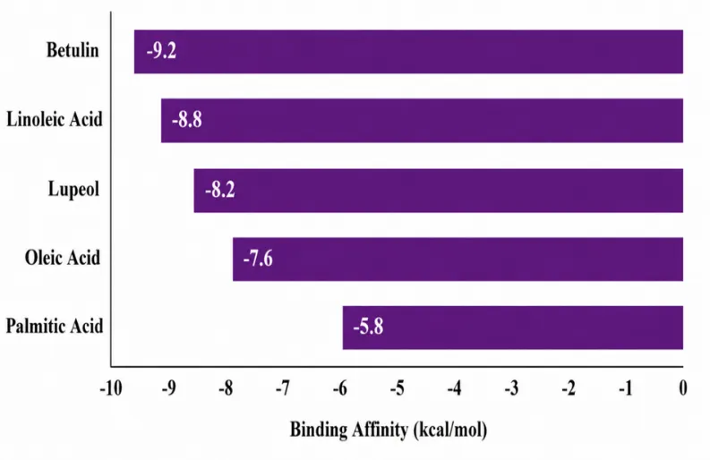
Fig 2- Graphical Representation of the Binding Affinity of the Ligands with Human 5-Lipoxygenase (Id: 3V99).
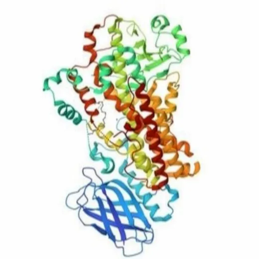
Fig. 4. Structure of Protein 5-Lipoxygenase (Id: 3V99).
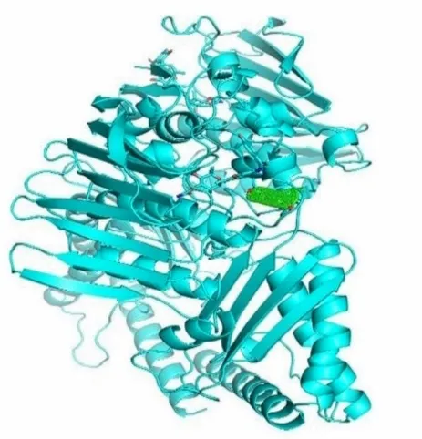
Fig 5: Docking Analysis of the 5-lipoxygenase target protein associated with psoriasis (PDB Id: 3V99) with Betulin, a phytocompound present in Melia dubia
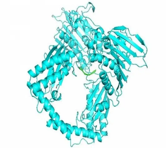
Fig 6: Docking Analysis of the 5-lipoxygenase target protein associated with psoriasis (PDB Id: 3V99) with Linoleic Acid, a phytocompound present in Melia dubia.
3.4 ADMET Analysis
ADMET Analysis of phytocompounds with high binding energy was performed. SwissADME tool was used to perform the ADMET analysis. The pharmacokinetic properties of betulin and linoleic acid were analysed using their SMILES formats. The results showed that both betulin and linoleic acid have favorable drug-likeness characteristics according to Lipinski's Rule of Five, indicating their potential for further investigation against the target protein associated with psoriasis. The molecular weights of the compounds were within the acceptable range of <500 Da and hydrogen-bond donors and acceptors were within suitable limits (Diana et al., 2017). The lipophilicity values of these compounds indicate favorable properties for permeability. Based on these results, the phytocompounds betulin and linoleic acid from Melia dubia may have potential for further investigation in the context of psoriasis.
Table 3: ADMET Analysis and Interpretation of Betulin and Linoleic Acid
|
ADMET Parameters |
Betulin (C₃₀H₅₀O₂) |
Linoleic Acid (C₁₈H₃₂O₂) |
|
Molecular Weight (g/mol) |
442.72 |
280.45 |
|
GI Absorption |
High |
High |
|
BBB Permeability |
No |
No |
|
Lipinski Rule of Five |
Passed |
Passed |
|
Hydrogen Bond Donors |
2 |
1 |
|
Hydrogen Bond Acceptors |
2 |
2 |
|
Bioavailability Score |
Good |
Moderate |
|
CYP Inhibition |
No Significant Inhibition |
No Significant Inhibition |
|
Toxicity Prediction |
Low Toxicity |
Low Toxicity |
|
Drug-Likeness |
Acceptable |
Acceptable |
DISCUSSION
Qualitative phytochemical screening and GC–MS profiling of the pigment extract obtained from Staphylococcus arlettae revealed the presence of several biologically relevant metabolites, including n-hexadecanoic acid, conjugated linoleic acid, octadecanoic acid, oleic acid derivatives, lanostane-type triterpenoids, lupeol derivatives, and phytol-related compounds. Similar classes of metabolites have previously been identified from microbial pigments and other natural products, where they have been associated with antioxidant, antimicrobial, and anti-inflammatory activities (Sasidharan et al., 2011; Bhardwaj et al., 2020). However, unlike previous studies that primarily investigated plant-derived metabolites, the present study demonstrates that pigment-producing bacteria isolated from the oral microbiota of Naja naja also constitute a promising source of structurally diverse bioactive compounds. The predominance of fatty acids and triterpenoid derivatives observed in the present investigation is consistent with earlier reports describing these metabolites as important contributors to microbial bioactivity. Similar occurrence of n-hexadecanoic acid has been reported in several bacterial and fungal extracts, where it has been associated with antimicrobial and anti-inflammatory properties (Bharathidhasan et al., 2015). Likewise, conjugated linoleic acid and oleic acid derivatives have previously been reported as naturally occurring lipid metabolites capable of modulating inflammatory responses and oxidative stress (Benjamin and Spener, 2009). The identification of these metabolites in the present pigment extract therefore supports the possibility that they collectively contribute to the observed biological activities rather than acting as isolated constituents. The detection of lanostane-type triterpenoids and a lupeol-related derivative further strengthens the pharmacological significance of the pigment extract. Similar triterpenoid compounds have been extensively reported from medicinal fungi and higher plants, where they exhibit anti-inflammatory, antioxidant, immunomodulatory, and anticancer properties through regulation of multiple inflammatory signalling pathways (Wasser, 2011; Saleem, 2009). Although lupeol has traditionally been described as a plant-derived pentacyclic triterpenoid, its identification in the present microbial pigment extract expands the chemical diversity associated with Staphylococcus arlettae and suggests that microbial pigments may represent an underexplored source of triterpenoid-like metabolites. The occurrence of a phytol-related diterpenoid alcohol is also in agreement with previous investigations demonstrating antioxidant, antimicrobial, and anti-inflammatory activities of phytol-containing natural extracts (Santos et al., 2013). The coexistence of fatty acids, diterpenoids, and triterpenoids within the same extract indicates a chemically diverse metabolite profile capable of producing complementary biological effects through multiple mechanisms rather than through a single dominant compound. Although phthalate ester derivatives were identified during GC–MS analysis, similar compounds have frequently been reported as contaminants originating from laboratory plasticware, solvents, or analytical instrumentation rather than authentic microbial metabolites (Biedermann et al., 2013). Therefore, these compounds should be interpreted cautiously and excluded from biological interpretation unless confirmed by additional analytical techniques. The present findings should also be interpreted within the limitations of GC–MS-based metabolite identification. Compound assignments were generated through comparison with the NIST spectral library and therefore represent putative identifications rather than definitive structural confirmation. Confirmation of the identified metabolites would require complementary analytical approaches such as LC–MS/MS, high-resolution mass spectrometry, or nuclear magnetic resonance spectroscopy. Furthermore, although many of the identified compounds have been individually reported to possess antioxidant, antimicrobial, and anti-inflammatory activities, the contribution of each metabolite within the complex pigment extract remains to be experimentally validated through purification, quantitative analysis, and mechanistic biological assays. Taken together, the present investigation demonstrates that the pigment-producing bacterium Staphylococcus arlettae isolated from the oral microbiota of Naja naja produces a chemically diverse metabolite profile enriched in biologically relevant fatty acids, triterpenoid derivatives, and diterpenoid compounds. While previous studies have largely focused on plant-derived sources of these metabolites, the present work identifies snake oral microbiota as a novel microbial reservoir of pigment-associated bioactive compounds. This expands current knowledge regarding microbial pigment chemistry and provides a scientific basis for future studies aimed at isolating individual metabolites and evaluating their therapeutic potential in antioxidant, anti-inflammatory, and antimicrobial applications.Bottom of Form
CONCLUSION
The present study suggests that Melia dubia contains bioactive phytocompounds that may have therapeutic potential against the target protein associated with psoriasis. Phytochemical screening and GC-MS analysis revealed the presence of bioactive compounds with reported medicinal properties, including anti-inflammatory, anti-cancer and immunomodulatory activities. 5-lipoxygenase was selected as the target protein, and the identified ligands obtained through GC-MS were docked against it (Betulin, Oleic Acid, Lupeol, Linoleic acid, Palmitic Acid). The molecular docking results showed that betulin and linoleic acid exhibited favorable binding affinities. ADMET analysis suggested that both betulin and linoleic acid may have potential for further investigation as antipsoriatic candidates. This work helps to understand the basic and therapeutic potential of Melia dubia. As a future perspective, formulations such as topical creams, gels, and nanoformulations can be developed for drug delivery purposes. Molecular dynamics simulations can be carried out to determine the stability and behaviour of protein–ligand complexes over time. In vitro and in vivo studies can be performed to evaluate the efficacy and therapeutic applications of phytocompounds from Melia dubia in keratinocytes and immune cells under physiological conditions. From this we can conclude that, this work acts as the starting point for the development of drug by a phytocompound present in Melia dubia for psoriasis.
REFERENCES
DOI:10.1056/NEJMra071371
Advances, Vol.33, No.8, pp.1582–1614. DOI:10.1016/j.biotechadv.2015.08.001
Vol.4, pp.177. DOI:10.3389/fphar.2013.00177
DOI:10.1124/pr.52.4.673
DOI:10.1056/NEJMra071371
Advances, Vol.33, No.8, pp.1582–1614. DOI:10.1016/j.biotechadv.2015.08.001
Vol.4, pp.177. DOI:10.3389/fphar.2013.00177
DOI:10.1124/pr.52.4.673


Sabarish M P, Akash M, Sandhiya M, Gowri Shankar Bagavanatham Anadavan, Targeting Human 5-Lipoxygenase: An in-Silico Study on The Antipsoriatic Potential of Melia Dubia Phytocompounds, Int. J. of Pharm. Sci., 2026, Vol 4, Issue 8, 5211-5223, https://doi.org/10.5281/zenodo.22204713
 10.5281/zenodo.22204713