

We use cookies to ensure our website works properly and to personalise your experience. Cookies policy
Department of Pharmacology, College of Pharmaceutical Sciences, Government Medical College Kannur, Kerala, India.
Rheumatoid arthritis (RA) is a chronic autoimmune inflammatory disease characterized by persistent synovial inflammation, progressive cartilage degradation, and bone erosion. Although genetic and environmental factors contribute to disease susceptibility, the molecular mechanisms linking genetic risk to sustained inflammatory signaling remain incompletely understood. The cluster of differentiation 40 (CD40)/tumor necrosis factor receptor-associated factor 1 (TRAF1)/nuclear factor kappa B (NF-?B) signaling axis has emerged as an important molecular network connecting genetic susceptibility, immune-cell activation, and chronic synovial inflammation in RA. Genetic variants within the CD40 and TRAF1/C5 loci have been associated with RA susceptibility, while dysregulated CD40 signaling and altered TRAF1 activity influence inflammatory responses and immune-cell survival. CD40 engagement recruits TRAF adaptor proteins and activates downstream signaling pathways, including NF-?B, a central transcriptional regulator of inflammatory gene expression. Persistent NF-?B activation promotes the production of pro-inflammatory cytokines, chemokines, adhesion molecules, and matrix-degrading enzymes, contributing to synovial hyperplasia, leukocyte recruitment, cartilage degradation, and osteoclast-mediated bone destruction. This review summarizes the biological functions of CD40 and CD40 ligand (CD40L), examines the regulatory role of TRAF1 in CD40-associated signaling, and discusses the contribution of the CD40/TRAF1/NF-?B axis to RA pathogenesis. Current and emerging therapeutic approaches targeting this network, including CD40/CD40L blockade, modulation of TRAF-dependent signaling, NF-?B pathway inhibition, and emerging molecular and RNA-based strategies, are also discussed. Particular attention is given to the limitations of broad pathway inhibition and the potential for selective, biomarker-guided therapeutic strategies. A better understanding of the cell-specific and context-dependent functions of this signaling network may facilitate the development of precision approaches that suppress pathogenic inflammation while preserving essential immune functions in patients with rheumatoid arthritis.
Rheumatoid arthritis (RA) is a chronic, systemic autoimmune disease characterized by persistent synovial inflammation, progressive cartilage destruction, bone erosion, and irreversible joint deformity.1 Unlike osteoarthritis, which primarily results from mechanical degeneration of articular cartilage, RA arises from dysregulated immune responses that target the synovial membrane, leading to chronic inflammation and structural joint damage.2,3 Beyond articular manifestations, RA is associated with several extra-articular complications, including cardiovascular disease, interstitial lung disease, vasculitis, osteoporosis, and anemia, all of which contribute significantly to morbidity and mortality.4,5
RA affects approximately 0.5–1% of the global population, with a higher prevalence in women than in men. Disease onset typically occurs between 30 and 60 years of age, although it can develop at any stage of adulthood.4 The pathogenesis of RA is multifactorial, resulting from a complex interplay between genetic predisposition, environmental factors, and immune dysregulation. Genetic susceptibility is largely attributed to polymorphisms in immune-related genes, particularly HLA-DRB1, PTPN22, and the TRAF1/C5 locus, whereas environmental factors such as cigarette smoking, microbial infections, hormonal influences, and oxidative stress contribute to disease initiation and progression.6,7,8
The hallmark pathological feature of RA is chronic synovitis, in which infiltration of macrophages, dendritic cells, T lymphocytes, B lymphocytes, and fibroblast-like synoviocytes (FLS) transforms the normally thin synovial membrane into an invasive pannus. This hyperplastic tissue actively invades adjacent cartilage and bone through continuous production of inflammatory cytokines, chemokines, matrix metalloproteinases (MMPs), and osteoclast-activating factors, ultimately leading to irreversible joint destruction.9,10,11
Among the intracellular signaling pathways involved in RA, the CD40/TRAF1/NF-κB signaling axis has emerged as a critical regulator of immune activation and chronic inflammation. CD40, a member of the tumor necrosis factor receptor (TNFR) superfamily, interacts with its ligand CD40L to recruit TNF receptor-associated factors (TRAFs), particularly TRAF1 and TRAF2, initiating signaling cascades that activate nuclear factor-kappa B (NF-κB), mitogen-activated protein kinase (MAPK), and phosphatidylinositol-3 kinase (PI3K)/Akt pathways.12,13 These signaling events stimulate the production of pro-inflammatory cytokines such as tumor necrosis factor-alpha (TNF-α), interleukin (IL)-1β, and IL-6, while promoting immune cell survival, synovial fibroblast proliferation, angiogenesis, and osteoclastogenesis.13 Persistent activation of this pathway establishes a self-sustaining inflammatory microenvironment that drives both synovial inflammation and structural joint damage.14
Current therapeutic approaches, including conventional disease-modifying antirheumatic drugs (DMARDs), biological agents, and Janus kinase (JAK) inhibitors, have substantially improved disease management by suppressing inflammatory pathways. However, many patients exhibit incomplete responses, treatment resistance, or adverse effects, highlighting the need for more selective molecular targets.15,16 Increasing evidence indicates that the CD40/TRAF1/NF-κB axis represents a mechanistic link between genetic susceptibility and chronic inflammation, making it an attractive target for precision medicine strategies in RA.
2. IMMUNOPATHOGENESIS OF RHEUMATOID ARTHRITIS
Rheumatoid arthritis (RA) is a systemic autoimmune disease in which genetic susceptibility and environmental factors contribute to the loss of immune tolerance and the development of persistent synovial inflammation. The disease involves coordinated interactions among innate and adaptive immune cells, including dendritic cells, macrophages, T lymphocytes, B lymphocytes, and fibroblast-like synoviocytes, resulting in a chronic inflammatory environment that progressively damages cartilage and bone.3,10
Antigen-presenting cells, particularly dendritic cells, initiate adaptive immune responses by presenting autoantigens to CD4⁺ T lymphocytes. Activated T cells subsequently promote macrophage and B-cell activation through cytokine secretion and cell–cell interactions, amplifying inflammation within the synovial microenvironment.3,7,10
B lymphocytes further contribute to disease progression through production of rheumatoid factor and anti-citrullinated protein antibodies, antigen presentation, and secretion of inflammatory mediators.7
Macrophages and other inflammatory cells produce cytokines, particularly tumour necrosis factor-α (TNF-α), interleukin (IL)-1β, and IL-6, which sustain synovial inflammation and activate resident stromal cells.¹¹ Persistent inflammatory stimulation induces fibroblast-like synoviocytes to acquire an aggressive phenotype characterized by increased proliferation and production of matrix metalloproteinases (MMPs) and other inflammatory mediators, promoting extracellular matrix degradation and invasion of adjacent cartilage.9
In parallel, inflammatory signaling enhances receptor activator of nuclear factor-κB ligand (RANKL)-dependent osteoclast differentiation, contributing to progressive bone erosion.17 Thus, the interaction between immune-cell activation, inflammatory cytokine networks, synovial fibroblast responses, and osteoclastogenesis drives the transition from persistent synovitis to irreversible structural joint damage.
Although multiple inflammatory pathways contribute to RA pathogenesis, intracellular signaling networks provide an important link between immune activation and chronic tissue destruction. Among these, the CD40/TRAF1/NF-κB axis is particularly relevant to the present review because it connects genetic susceptibility and immune-cell activation with persistent inflammatory signaling and progressive joint damage.
3. BIOLOGY OF CD40 AND CD40L
3.1 Structure and Expression of CD40
CD40 is a type I transmembrane glycoprotein belonging to the tumor necrosis factor receptor (TNFR) superfamily and is expressed predominantly on B lymphocytes, dendritic cells, monocytes, macrophages, and thymic epithelial cells. Under inflammatory conditions, CD40 expression is also induced on non-hematopoietic cells, including endothelial cells, epithelial cells, and fibroblast-like synoviocytes (FLS). Unlike several members of the TNFR family, CD40 lacks intrinsic kinase activity and therefore depends on cytoplasmic adaptor proteins to initiate intracellular signaling.19
3.2 CD40 Ligand (CD40L/CD154)
CD40 ligand (CD40L, also known as CD154) is a type II transmembrane protein primarily expressed on activated CD4⁺ T lymphocytes. It is also detected on activated platelets, B cells, macrophages, natural killer cells, mast cells, and endothelial cells, allowing CD40-mediated communication between innate and adaptive immune cells. Binding of CD40L to CD40 is essential for B-cell activation, immunoglobulin class switching, germinal center formation, dendritic-cell maturation, and the generation of long-term humoral immunity.20
In addition to its membrane-bound form, soluble CD40L retains biological activity and contributes to inflammatory responses by activating CD40-expressing target cells. Elevated CD40L expression has been reported in several autoimmune diseases, including rheumatoid arthritis, where persistent CD40–CD40L interactions contribute to chronic immune activation.20
3.3 CD40 Signal Transduction
Engagement of CD40 by CD40L induces receptor trimerization, exposing binding sites within the cytoplasmic domain for members of the TNF receptor-associated factor (TRAF) family. Because CD40 lacks catalytic activity, recruitment of TRAF adaptor proteins is essential for transmitting intracellular signals. TRAF1, TRAF2, TRAF3, TRAF5, and TRAF6 associate with CD40 to regulate downstream signaling events controlling immune-cell activation, proliferation, survival, and inflammatory gene expression.21
Following TRAF recruitment, CD40 signaling activates several intracellular pathways, including NF-κB, mitogen-activated protein kinase (MAPK), phosphatidylinositol-3 kinase (PI3K)/Akt, and c-Jun N-terminal kinase (JNK). These signaling cascades coordinate transcriptional programs responsible for cytokine production, co-stimulatory molecule expression, and cellular survival during immune responses.21
3.4 Physiological Functions of CD40 Signaling
CD40 signaling is fundamental for coordinating adaptive immune responses. In B lymphocytes, CD40 activation promotes proliferation, immunoglobulin class switching, plasma-cell differentiation, and memory B-cell formation. In dendritic cells, CD40 enhances antigen presentation through increased expression of major histocompatibility complex (MHC) molecules and co-stimulatory proteins, thereby facilitating efficient T-cell activation.20
Beyond immune-cell activation, CD40 regulates inflammatory responses by inducing adhesion molecules, chemokines, and inflammatory mediators that promote leukocyte recruitment and tissue inflammation. Consequently, tight regulation of CD40 signaling is essential for maintaining immune homeostasis while preventing excessive inflammatory responses.22
3.5 Dysregulation of CD40 Signaling in Rheumatoid Arthritis
In rheumatoid arthritis, CD40 expression is markedly increased within the inflamed synovium, particularly on fibroblast-like synoviocytes and macrophages. Ligation of CD40 stimulates synovial fibroblasts to proliferate and upregulate intercellular adhesion molecule-1 (ICAM-1), vascular cell adhesion molecule-1 (VCAM-1), and IL-6, thereby enhancing leukocyte recruitment and sustaining synovial inflammation.18
Activation of CD40 on synovial macrophages further amplifies inflammatory responses by stimulating intracellular signaling pathways that increase production of pro-inflammatory cytokines and perpetuate chronic synovitis. These findings identify CD40 as an important upstream regulator of inflammatory signaling within the rheumatoid joint.23
Because CD40 signaling is mediated primarily through recruitment of TRAF adaptor proteins, particularly TRAF1, alterations in TRAF-dependent signaling directly influence the magnitude and duration of inflammatory responses. The following section therefore focuses on the biological functions of TRAF1 and its contribution to rheumatoid arthritis susceptibility and disease progression.
4. TRAF1: STRUCTURE, SIGNALING, AND ROLE IN RHEUMATOID ARTHRITIS
4.1 Structure and Biological Characteristics of TRAF1
Tumor necrosis factor receptor-associated factor 1 (TRAF1) is a member of the TRAF adaptor protein family that mediates intracellular signaling downstream of multiple members of the TNF receptor superfamily. Unlike other TRAF proteins, TRAF1 lacks an N-terminal RING finger domain and therefore possesses no intrinsic E3 ubiquitin ligase activity. Instead, TRAF1 functions primarily as a regulatory adaptor that forms heterotrimers with TRAF2, thereby modulating receptor-mediated signaling rather than directly initiating it.21
Structurally, TRAF1 contains a conserved C-terminal TRAF domain responsible for receptor binding and protein oligomerization, enabling interactions with several TNFR superfamily members including CD40, TNFR2, 4-1BB, CD30, and lymphotoxin-β receptor. Through these interactions, TRAF1 regulates signaling complexes involved in immune activation, cell survival, and inflammatory responses.21
4.2 Recruitment of TRAF1 Following CD40 Activation
Upon CD40 ligation, adaptor proteins are recruited to the receptor's cytoplasmic domain to assemble signaling complexes. Although TRAF2 is the principal adaptor recruited to CD40, TRAF1 is rapidly induced during immune activation and associates with TRAF2 to stabilize receptor-associated signaling complexes. Formation of TRAF1–TRAF2 heterocomplexes enhances signal persistence by preventing TRAF2 degradation and facilitating sustained downstream signaling.24
Unlike TRAF2 and TRAF6, which directly initiate signaling cascades, TRAF1 primarily regulates the magnitude and duration of receptor signaling by controlling the stability and localization of signaling proteins. Consequently, TRAF1 serves as an important molecular modulator rather than a direct signaling initiator.25
4.3 Physiological Functions of TRAF1
Under physiological conditions, TRAF1 expression is low in resting immune cells but is rapidly induced following activation by TNF-α, CD40, and other inflammatory stimuli. Increased TRAF1 expression promotes survival of activated T cells and B cells by limiting apoptosis and supporting long-term immune responses. This regulatory function is particularly important during chronic immune activation, where prolonged lymphocyte survival facilitates effective adaptive immunity.26
In addition to regulating lymphocyte survival, TRAF1 influences macrophage activation, cytokine production, and receptor signaling through its interactions with TRAF2 and cellular inhibitor of apoptosis proteins (cIAP1/2). These interactions help coordinate inflammatory responses while maintaining controlled activation of downstream signaling pathways.26
4.4 Genetic Susceptibility of TRAF1 in Rheumatoid Arthritis
Genome-wide association studies have consistently identified the TRAF1/C5 locus on chromosome 9 as one of the major non-HLA susceptibility loci for rheumatoid arthritis. Multiple single nucleotide polymorphisms (SNPs), including rs3761847 and rs7034653, are associated with increased disease susceptibility and greater disease severity across different populations.27
Functional studies indicate that disease-associated variants reduce TRAF1 expression in monocytes and T lymphocytes. Although reduced TRAF1 expression diminishes cytokine production in activated T cells, it simultaneously enhances inflammatory responses in monocytes and macrophages by disrupting normal regulation of receptor signaling. The resulting imbalance favors excessive production of inflammatory cytokines and contributes to persistent synovial inflammation characteristic of rheumatoid arthritis.27
4.5 Dual Regulatory Role of TRAF1
Unlike many signaling adaptors that function exclusively as positive regulators, TRAF1 exerts context-dependent effects on inflammatory signaling. During TNF receptor signaling, TRAF1 stabilizes TRAF2-containing complexes and promotes cell survival by facilitating recruitment of cellular inhibitor of apoptosis proteins. Conversely, during Toll-like receptor and NOD-like receptor signaling, TRAF1 negatively regulates inflammation by sequestering linear ubiquitin chain assembly complex (LUBAC), thereby limiting excessive inflammatory responses.26
This dual regulatory function enables TRAF1 to maintain immune homeostasis under physiological conditions while simultaneously influencing the intensity of inflammatory responses during chronic autoimmune disease. Dysregulation of this balance contributes to persistent immune activation and progression of rheumatoid arthritis.26
4.6 Clinical Significance of TRAF1
The identification of TRAF1 as both a genetic susceptibility locus and a critical regulator of immune signaling highlights its importance in rheumatoid arthritis pathogenesis. Altered TRAF1 expression influences immune-cell survival, inflammatory cytokine production, and responsiveness to receptor-mediated signaling, making TRAF1 an attractive biomarker for disease susceptibility and a potential therapeutic target.
However, because TRAF1 primarily functions by regulating intracellular signaling rather than acting as an independent effector molecule, its biological effects are largely mediated through downstream activation of the NF-κB pathway. Understanding the mechanisms by which TRAF1 controls NF-κB activation is therefore essential for explaining persistent synovial inflammation and identifying novel therapeutic strategies in rheumatoid arthritis.
5. THE CD40–TRAF1–NF-ΚB SIGNALING AXIS IN RHEUMATOID ARTHRITIS
5.1 CD40–TRAF1-Mediated Activation of NF-κB
Binding of CD40L to CD40 promotes recruitment of TRAF adaptor proteins, particularly TRAF2 and TRAF1, forming signaling complexes that activate both the canonical and non-canonical NF-κB pathways. TRAF1 enhances the stability of TRAF2-containing complexes, thereby regulating the magnitude and duration of CD40-mediated signaling. Activation of these pathways results in nuclear translocation of NF-κB transcription factors, which initiate expression of genes involved in inflammation, immune-cell survival, and tissue remodeling.24 The major downstream signaling pathways and biological effects of CD40/CD40L engagement are summarized in Figure 1.
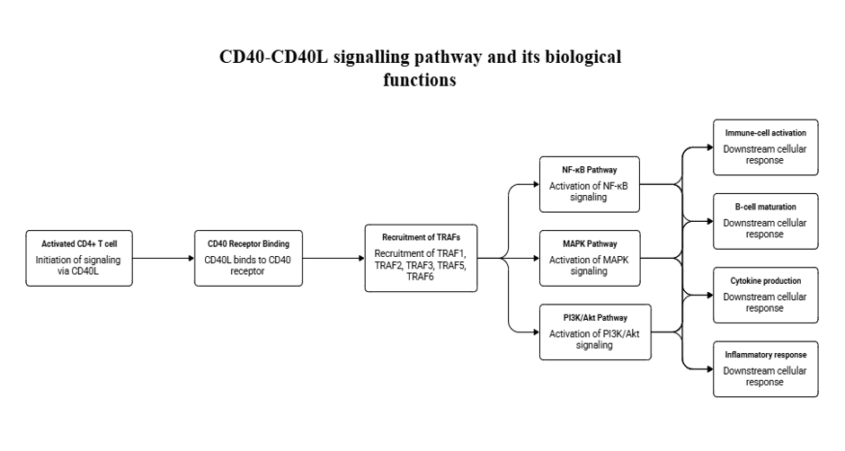
Figure 1: CD40/CD40L signaling pathway and its biological functions.
Schematic representation of CD40/CD40L-mediated signaling involving recruitment of TRAF adaptor proteins and activation of downstream NF-κB, MAPK, and PI3K/Akt pathways, contributing to immune-cell activation, B-cell responses, cytokine production, and inflammatory responses. Created by the authors based on published evidence.19–25
5.2 NF-κB as the Central Regulator of Synovial Inflammation
NF-κB functions as a master regulator of inflammatory gene expression in rheumatoid arthritis. Persistent activation of NF-κB induces transcription of pro-inflammatory cytokines, chemokines, adhesion molecules, cyclooxygenase-2 (COX-2), inducible nitric oxide synthase (iNOS), and matrix metalloproteinases (MMPs), thereby promoting leukocyte recruitment, synovial hyperplasia, cartilage degradation, and osteoclast-mediated bone erosion.28
In addition to amplifying inflammation, NF-κB promotes resistance to apoptosis in fibroblast-like synoviocytes, allowing persistent pannus formation and progressive joint destruction.29
5.3 Dysregulation of the CD40–TRAF1–NF-κB Axis in Rheumatoid Arthritis
In rheumatoid arthritis, sustained CD40 stimulation together with altered TRAF1 regulation results in persistent NF-κB activation within synovial tissues. Continuous NF-κB signaling maintains chronic production of inflammatory mediators and establishes a positive feedback loop that perpetuates synovial inflammation and structural joint damage.30
Collectively, the CD40–TRAF1–NF-κB axis links extracellular immune activation with intracellular inflammatory signaling, making it a central pathway in RA pathogenesis and an attractive target for therapeutic intervention. An integrated overview of the relationship between genetic susceptibility, CD40/TRAF1/NF-κB signaling, persistent synovial inflammation, joint destruction, and potential therapeutic intervention is presented in Figure 2.
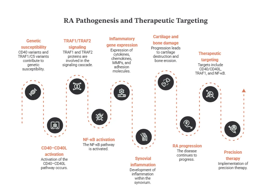
Figure 2: Integrated model of genetic susceptibility, CD40/TRAF1/NF-κB signaling, and rheumatoid arthritis pathogenesis.
Conceptual overview illustrating the proposed relationship between genetic susceptibility, CD40/CD40L signaling, TRAF1-associated signaling, NF-κB activation, inflammatory gene expression, synovial inflammation, cartilage degradation, bone destruction, and potential therapeutic intervention in rheumatoid arthritis. Created by the authors based on information synthesized from published literature.23, 24, 27–30, 33, 39
6. CURRENT THERAPEUTIC STRATEGIES TARGETING THE CD40/TRAF1/NF-ΚB PATHWAY IN RHEUMATOID ARTHRITIS
The therapeutic relevance of the CD40/TRAF1/NF-κB axis arises from its position at the intersection of immune activation, inflammatory signaling, and tissue destruction. Current approaches can be broadly categorized into strategies targeting the upstream CD40–CD40L interaction, indirect modulation of TRAF1 signaling, inhibition of NF-κB activation, and selective targeting of downstream pathway components. The principal therapeutic intervention points within the CD40/TRAF1/NF-κB signaling network are illustrated in Figure 3.
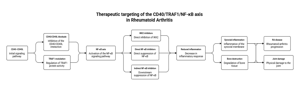
Figure 3: Therapeutic targeting of the CD40/TRAF1/NF-κB axis in Rheumatoid Arthritis.
Schematic representation of potential therapeutic intervention points within the CD40/TRAF1/NF-κB signaling network, including CD40/CD40L blockade, potential modulation of TRAF1-associated signaling, inhibition of IKK2, and direct or indirect modulation of NF-κB signaling. These approaches may reduce inflammatory signaling, synovial inflammation, and joint destruction. Created by the authors based on published evidence. 26, 30–35
6.1 Therapeutic Targeting of the CD40–CD40L Axis
The CD40–CD40L interaction represents an attractive upstream therapeutic target because its inhibition can simultaneously reduce immune-cell activation and downstream inflammatory signaling. Anti-CD40L monoclonal antibodies were initially developed to block CD40-mediated immune activation; however, early clinical development was limited by thromboembolic complications associated with Fc-mediated platelet activation.31
Subsequent approaches have focused on Fc-modified or Fc-silent CD40/CD40L inhibitors designed to retain pathway blockade while reducing platelet activation and thrombotic risk. Agents including BI 655064, iscalimab, bleselumab, and VIB4920 represent newer strategies aimed at modulating CD40-dependent immune activation and have been investigated in autoimmune diseases.31
Although direct CD40/CD40L blockade remains an attractive strategy, the clinical development of these agents highlights the challenge of selectively inhibiting pathogenic immune activation while preserving the physiological functions of CD40 signaling.
6.2 Therapeutic Strategies Targeting TRAF1
In contrast to CD40 and NF-κB, TRAF1 currently lacks a clinically established TRAF1-specific inhibitor or activator. This reflects the complex and context-dependent role of TRAF1 in immune regulation, in which both excessive and insufficient TRAF1 activity may produce different biological outcomes.32
Potential therapeutic approaches include modulation of TRAF1 stability or function, disruption of TRAF1–TRAF2–cIAP signaling complexes, and manipulation of TRAF1 interactions with regulatory components such as LUBAC. These strategies remain experimental but could provide greater pathway selectivity than broad NF-κB inhibition.26
At present, TRAF1 signaling is therefore targeted primarily indirectly, through inhibition of upstream CD40 signaling or downstream inflammatory pathways. Further clarification of the cell-specific functions of TRAF1 will be necessary before TRAF1-directed therapies can be translated into clinical applications.
6.3 Indirect Targeting of NF-κB by Current RA Therapies
Most established RA therapies do not directly inhibit NF-κB but instead suppress upstream inflammatory signals that converge on this pathway. Methotrexate reduces inflammatory cytokine production and consequently limits NF-κB activation, whereas TNF inhibitors such as etanercept, infliximab, and adalimumab reduce TNF-dependent inflammatory signaling. Similarly, IL-6 pathway inhibition and JAK inhibition indirectly attenuate inflammatory pathways that contribute to NF-κB-driven responses.34
Other therapies act through more specific inflammatory pathways. Tacrolimus reduces T-cell activation and thereby indirectly limits NF-κB-mediated inflammation, while denosumab blocks RANKL-dependent signaling and reduces osteoclast formation and bone erosion.34
These observations indicate that part of the clinical efficacy of existing RA treatments may arise from their ability to reduce the inflammatory signals that sustain NF-κB activation, even though NF-κB itself is not their primary molecular target.
6.4 Direct Inhibition of NF-κB Signaling
Direct inhibition of NF-κB has been investigated using several experimental strategies. Iguratimod and tetrandrine have been reported to suppress NF-κB-dependent inflammatory signaling, while proteasome inhibitors such as bortezomib and MG132 prevent IκB degradation and consequently inhibit NF-κB activation.34
Experimental gene-based approaches have also been explored. A degradation-resistant IκBα super-repressor can maintain NF-κB in an inactive state, whereas NF-κB decoy oligodeoxynucleotides competitively interfere with NF-κB binding to target DNA. Similarly, siRNA approaches directed against NF-κB subunits have demonstrated potential for selectively reducing inflammatory signaling.30
Despite promising experimental findings, broad NF-κB inhibition presents a major therapeutic challenge because NF-κB is also essential for normal immune responses and cellular homeostasis. Therefore, selective inhibition of specific pathway components may provide a better balance between efficacy and safety.
6.5 Targeting IKK2 as a Selective Therapeutic Strategy
The IκB kinase 2 (IKK2/IKKβ) complex represents a potential intermediate target for more selective inhibition of canonical NF-κB signaling. IKK2 contributes to inflammatory responses induced by CD40L, IL-1β, and TNF in synovial fibroblasts and endothelial cells. Its inhibition has been associated with reduced production of inflammatory cytokines, chemokines, vascular endothelial growth factor, and matrix metalloproteinases.31
By selectively targeting IKK2 rather than completely suppressing NF-κB activity, this approach may reduce synovial inflammation and angiogenesis while minimizing disruption of essential NF-κB-dependent physiological functions.35
6.6 Emerging Therapeutic Targets within the CD40/TRAF1/NF-κB Network
Several upstream and downstream regulators of NF-κB are being investigated as potential therapeutic targets in RA. These include OX40/OX40L-mediated co-stimulation, IRAK4-dependent signaling downstream of IL-1 and Toll-like receptors, GM-CSF-mediated macrophage activation, and emerging targets such as TYK2 and the bradykinin B1 receptor. These approaches may provide additional opportunities to modulate inflammatory signaling without directly inhibiting the entire NF-κB system.31
6.7 Natural Compounds Targeting NF-κB
Natural compounds have also attracted considerable interest as potential modulators of NF-κB signaling. Celastrol, resveratrol, curcumin, quercetin, vitamin D, and glucosamine have been investigated for their ability to suppress inflammatory signaling through mechanisms involving NF-κB and related pathways.34
Additional compounds, including sulforaphane, dihydromyricetin, oleuropein, sinomenine, and gedunin, have been reported to modulate NF-κB-associated inflammation, in some cases through activation of antioxidant pathways such as Nrf2/HO-1.49 However, most evidence remains preclinical, and further studies are required to establish their therapeutic efficacy, pharmacokinetic properties, and safety in RA.
6.8 RNA-Based Therapeutic Strategies
Non-coding RNAs represent an emerging approach for selectively regulating the CD40/TRAF1/NF-κB network. Anti-inflammatory microRNAs, including miR-23b, miR-22, miR-27a/b, miR-7-5p, miR-20a, and miR-146a, have been associated with suppression of NF-κB activity and inflammatory cytokine production. Conversely, miR-128-3p, miR-19b, miR-145-5p, and miR-34a may promote inflammatory signaling and joint damage.37
Long non-coding RNAs and circular RNAs further regulate inflammatory pathways through transcriptional mechanisms and microRNA-sponging activity. These findings suggest that RNA-based interventions could offer highly selective modulation of inflammatory signaling, although delivery, stability, and target specificity remain major challenges for clinical translation.37
6.9 Targeting NF-κB-Dependent Bone Destruction
Therapeutic inhibition of NF-κB-dependent osteoclastogenesis represents another potential strategy for limiting structural joint damage. Experimental compounds including pirfenidone, safranal, EPZ015866, eltanexor, isopsoralen, avicularin, dictamnine, and strontium ranelate have been investigated for their ability to reduce bone destruction through modulation of NF-κB-dependent pathways.38
TRAF6-directed approaches, including TRAF-STOP and RANK-Tet peptides, have also been explored to interfere with RANK/TRAF6/NF-κB signaling and inhibit osteoclast differentiation. Although these strategies primarily target pathways distinct from CD40/TRAF1, they demonstrate the broader therapeutic potential of selectively modulating NF-κB-dependent mechanisms involved in bone destruction.38
Overall therapeutic perspective
Current RA treatments primarily suppress the CD40/TRAF1/NF-κB axis indirectly, whereas emerging approaches aim to selectively interfere with CD40/CD40L interactions, TRAF1-associated signaling complexes, IKK2, NF-κB transcriptional activity, non-coding RNAs, and NF-κB-dependent osteoclastogenesis. The major challenge is to achieve sufficient suppression of pathogenic inflammation without compromising the physiological functions of NF-κB and CD40 signaling. Future therapies that target specific molecular nodes within this network may therefore offer greater precision and improved therapeutic selectivity
7. FUTURE PERSPECTIVES AND RESEARCH GAPS
The precise functional consequences of TRAF1 genetic variants, the cell-specific effects of TRAF1 signaling, and the development of selective pathway-directed therapies require further investigation.
7.1 Clarifying the Functional Consequences of TRAF1 Genetic Variants
Although polymorphisms within the TRAF1/C5 locus are consistently associated with RA susceptibility, the mechanisms through which these variants alter TRAF1 expression and downstream inflammatory signaling remain incompletely defined. Future studies should determine whether specific genetic variants directly influence TRAF1 expression, protein stability, or interactions with other signaling adaptors and whether these effects differ between immune-cell populations.33,39
7.2 Defining the Cell-Specific Functions of TRAF1
The effects of TRAF1 appear to vary according to cellular context, with potentially distinct functions in T cells, B cells, monocytes, macrophages, and synovial fibroblasts. A better understanding of these cell-specific effects is essential because broad manipulation of TRAF1 could potentially suppress beneficial immune responses while failing to adequately control pathogenic inflammation.26
7.3 Development of Selective Pathway-Directed Therapies
The absence of a clinically established TRAF1-specific therapy remains a major gap in the field. Future research should focus on identifying strategies that selectively modulate TRAF1-dependent signaling without disrupting its physiological functions. Similarly, selective inhibition of specific components of the NF-κB pathway may provide greater therapeutic precision than global NF-κB suppression, which can interfere with normal immune homeostasis.26,32
7.4 Integration of Genetics with Precision Medicine
The association between TRAF1/C5 variants and RA susceptibility provides an opportunity to develop genetically informed approaches to disease prediction and treatment. Future studies should investigate whether TRAF1 genetic profiles can identify patients at increased risk of severe disease or predict responses to specific biological or pathway-directed therapies. Such approaches could facilitate patient stratification and support personalized treatment strategies.33
7.5 Translational Challenges
Many emerging interventions targeting CD40/CD40L, TRAF1, NF-κB, and related signaling pathways remain at the preclinical or early clinical stage. Future research should prioritize validation in relevant human disease models, assessment of long-term safety, optimization of drug delivery, and identification of biomarkers that reflect pathway activity. Particular attention should be given to distinguishing pathogenic signaling from the physiological functions of CD40 and NF-κB, thereby minimizing systemic immunosuppression and other adverse effects.31
CONCLUSION
The CD40/TRAF1/NF-κB signaling axis represents an important molecular link between genetic susceptibility, immune activation, and persistent synovial inflammation in rheumatoid arthritis. Genetic variation within the CD40 and TRAF1/C5 loci may influence individual susceptibility to RA, while dysregulated CD40 signaling and TRAF1-dependent modulation of downstream pathways contribute to sustained inflammatory responses. Activation of NF-κB subsequently promotes the expression of inflammatory mediators and genes involved in synovial proliferation, cartilage degradation, and bone destruction, thereby linking immune dysregulation with progressive joint damage.
Although current RA therapies effectively target major inflammatory mediators, most do not directly address the CD40/TRAF1/NF-κB network as an integrated pathogenic axis. Emerging approaches targeting CD40/CD40L interactions, TRAF-dependent signaling, NF-κB pathway components, and associated regulatory mechanisms may therefore provide opportunities for more precise therapeutic intervention. However, the complex and context-dependent functions of TRAF1 and NF-κB highlight the need for selective rather than broad pathway inhibition.
Future research integrating genetic profiling, cell-specific molecular characterization, and biomarker-guided treatment may help identify patients in whom dysregulation of the CD40/TRAF1/NF-κB axis has a particularly important pathogenic role. Such approaches could support the development of personalized therapeutic strategies that selectively suppress pathogenic inflammation while preserving essential immune functions. Overall, continued investigation of this signaling network may contribute to improved understanding of RA pathogenesis and facilitate the development of more targeted and effective treatments for patients with rheumatoid arthritis.
REFERENCES



Bijesh Vatakkeel, Nesla P P, Haritha M S, The Cluster of Differentiation 40/Tumor Necrosis Factor Receptor Associated Factor 1/Nuclear Factor Kappa B Axis in Rheumatoid Arthritis: from Genetic Susceptibility and Synovial Inflammation to Therapeutic Opportunities, Int. J. of Pharm. Sci., 2026, Vol 4, Issue 9, 1662-1676. https://doi.org/10.5281/zenodo.22742303
 10.5281/zenodo.22742303