
We use cookies to ensure our website works properly and to personalise your experience. Cookies policy
Srinath college of pharmacy Chhatrapati Sambhajinagar
4D bioprinting is an emerging frontier in biomedical science that builds upon conventional 3D bioprinting by introducing dynamic, stimuli-responsive capabilities1,2 Unlike traditional 3D scaffolds, 4D constructs can change shape, properties, or functions over time in response to environmental cues, enabling more realistic tissue regeneration, organ-specific therapeutics, and advanced drug delivery strategies. This technology utilizes smart biomaterials, including shape-memory polymers, hydrogels, and composite bioinks, which respond to stimuli such as temperature, pH, light, and magnetic or electric fields .Through precise design of scaffold architecture and material gradients, these constructs can achieve predictable deformations and functional adaptations, enhancing cellular integration and therapeutic efficacy3,4. Current research demonstrates potential applications in preclinical tissue engineering, organ-targeted therapies, and controlled drug delivery, although clinical translation is still limited 5,6. By integrating material science, bioprinting techniques, and stimulus-driven programming,. 4D bioprinting has shown promise in addressing several limitations. This review summarizes the mechanistic insights, preparation methods, biomedical applications, and future prospects of 4D bioprinting, emphasizing its relevance in advanced drug delivery and organ-specific therapeutics. It aims to provide a comprehensive understanding of this innovative technology, highlighting ongoing research trends, challenges, and potential directions for clinical translation 7,8
3D Bioprinting: A Brief Overview
3d bioprinting is a revolutionary technique in biomedical sciences that allows the layer-by-layer construction of biological structures using living cells, biomaterials, and growth factors. This technology has transformed the way researchers approach tissue engineering, regenerative medicine, and organ modeling. The primary principle is to fabricate complex tissue constructs that mimic the architecture and functionality of natural tissues9.
Various bioinks, such as natural polymers (gelatin, alginate, collagen), synthetic polymers (PEG, PCL), and composite materials, are used in 3d bioprinting. These bioinks are designed to support cell viability, proliferation, and differentiation 10. Over the past decade, 3d bioprinting has been successfully applied to create skin grafts, cartilage constructs, vascular networks, and organ-on-chip models for drug screening . It has enabled more accurate in vitro models compared to traditional 2d cultures, improving our understanding of tissue responses and therapeutic testing 11.
Despite its success, 3d bioprinting remains primarily static. Once printed, the structures maintain a fixed shape and functionality, which limits their ability to mimic the dynamic nature of living tissues. Additionally, replicating the intricate heterogeneity and vascularization of natural tissues is challenging. While 3d bioprinting has made progress in creating functional tissue models, scalability and long-term integration in clinical applications remain limited. Bioinks often struggle to balance mechanical strength with biocompatibility, affecting tissue maturation and stability 12
Limitations of 3D Bioprinting
Some of the main limitations that have emerged in the 3D bioprinting field include:
These challenges have motivated researchers to explore next-generation bioprinting technologies that can introduce responsiveness and adaptability into printed constructs 9,14.
4D Bioprinting: Introducing Time-Responsive Constructs
4D bioprinting is the natural evolution of 3D printing, where the fourth dimension—time—is incorporated into the design of printed structures. Unlike conventional 3D constructs, 4D-printed materials can undergo programmed shape or functional changes in response to external stimuli such as temperature, pH, light, or magnetic fields 1,15. This transformative approach allows the creation of smart, adaptive biomedical devices capable of interacting dynamically with biological environments.
The success of 4D bioprinting depends largely on stimuli-responsive bioinks. These include shape-memory polymers (SMPs), hydrogels, and composite materials that can reversibly transform in response to specific triggers 10,12,14. For example, a hydrogel scaffold may expand or contract depending on pH changes, or a shape-memory polymer implant may alter its conformation at body temperature. These adaptive behaviors open new avenues in drug delivery, tissue regeneration, and personalized implants .
4D bioprinting is particularly exciting for drug delivery applications. Drugs can be embedded in responsive matrices, allowing controlled, on-demand release at targeted sites, improving therapeutic efficacy while minimizing side effects. In tissue engineering, 4D constructs can mimic natural tissue remodeling, responding to environmental cues like mechanical stress or chemical gradients, which is essential for functional integration 11,15,16.
Recent Innovations and Potential
Recent studies have introduced dual-stimuli responsive materials capable of changing shape in response to both temperature and pH variations, providing higher control over structure and function . Preclinical studies demonstrate the potential of 4D-printed constructs in vascular grafts, cartilage repair, and smart implants. While clinical translation is still emerging, ongoing research highlights the significant potential of 4D bioprinting to overcome the limitations of traditional 3D methods and revolutionize regenerative medicine and drug delivery .
In summary, while 3D bioprinting has paved the way for tissue engineering and biomedical applications, 4D bioprinting adds a dynamic, adaptive dimension that holds promise for creating more effective, responsive, and clinically relevant solutions. This review will explore the mechanisms, preparation methods, biomedical applications, and future prospects of 4D bioprinting, providing insights into its role in drug delivery and tissue therapeutics 17–19.
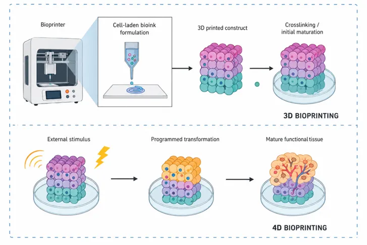
Figure 1: Comparison of 3D and 4D Bioprinting processes for Tissue Engineering
3.MECHANISMS OF 4D BIOPRINTED SCAFFOLDS
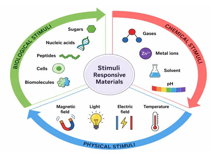
Figure 2: Stimuli responsive polymers
1. Thermal-Responsive Mechanism
2. pH-Responsive Mechanism
Polymers containing acidic (–COOH) or basic (–NH₂) groups ionize differently depending on environmental pH
3. Light-Responsive Mechanism
4. Magnetic-Responsive Mechanism
5. Enzyme-Responsive Mechanism
These mechanisms do not always rely on external stimuli but represent intrinsic smart material responses crucial in 4D bioprinting:
a) Shape Memory Response
b) Swelling–Shrinking Mechanism
c) Folding/Unfolding & Twisting
|
Type |
Key Features |
Examples |
|
Natural Polymers |
ECM-like, biocompatible |
Collagen, Alginate, Gelatin, HA, Fibrin |
|
Synthetic Polymers |
Strong, tunable degradation |
PLA, PGA, PLGA, PCL, PEG |
|
Hydrogels |
Soft tissue–like, cell-friendly |
Alginate hydrogel, PEG-hydrogel, GelMA |
|
Composite Bioinks |
Natural + synthetic blends, improved strength |
Collagen–PCL, Alginate–PEG |
|
Smart Materials |
Stimuli-responsive, shape-changing |
Shape-memory polymers, Responsive hydrogels |
Comparative Table: Mechanisms of 4D Bioprinted Scaffolds
Table 1: Mechanism of 4D Bioprinted Scaffolds
|
Mechanism |
Trigger/Stimulus |
Response |
Applications |
|
Thermal-Responsive |
Temperature change |
Shrinkage/expansion of polymer chains |
Drug delivery, vascular grafts, tissue folding |
|
pH-Responsive |
Local acidic/alkaline pH |
Swelling/shrinking, degradation |
Tumor therapy, GI tract targeting |
|
Light-Responsive |
UV/Visible light |
Folding, bending, conformational shift |
Nerve regeneration, patterned drug release |
|
Magnetic-Responsive |
External magnetic field |
Movement, fiber alignment, heating |
Targeted therapy, stem cell guidance |
|
Enzyme-Responsive |
Enzyme presence (MMPs, lipases) |
Degradation/remodeling of scaffold |
Cancer therapy, wound healing, adaptive implants |
|
Shape Memory Response |
Heat, hydration, or light |
Recovery to original shape |
Self-deployable stents, implants |
|
Swelling–Shrinking |
Hydration/dehydration |
Volume expansion/contraction |
Soft tissue mimicry, drug release |
|
Folding/Unfolding/Twisting |
Stress anisotropy, external cue |
Origami-like dynamic reconfiguration |
Vascular scaffolds, organ-on-chip |
4. Conventional Bioprinting Technologies Adapted for 4D Bioprinting
4D bioprinting extends the principles of 3D bioprinting by integrating the dimension of time, which allows the fabricated construct to transform its properties, structure, or function in response to specific stimuli .The success of this approach depends heavily on the printing technologies that can process stimuli-responsive bioinks while maintaining fidelity, mechanical integrity, and biological viability . Unlike conventional 3D printing that ends at static fabrication, 4D approaches require methods that can incorporate smart programming during or after fabrication 15,18.
The fabrication technologies commonly adapted for 4D bioprinting include:
Each method has unique advantages and limitations, and their application is often determined by the type of bioink, the scale of construct, and the targeted biological funct:
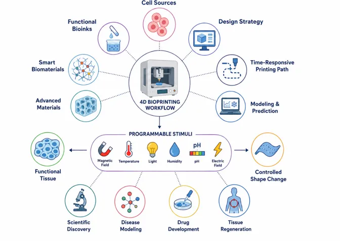
Figure 3:4D bioprinting Process
4.1. Extrusion-Based Bioprinting (EBB)
1.1 Working Principle
Extrusion-based bioprinting is the most widely used technology for 4D applications. It relies on the mechanical or pneumatic extrusion of bioink through a nozzle, depositing continuous filaments that are layered to form 3D structures .
1.2 Suitability for 4D Applications
Compatible with high-viscosity bioinks such as hydrogels, polymer blends, and nanocomposites .
Enables the incorporation of shape-memory polymers (SMPs) and hydrogels that swell, shrink, or fold in response to stimuli .
Multi-nozzle systems allow gradient or multi-material constructs, essential for programming shape transformation 4,20,24.
1.3 Example Applications
Bone tissue scaffolds that expand with hydration .23
Cardiac patches printed with SMP composites that beat in synchronization with electrical signals 22.
Self-folding drug carriers programmed by differential crosslinking density .
1.4 Advantages and Challenges
Advantages: Versatile, scalable, able to handle cell-laden viscous inks.
4.2. Inkjet Bioprinting
2.1 Working Principle
Inkjet printing dispenses droplets of bioink onto a substrate, similar to an office inkjet printer. Droplets are controlled by thermal, piezoelectric, or electrostatic mechanisms .
2.2 Role in 4D Bioprinting
2.3 Applications
Smart hydrogels that fold into tubular structures upon hydration
2.4 Pros and Cons
4.3. Stereolithography (SLA)
3.1 Working Principle
SLA uses light (UV/laser) to selectively cure photosensitive polymers layer by layer. The method can achieve microscale precision and highly complex architectures .
3.2 4D Integration
3.3 Applications
3.4 Advantages and Limitations
4.4.Laser-Assisted Bioprinting (LAB)
4.1 Principle
LAB employs focused laser pulses to generate droplets of bioink from a donor layer onto a receiving substrate. This non-contact process allows ultra-high precision in positioning .
4.2 Application in 4D Bioprinting
Ideal for printing smart constructs where small-scale cell distribution drives overall transformation
4.3 Use Cases
4.4 Pros and Cons
4.5. Direct Ink Writing (DIW) and Hybrid Approaches
5.1 Direct Ink Writing (DIW)
DIW involves directly writing bioinks with controlled deposition patterns It is highly versatile for incorporating multi-materials in a single construct.
5.2 Hybrid Bioprinting
Hybrid systems combine extrusion, SLA, and inkjet in a single platform This integration allows simultaneous use of cell-laden hydrogels and structural polymers, programming each layer with distinct responsiveness.
4.6. Programming Mechanisms in 4D Bioprinting
While technologies define the printing step, programming defines transformation. Key mechanisms include
These mechanisms are integrated with the printing technologies to create living constructs that adapt dynamically in vivo 11,20
4.7. Stepwise Preparation Framework
CAD tools generate 3D/4D scaffold models incorporating microchannels, gradients, or folding zones
Bioinks chosen based on responsiveness (thermal, pH, magnetic, photothermal)
Extrusion, inkjet, SLA, or LAB methods are employed depending on viscosity, cell content, and resolution
Micro-architectures such as pores, channels, and aligned fibers are formed during deposition or curing
Drugs, growth factors, or nanoparticles are encapsulated within bioink or post-loaded into porous regions
Crosslinking (thermal, UV, or ionic) stabilizes the construct. Smart programming introduced at this stage
Structural fidelity, cell viability, drug release kinetics, and transformation efficiency are tested before in vivo application
4.8. Integration Across Technologies
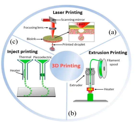
Figure 4: Conventional Bioprinting Technologies Adapted for 4D Bioprinting
4.9.Comparative Summary of Methods
Table 2:Comparative Summary of Methods
|
Technology |
Resolution |
Bioink Compatibility |
Stimuli Integration |
Applications |
Limitations |
|
Extrusion-based |
100–200 µm |
High-viscosity hydrogels, SMPs |
Thermal, swelling, shape memory |
Bone, cartilage, stents |
Lower resolution, shear stress |
|
Inkjet |
20–100 µm |
Low-viscosity inks |
pH, hydration, drug delivery |
Drug carriers, organ-on-chip |
Poor mechanical strength |
|
SLA |
<50 µm |
Photopolymers, hydrogels |
Light, heat |
Microtissues, microneedles |
Limited bioink options |
|
LAB |
<10 µm |
Hydrogel-cell suspensions |
Electrical, magnetic |
Neural scaffolds, micro-tissues |
Expensive, low throughput |
|
DIW/Hybrid |
50–150 µm |
Multi-material systems |
Multi-stimuli |
Complex tissues, vascular grafts |
Complex setup |
It is important to note that technologies such as extrusion, inkjet, SLA, and laser-assisted printing are fundamentally derived from conventional 3D bioprinting platforms. In 4D bioprinting, these fabrication techniques are integrated with stimuli-responsive biomaterials and programmable design strategies to enable dynamic post-print transformation over time
5.APPLICATIONS OF 4D BIOPRINTING IN BIOMEDICAL SCIENCES
4D bioprinting represents a natural evolution of 3D bioprinting, moving from static constructs to dynamic, adaptive systems that respond to biological or environmental stimuli 1,11. This adaptability enables a wide spectrum of biomedical applications ranging from tissue regeneration to controlled drug delivery and the design of smart implants. The following sections highlight key areas where 4D bioprinting is being explored, alongside its potential to transform healthcare solutions 12,14,21
5.1. Tissue Engineering and Regenerative Medicine
One of the most significant applications of 4D bioprinting is in the fabrication of scaffolds for tissue regeneration Traditional 3D scaffolds provide mechanical support and spatial architecture but remain static once implanted. In contrast, 4D bioprinted scaffolds are designed with stimuli-responsive materials such as hydrogels and shape-memory polymers, enabling them to adapt over time 18,20
For instance, a scaffold implanted for bone repair can swell or contract in response to environmental changes, ensuring better integration with host tissue
Dynamic scaffolds can also be programmed to release growth factors or bioactive molecules at specific time points, promoting cellular differentiation and vascularization 9,11Cartilage repair is another field where 4D scaffolds hold promise, as they can adapt to cyclic loading and mechanical strain experienced in joints 18,19. Similarly, soft tissues such as skin and muscle benefit from scaffolds that can alter their structure during the healing process, ensuring a closer mimicry of natural tissue dynamics 4,10
5.2. Organ-on-Chip and Disease Modeling
4D bioprinting is also advancing organ-on-chip technology, which provides miniature models of human physiology for studying disease mechanisms and drug responses 17,19 Conventional chips are static, but 4D printed platforms incorporate dynamic features such as folding, bending, or swelling channels to replicate the changing microenvironment of real organs 27,28
For example, vascular-on-chip systems can simulate the pulsatile flow of blood by adjusting the geometry of the scaffold under fluidic forces
This adaptive nature is especially valuable for disease modeling, where microenvironments must change over time to replicate progression.
Cancer-on-chip or heart-on-chip devices developed with 4D scaffolds enable real-time tracking of cellular behavior under dynamic conditions, offering better predictive power for drug testing compared to conventional static platforms
5.3. Drug Delivery and Controlled Release Systems
Drug delivery is an area where the dynamic capabilities of 4D bioprinting are particularly impactful.
By incorporating stimuli-responsive materials into printed scaffolds, it is possible to create systems that release drugs in response to environmental triggers such as temperature, pH, enzymes, or light 18,20
For example, a hydrogel scaffold can be programmed to shrink and expel its cargo when exposed to a change in pH, which is useful in tumor environments 19,27
Moreover, 4D systems allow spatiotemporal control over release profiles, enabling both immediate and sustained delivery depending on clinical needs11,16. Organ-specific targeting is another advantage, as scaffolds can be shaped or triggered to adapt to particular anatomical niches 10,29
Recent experimental work has explored combining therapeutic and diagnostic (“theranostic”) functions within the same scaffold, where a single construct can release drugs and simultaneously provide imaging or monitoring capability 19
5.4. Medical Devices and Smart Implants
Beyond scaffolds, 4D bioprinting is being used to create medical devices and implants with enhanced functionality .Shape-memory polymers enable the fabrication of stents that can be inserted in a compact form and expand when triggered by body temperature, reducing the need for invasive procedures .
Similarly, orthopedic implants can be designed to adapt their stiffness or geometry during bone healing, ensuring better long-term integration.
Smart prosthetics, which adjust to mechanical strain or patient activity, also fall under this category Unlike conventional static implants, these devices provide a dynamic interface with the body, potentially reducing complications such as implant rejection or poor fit 29,30
5.5. Neural and Cardiovascular Applications
4D bioprinting offers opportunities for regenerating highly sensitive and complex tissues such as neural and cardiovascular systems
In neural repair, dynamic scaffolds guide axonal growth by altering their geometry or releasing neurotrophic factors in a controlled manner 4,5,18These approaches show promise for spinal cord injuries and neurodegenerative diseases .
Cardiovascular applications include vascular grafts that can change diameter in response to blood pressure, reducing the risk of thrombosis or rupture 19,22Similarly Preclinical studies have demonstrated rhythm-responsive contraction behavior in 4D printed hydrogel-based cardiac patch models.
5.6. Wound Healing and Soft Tissue Repair
The dynamic properties of 4D scaffolds are especially beneficial for wound healing, where tissue environments are constantly changing .
Hydrogels incorporating antimicrobial agents can swell to maintain a moist environment, while shrinking during later stages of healing to promote closure
Shape-adaptive patches can be applied to irregular wounds, conforming to the site and ensuring intimate contact with tissue 19,20,25
Such responsive wound dressings not only accelerate healing but can also deliver bioactive compounds or drugs at required intervals .This dual functionality offers a major improvement over conventional dressings, which often need to be changed frequently and provide limited therapeutic benefits .
5.7. Cancer Research and Targeted Therapeutics
In oncology, 4D bioprinting is being leveraged to build dynamic tumor models for preclinical drug testing. These scaffolds replicate the evolving tumor microenvironment more accurately than static 3D systems, enabling researchers to study drug resistance and metastatic behavior
On the therapeutic side, 4D printed scaffolds are being investigated for localized delivery of anticancer agents By responding to tumor-specific cues such as acidic pH or high enzyme activity, scaffolds can release drugs precisely at the tumor site, minimizing systemic toxicity
Integration with immunotherapeutics is also being studied, where scaffolds act as delivery vehicles for cytokines or engineered immune cells 19,31
5.8. Future Integrations and Emerging Trends
While most current applications are still in preclinical stages, the potential of 4D bioprinting continues to expand with interdisciplinary integration
Artificial intelligence and computational modeling are being incorporated to design patient-specific constructs with predictive accuracy 30
Nanotechnology integration allows for improved responsiveness and multifunctionality, while robotics and automated systems are enhancing reproducibility and scalability 18,32
The combination of these fields could lead to transformative healthcare applications where scaffolds, devices, or implants not only adapt passively but are actively programmed to monitor, respond, and heal in real time
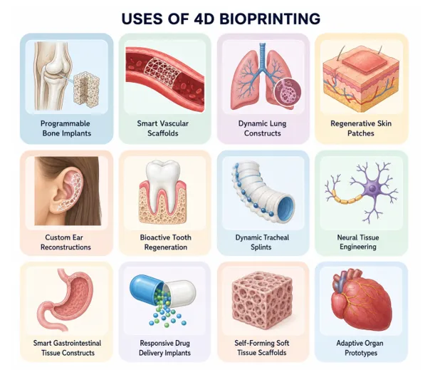
Figure 5:Various Organ Specific Application of 4D Bioprinting
6.FUTURE PROSPECTS
4D bioprinting represents a transformative leap in biomedical engineering, moving beyond the static nature of 3D scaffolds to dynamic, stimuli-responsive constructs that can mimic real tissue behavior. Its future is promising, with opportunities in organ-specific therapeutics, advanced drug delivery, tissue regeneration, and personalized medicine 5,16
6.1. Clinical Translation Potential
Personalized Medicine
4D bioprinting allows the creation of patient-specific scaffolds that adapt to the target tissue environment, improving integration and therapeutic outcomes .Computational modeling and medical imaging data (MRI/CT) can guide scaffold design, ensuring customized geometry, porosity, and mechanical properties for each patient .7Such personalization will enable more predictable regenerative outcomes and optimized biomechanical performance.
Dynamic Organ Mimicry:
Future scaffolds are expected to respond to physiological cues, such as body temperature, pH, enzymatic activity, or mechanical stress, enabling functional organ regeneration in situ .For instance, cardiac patches may contract synchronously with myocardial motion, and bone scaffolds could expand to fill osteogenic defects as healing progresses .8,25These “living implants” could mark a paradigm shift in regenerative and reconstructive surgery.
Stimuli-Responsive Drug Delivery:
The integration of therapeutic molecules within smart, stimuli-responsive scaffolds enables spatiotemporal control over drug release 28Future 4D systems may combine multi-stimuli responses—such as pH, temperature, enzymatic activity, or magnetic fields—to deliver therapeutics selectively to pathological sites like tumors or ischemic tissues
Such precision-controlled release minimizes systemic toxicity while enhancing localized efficacy.
6.2. Emerging Research Trends
Multimaterial Printing:
Recent advances in composite bioinks enable the integration of hydrogels, polymers, nanoparticles, and bioactive molecules to achieve multifunctionality 6These hybrid inks can mimic the complexity of native tissues by offering tunable mechanical and biochemical properties. Researchers are exploring blends of natural polymers like gelatin methacrylate (GelMA) and synthetic materials such as polycaprolactone (PCL) to achieve both elasticity and durability 27
Shape-Memory and Self-Folding Materials:
Shape-memory polymers and hydrogels with self-folding capabilities represent a major leap forward in minimally invasive bioprinting 8These materials can transform shape post-implantation, improving tissue integration and allowing for on-demand functionality. Such innovation is particularly valuable for vascular grafts, stents, and neural conduits 18,31
Integration with AI and Computational Modeling:
Artificial intelligence and computational modeling are increasingly used to simulate the dynamic behavior of 4D scaffolds and optimize printing parameters 7Predictive algorithms can model degradation kinetics, swelling ratios, and drug release profiles, reducing preclinical trial time and improving reproducibility 26,28This synergy between biofabrication and data science accelerates translation from lab-scale prototypes to clinical-ready implants.
Preclinical Case Studies:
Recent preclinical research highlights successful outcomes in bone, cartilage, cardiac, and neural regeneration using 4D scaffolds .These models demonstrate superior tissue remodeling, vascularization, and mechanical adaptation compared to static 3D constructs .Drug delivery studies using pH- and temperature-responsive scaffolds have also achieved sustained and localized therapeutic action 20,22
6.3. Challenges and Roadmap for Clinical Adoption
Despite remarkable laboratory success, several translational challenges remain before clinical implementation.
Material Biocompatibility: Ensuring non-immunogenicity, tunable degradation, and consistent bioactivity remains essential
Scaffold Complexity: Balancing dynamic behavior with mechanical integrity for large organ structures requires optimization in design and material formulation
Regulatory Pathways: As 4D bioprinted constructs represent a new category of adaptive biomedical devices, standard regulatory frameworks must evolve to evaluate time-dependent safety and efficacy 8,17,19
Cost and Scalability: Achieving mass production while maintaining precision and patient customization remains an economic and technical challenge 30
7.CONCLUSION
4D bioprinting has emerged as a emerging technology in biomedical science, bridging the gap between static 3D constructs and dynamic, responsive tissue scaffolds. By integrating smart biomaterials, stimuli-responsive mechanisms, and advanced fabrication techniques, 4D scaffolds can change shape, properties, or functionality over time, offering unprecedented possibilities in tissue regeneration, organ-specific therapeutics, and targeted drug delivery.
The review highlights that while 3D bioprinting laid the foundation for creating precise, patient-specific tissue constructs, it is limited by its static nature, lack of adaptability, and inability to respond to dynamic biological environments .4D bioprinting addresses these limitations by incorporating environmentally responsive elements that enable scaffolds to adapt to physiological cues such as temperature, pH, mechanical stress, and enzymatic activity, improving cellular integration and therapeutic outcomes.
Preclinical studies have demonstrated promising results across multiple biomedical applications, including bone and cartilage regeneration, cardiac patches, neural repair, and controlled drug delivery systems. Additionally, the incorporation of shape-memory polymers, hydrogels, and composite bioinks has facilitated precise spatial and temporal control over scaffold behavior, supporting the feasibility of patient-specific, minimally invasive, and multifunctional therapeutic platforms 8,22,28
Despite its potential, several challenges remain, including material biocompatibility, regulatory hurdles, scaffold complexity, and scalability for clinical applications 26,27,29However, ongoing research, advancements in computational modeling, AI-driven design, and multi-material printing, as well as increasing preclinical successes, indicate that these barriers can be overcome in the near future .
In conclusion, 4D bioprinting is poised to transform regenerative medicine and drug delivery, offering dynamic, patient-tailored solutions that were previously unattainable with traditional 3D constructs .As research progresses and clinical translation accelerates, this technology is likely to become a cornerstone of personalized therapeutics, driving innovation and improving patient outcomes across a broad spectrum of diseases and organ-specific treatments 26,30
REFERENCES


Tanaya Markande*, Sumeet Bhilwade, Sanika Nalande, Aarti More, Pranjali Narawade, Snehal Nage, 4D Bioprinting: Shaping the Future of Dynamic and Smart Tissue Regeneration, Int. J. of Pharm. Sci., 2026, Vol 4, Issue 6, 7069-7086. https://doi.org/10.5281/zenodo.20967558
 10.5281/zenodo.20967558