
View Article

  • Inflammation And Anti-Inflammatory Therapeutics: Molecular Mechanisms, Clinical Burden, Therapeutic Limitations, And Emerging Safer Alternatives

  • M.Pharm Department of Pharmacy, M.J.P. Rohilkhand University, Bareilly (243006), Uttar Pradesh, India

Abstract

Inflammation is a tightly regulated biological response that protects the host, clears injurious stimuli, and restores tissue homeostasis, yet its dysregulation underlies a large share of acute and chronic human disease. This review consolidates current understanding of the molecular architecture of inflammation, spanning pattern-recognition receptor signalling, cellular and chemical mediators, and the intracellular cascades (NF-?B, MAPK, JAK–STAT, and the NLRP3 inflammasome) that convert an initiating stimulus into a coordinated inflammatory programme. It examines the clinical footprint of inflammation, using C-reactive protein as a representative biomarker linking systemic inflammation to cardiovascular disease, type-2 diabetes, neurodegeneration, renal failure, and rheumatoid arthritis, and summarises the substantial and unevenly distributed global burden of immune-mediated inflammatory diseases. Conventional pharmacological strategies — NSAIDs, corticosteroids, DMARDs, biologic agents, and JAK inhibitors — are reviewed alongside their well-documented gastrointestinal, renal, cardiovascular, immunological, and economic costs, which together constrain long-term management. Against this backdrop, the review surveys plant-derived anti-inflammatory agents (curcumin, gingerols, eugenol, aloe vera constituents, epigallocatechin gallate, and Portulaca oleracea phytochemicals) as multi-targeted, comparatively safer alternatives, while acknowledging persistent barriers of bioavailability, standardisation, and clinical validation. Finally, it considers emerging strategies — nanocarrier-based delivery, rational combination regimens, biomarker-guided personalised therapy, and computational/AI-assisted drug discovery — that may narrow the gap between efficacy and safety. Taken together, the evidence indicates that no single therapeutic class offers both maximal efficacy and minimal risk, and that future progress will depend on integrating mechanistic pharmacology with safer delivery platforms and individualised treatment selection.

Keywords

Inflammation; NF-?B; Cytokines; NSAIDs; Biologics; Herbal anti-inflammatory agents;NLRP3 inflammasome

Introduction

× Popup Image

1.1 Definition and Historical Perspective

Inflammation is classically understood as the reaction of vascularised living tissue to harmful stimuli — pathogens, physical injury, or chemical irritants — and forms a core component of the innate, non-specific arm of host defence 1,2. Biochemically, it can also be viewed as a state of heightened tissue catabolism: the accumulation of low-molecular-weight breakdown products raises local osmotic pressure, drawing fluid into the tissue and producing the oedema that is one of its most visible hallmarks 3.

Awareness of inflammatory disease is ancient. Egyptian medical papyri dating to roughly 1650 BCE already record descriptions consistent with tissue injury and its visible sequelae 4. Centuries later, the Roman encyclopaedist Aulus Cornelius Celsus, writing in his treatise De Medicina in the first century CE, distilled the clinical presentation of inflammation into four cardinal signs — rubor (redness), calor (heat), tumor (swelling), and dolor (pain) — arising respectively from local vasodilation, increased regional blood flow and metabolism, fluid and cellular accumulation, and stimulation of sensory nerve endings by inflammatory mediators 5,6. A fifth sign, functio laesa (loss of function), is traditionally attributed to Galen in the second century CE, although historians note that this attribution may be a later retrospective addition rather than a documented original contribution 7.

Between antiquity and the modern era, explanations of inflammation were dominated by the Hippocratic humoral theory, which framed disease as an imbalance among four bodily fluids 4. This view persisted for well over a millennium until autopsy-based pathology — beginning with Giovanni Morgagni's organ-level correlations in 1761 and maturing through nineteenth-century histology — relocated disease from diffuse humoral imbalance to discrete tissue and, eventually, cellular lesions 4,6. Rudolf Virchow's cellular pathology, published in 1858 and encapsulated in the principle omnis cellula e cellula ("every cell arises from another cell"), established the cell as the fundamental unit of both physiology and pathology and reframed inflammatory diseases, including atherosclerosis, as processes originating in cellular and vascular derangement 8. Alongside Virchow, Julius Cohnheim and Carl Weigert extended the microscopic study of vascular and leukocytic changes during inflammation. Late in the same century, Ilya Metchnikoff's work on phagocytosis and Paul Ehrlich's studies of antibodies and complement established that much of the tissue damage seen in infection arises from the host's own immune response rather than from direct pathogen action 9.

The twentieth century added a biochemical dimension to this cellular framework. The identification of cytokines, complement components, and reactive oxygen species clarified that immune cells cause tissue injury largely through secreted molecules rather than by direct contact alone 10, and more recent work has implicated regulated cell-death pathways — apoptosis, necroptosis, and pyroptosis — in shaping how inflammation initiates and resolves 11. Discovery of the prostaglandins in the 1930s by Ulf von Euler, followed by elucidation of their arachidonic-acid/cyclooxygenase biosynthetic pathway in the 1960s–70s and Sir John Vane's demonstration that aspirin-like drugs act by inhibiting prostaglandin synthesis, provided the molecular rationale for NSAID pharmacology and earned Vane, Bengt Samuelsson, and Sune Bergström the 1982 Nobel Prize in Physiology or Medicine 12. Thus, the understanding of inflammation has evolved from a purely descriptive clinical phenomenon into a multi-layered biological process involving receptor recognition, cellular recruitment, chemical signalling, and regulated cell death — the mechanistic layers addressed in Section 2.

1.2 Acute versus Chronic Inflammation

Acute inflammation is typically studied through models of microbial infection, in which monocytes are recruited to the injured or infected site after an initial wave of neutrophil infiltration, a process that can continue for several days 13. Infection with Listeria monocytogenes, for example, drives strong monocytosis and Toll-like-receptor-dependent monocyte production in the bone marrow, leading to CCR2⁺ classical monocyte recruitment that contributes both to pathogen clearance and to initiation of adaptive immunity 14,15,16. This recruitment is coordinated chiefly by the chemokines CCL2 and CCL7 acting on bone-marrow monocytes 17,18.

Chronic inflammation, in contrast, persists over extended periods and underlies diseases such as atherosclerosis, in which lipid deposition within the arterial wall attracts monocytes that differentiate into macrophages and accumulate lipid to become foam cells within the developing plaque 19. Both classical and non-classical monocyte subsets are recruited into plaques through chemokine-receptor signalling involving CCR2, CCR5, and CX3CR1 20, and hypercholesterolaemia-associated monocytosis in animal models correlates with plaque burden and cardiovascular risk 21,22,23.

Materials and Methods

This review was compiled through a structured, non-systematic literature search of PubMed, Scopus, and Google Scholar, using combinations of the terms “inflammation,” “NF-κB,” “NLRP3 inflammasome,” “NSAIDs,” “corticosteroids,” “biologic therapy,” “JAK inhibitors,” and “plant-derived anti-inflammatory agents,” restricted primarily to peer-reviewed articles published in English. Reference lists of retrieved articles were hand-searched for additional relevant sources. Preclinical, clinical, and epidemiological studies were included where they contributed mechanistic insight, clinical relevance, or comparative safety data; conference abstracts and non-peer-reviewed sources were excluded except where no peer-reviewed alternative was available.

2. Molecular Mechanisms of Inflammation

The inflammatory response is initiated by receptor-mediated recognition of danger signals, propagated by cellular and chemical mediators, and executed through a small number of convergent intracellular signalling pathways. This section addresses each layer in turn; Figure 1 summarises how the components discussed in Sections 2.1–2.5 fit together.
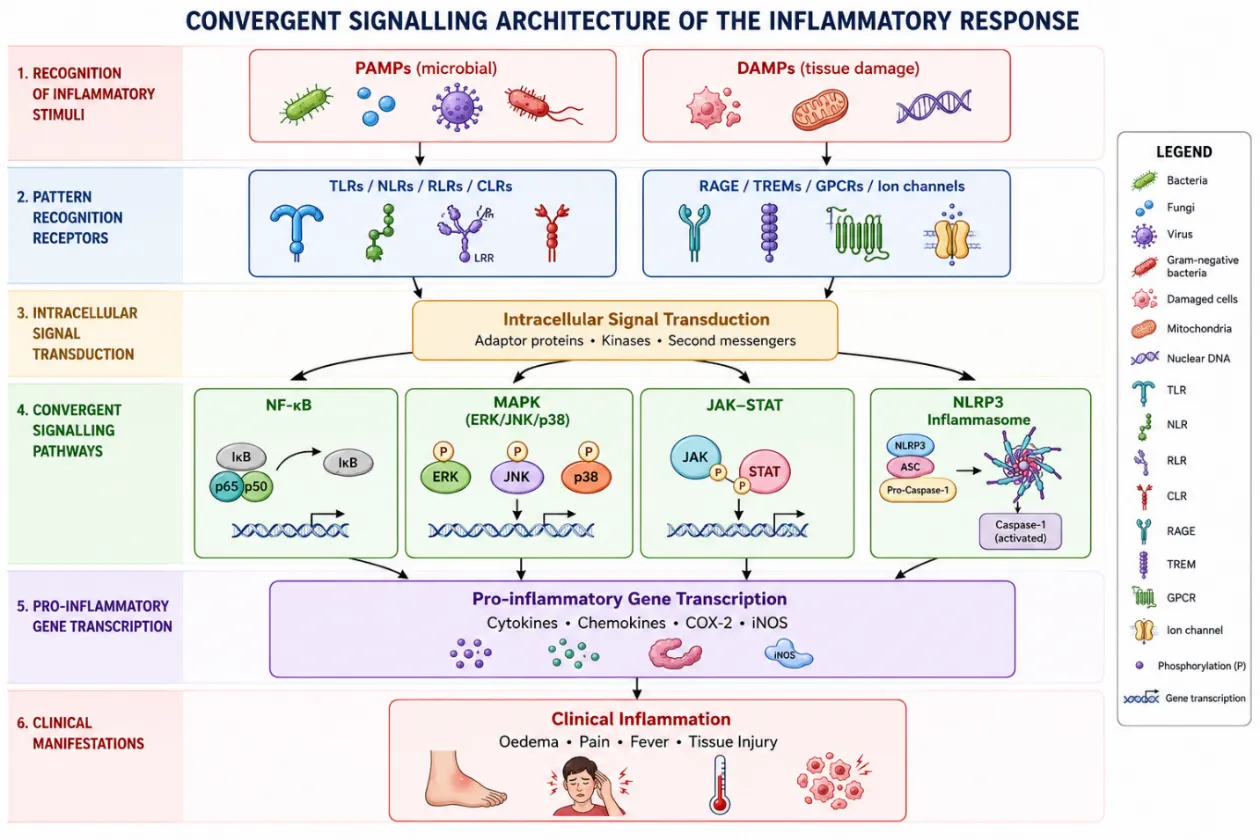
 

Figure 1. Convergent signalling architecture of the inflammatory response: PAMP/DAMP recognition converges, via NF-κB, MAPK, JAK–STAT, and NLRP3 inflammasome signalling, on pro-inflammatory gene transcription and the clinical manifestations of inflammation.

2.1 Recognition of Inflammatory Stimuli

Pathogen- and Damage-Associated Molecular Patterns

Innate immune cells detect microbial "non-self" through germline-encoded pattern recognition receptors (PRRs) that recognise conserved pathogen-associated molecular patterns (PAMPs) shared across microbial classes 24. A parallel surveillance system recognises damage-associated molecular patterns (DAMPs) — endogenous molecules such as nucleic acids, proteins, ions, and metabolites that are normally sequestered or inert but become immunostimulatory once their location, concentration, or chemical state changes following cellular stress or injury 25. Both PAMPs and DAMPs converge on classical PRR families, including Toll-like receptors (TLRs), NOD-like receptors (NLRs), RIG-I-like receptors (RLRs), and C-type lectin receptors (CLRs), while DAMPs are additionally sensed by non-PRR receptors such as the receptor for advanced glycation end-products (RAGE), TREMs, selected G-protein-coupled receptors, and ion channels 26,27. Table 1 summarises representative PRR families, their cellular localisation, and their principal ligands.

Table 1. Representative pattern recognition receptor (PRR) families and their ligands.

PRR family

Receptor

Cellular localisation

Representative ligand

Origin of ligand

TLR

TLR1

Plasma membrane

Triacyl lipoprotein

Bacteria

TLR

TLR2

Plasma membrane

Lipoprotein

Bacteria, viruses, parasites, self

TLR

TLR3

Endolysosome

dsRNA

Virus

TLR

TLR4

Plasma membrane

LPS

Bacteria, viruses, self

TLR

TLR5

Plasma membrane

Flagellin

Bacteria

TLR

TLR6

Plasma membrane

Diacyl lipoprotein

Bacteria, viruses

TLR

TLR7 (human TLR8)

Endolysosome

ssRNA

Virus, bacteria, self

TLR

TLR9

Endolysosome

CpG-DNA

Virus, bacteria, protozoa, self

RLR

RIG-I

Cytoplasm

Short dsRNA, 5′-triphosphate dsRNA

RNA/DNA viruses

RLR

MDA5

Cytoplasm

Long dsRNA

RNA viruses

NLR

NOD1 / NOD2

Cytoplasm

iE-DAP / MDP

Bacteria

CLR

Dectin-1 / Dectin-2

Plasma membrane

β-Glucan

Fungi

Non-PRR DAMP receptor

RAGE

Plasma membrane

HMGB1, AGEs, S100 proteins

Self (damaged tissue)

TLR, Toll-like receptor; RLR, RIG-I-like receptor; NLR, NOD-like receptor; CLR, C-type lectin receptor; RAGE, receptor for advanced glycation end-products. Adapted from Cao 26 and Hudson & Lippman 28

.RAGE illustrates how a single DAMP receptor can serve simultaneously as a disease biomarker and a druggable target. Genetic variants in RAGE, including the Gly82Ser coding polymorphism and two promoter variants, have been linked to chronic obstructive pulmonary disease and cardiovascular risk 29,30,31, while its soluble form (sRAGE) is inversely correlated with cardiovascular disease and several inflammatory states, acting as a natural ligand decoy 32,33,34. Because RAGE-knockout mice are developmentally normal — and in aged-mouse studies appear to live longer than wild-type littermates 35 — RAGE blockade is considered a comparatively low-risk therapeutic strategy, and small-molecule inhibitors targeting either its extracellular ligand-binding domain or intracellular signalling region are under active development 36.

2.2 Cellular Mediators of Inflammation

Innate immune defence is carried out chiefly by macrophages, neutrophils, mast cells, and dendritic cells, which respond non-specifically to injury, complemented by antigen-specific T- and B-lymphocytes.

Macrophages, derived from circulating monocytes and resident tissue populations, are activated at sites of injury where they clear debris and secrete cytokines, chemokines, and growth factors that both drive and later resolve the inflammatory response. A key regulatory function is the clearance of apoptotic neutrophils; when this clearance fails, neutrophils undergo necrosis and release their toxic contents, perpetuating tissue damage and pushing the response toward chronicity 37. Because of their longevity and re-activation potential, macrophages are considered at least as important as neutrophils in sustaining chemically induced or persistent tissue injury 38,39.

Table 2. Macrophage-driven cellular events across the phases of inflammation and tissue repair 37.

Phase

Key Cellular Events

Outcome

Injury

Tissue damage triggers release of danger signals and recruitment of neutrophils and monocytes/macrophages from the blood

Initiation of the inflammatory response

Inflammation

Neutrophils and activated macrophages accumulate at the site; macrophages phagocytose tissue debris and apoptotic neutrophils

Determines whether resolution or chronicity follows

Failure to resolve

Apoptotic neutrophils that are not cleared undergo necrosis, spilling their toxic contents

Chronic inflammation and progressive tissue damage

Successful resolution

Macrophages clear debris efficiently and signal recruitment of fibroblasts

Tissue repair and remodelling

Neutrophils constitute the first cellular line of defence; roughly 55–60% of bone-marrow output is devoted to their production, generating on the order of 100 billion neutrophils daily in a healthy adult and underscoring their centrality to antimicrobial defence. In chronic inflammatory states, however, neutrophils are frequently implicated in tissue damage, and autophagy has emerged as an important, still incompletely characterised, regulator of neutrophil differentiation, phagocytosis, cytokine release, degranulation, and extracellular trap formation 40.

Mast cells reside in essentially all vascularised tissues and, since their description in 1863, have been recognised chiefly for driving IgE-mediated allergic conditions such as asthma, allergic rhinitis, and urticaria. It is now appreciated that they also generate protective innate responses to bacterial and viral challenge and shape lymph-node architecture during adaptive immune induction, functioning as sentinel cells that couple pathogen sensing to downstream immunity 41,42,43.

Dendritic cells and T-lymphocytes complete the cellular circuit linking innate recognition to adaptive immunity: dendritic cells capture and present antigen to prime T-cell responses, while T-lymphocytes — through helper, cytotoxic, and regulatory subsets — determine the character, magnitude, and eventual resolution of the adaptive inflammatory response.

2.3 Chemical Mediators of Inflammation

Alongside cellular effectors, a family of small-molecule and peptide mediators shapes the vascular and functional changes of inflammation.

Histamine, released mainly from mast cells and basophils, is a vasodilator and smooth-muscle constrictor that increases vascular permeability, mucus secretion, and gastric acid output; through H2 receptors it can also exert anti-inflammatory feedback, including inhibition of neutrophil lysosomal enzyme release and activation of suppressor T-lymphocytes 44,45.

Bradykinin and related kinins (kallidin, met-lys-bradykinin) are generated proteolytically from kininogens by plasma and tissue kallikreins and produce smooth-muscle contraction, vasodilation, and increased capillary flow. Tissue kallikreins, which are biochemically distinct from plasma kallikrein, act on both high- and low-molecular-weight kininogen pools and are comparatively resistant to protease-inhibitor blockade, making the tissue kinin pathway an important, partly independent contributor to local inflammatory oedema 46,47.

Prostaglandins, principally PGD2 in mast-cell-driven reactions, are generated from arachidonic acid via the cyclooxygenase pathway and modulate smooth-muscle tone, vascular permeability, pruritus, pain, and platelet function 48,49,50. Leukotrienes (LTC4, LTD4, LTE4 — historically termed the slow-reacting substance of anaphylaxis) are produced by mast cells, basophils, eosinophils, neutrophils, and monocytes and reproduce many of histamine's biological effects, apparently in part through receptor-coupled G-protein signalling 51.

Cytokines released by mast cells and other leukocytes — including IL-1, IL-2, IL-4, IL-5, IL-6, TNF-α, and GM-CSF — orchestrate the late phase of allergic and inflammatory reactions by modulating macrophages, T- and B-lymphocytes, and eosinophils; IL-4 and TNF-α, for example, jointly up-regulate IgE receptor expression on antigen-presenting cells, while IL-5 supports eosinophil differentiation and survival 52,53,54.

2.4 Major Intracellular Signalling Pathways

2.4.1 NF-κB Pathway

Nuclear translocation of cytoplasmic NF-κB/Rel complexes is central to inflammation because it drives transcription of pro-inflammatory genes in response to pathogen- or stress-related stimuli 55. NF-κB is markedly activated at inflamed sites in diverse diseases: in rheumatoid arthritis its subunits p50 and p65 are enriched in synovial lining and sublining mononuclear cells, correlating with elevated IL-1, IL-6, IL-8, and TNF-α production relative to osteoarthritis controls 55; in asthmatic bronchial epithelium, its nuclear localisation parallels expression of cytokines, chemokines, iNOS, and COX-2 56; in Helicobacter pylori-associated gastritis, the density of NF-κB-positive gastric epithelial cells tracks disease severity; and in inflammatory bowel disease, antisense blockade of p65 in lamina propria macrophages reduces pro-inflammatory cytokine output 57. NF-κB activation also contributes to neurodegenerative and atherosclerotic inflammation 58,59, and its central, disease-spanning role makes it one of the most heavily pursued targets for novel anti-inflammatory agents.

2.4.2 MAPK Pathway

Mitogen-activated protein kinase (MAPK) cascades are highly conserved eukaryotic signalling modules that integrate diverse inputs — hormones, growth factors, cytokines, GPCR ligands, TGF-β family signals, PAMPs/DAMPs via PRRs, and environmental stress — into coordinated transcriptional, translational, cell-cycle, apoptotic, and differentiation responses 60,61. Three MAPK sub-families are of particular relevance to inflammation.

The extracellular signal-regulated kinases (ERK1/2) were the first mammalian MAPKs identified and are classically engaged by mitogenic and insulin signalling through the Ras–Raf–MEK cascade 62,63. The c-Jun N-terminal kinases (JNKs), first purified as a c-Jun-phosphorylating activity from cycloheximide-treated liver, are activated not only by mitogens but robustly by environmental stress (heat shock, ionising radiation, oxidants), genotoxins, ischaemia–reperfusion injury, mechanical shear, vasoactive peptides, pro-inflammatory cytokines, and PAMPs/DAMPs 64,65. p38 MAPK, identified as a stress- and IL-1-activated kinase closely related to the yeast osmosensor Hog1p, phosphorylates and activates MAPKAP kinase-2 (MK2), which in turn regulates the small heat-shock protein Hsp27 66,67.

2.4.3 JAK–STAT Pathway

The Janus kinase–signal transducer and activator of transcription (JAK–STAT) axis transmits signals from membrane cytokine receptors to the nucleus, governing proliferation, differentiation, and apoptosis 68. Its four kinase members — JAK1, JAK2, JAK3, and TYK2 — are differentially expressed across tissues and mediate immune modulation, antiviral defence, and haematopoiesis, but dysregulating mutations can cause severe immunodeficiency or malignant transformation 69,70. Because the pathway sits directly downstream of numerous pro-inflammatory cytokine receptors, it has become one of the principal targets of the newest generation of oral anti-inflammatory drugs, the JAK inhibitors discussed in Section 4.5 71,72.

2.4.4 NLRP3 Inflammasome

The NLRP3 inflammasome is an intracellular multiprotein platform that activates caspase-1 and drives secretion of IL-1β and IL-18 in response to infection and cellular damage; its aberrant activation has been implicated in cryopyrin-associated periodic syndromes, Alzheimer's disease, type-2 diabetes, and atherosclerosis 73,74,75. Assembly is triggered by a heterogeneous set of signals — ionic flux, mitochondrial dysfunction, reactive-oxygen-species generation, and lysosomal damage — the precise integration of which remains incompletely understood 76. Alongside NLRP3, several other PRR family members (NLRP1, NLRC4, AIM2, pyrin, and additionally NLRP2, NLRP6, NLRP7, NLRP12, and IFI16) can also nucleate inflammasomes, typically recruiting the adaptor ASC to activate pro-caspase-1 77,78.

2.5 The Cytokine Network in Acute and Chronic Inflammation

Cytokines are secreted polypeptides that largely determine the composition of the inflammatory infiltrate, the activation state of resident and recruited cells, and the systemic response to injury. Most are pleiotropic and act locally or systemically in autocrine or paracrine fashion, and they operate within extensive networks characterised by both synergistic and antagonistic cross-talk 79. Acute inflammation is dominated by IL-1, TNF-α, IL-6, IL-11, IL-8 and related chemokines, G-CSF, and GM-CSF, whereas chronic inflammation additionally recruits cytokines governing humoral immunity (IL-4, IL-5, IL-6, IL-7, IL-13) and cellular immunity (IL-1, IL-2, IL-3, IL-4, IL-7, IL-9, IL-10, IL-12, interferons, TGF-β, and TNF-α/β), with several mediators — IL-1 chief among them — contributing to both phases. Table 3 summarises this distribution.

Table 3. Principal cytokines associated with acute versus chronic inflammation.

Phase

Cytokines / chemokines

Acute inflammation

IL-1, IL-6, IL-8, IL-11, IL-16, IL-17, TNF-α, eotaxin, G-CSF, GM-CSF

Shared (acute + chronic)

IL-1, IL-6, IL-11, IL-17, TNF-α, eotaxin, GM-CSF

Chronic inflammation (humoral)

IL-4, IL-5, IL-6, IL-7, IL-13

Chronic inflammation (cellular)

IL-2, IL-3, IL-9, IL-10, IL-12, IL-14, IL-15, interferons, TGF-β, TNF-β

Several mediators (e.g., IL-1, IL-6, TNF-α) contribute to both phases. Adapted from Dinarello 79.

TNF-α and TNF-β share a common receptor family and overlapping biology. TNF-α (cachectin), a 17-kDa trimeric product of activated macrophages, monocytes, fibroblasts, mast cells, and selected T and NK cells, induces fever both directly (via hypothalamic PGE2 synthesis) and indirectly (via IL-1 release), stimulates synovial collagenase and PGE2 production implicated in rheumatoid joint damage, and — together with IL-1 — drives hepatic acute-phase protein synthesis via secondary induction of IL-6 80. IL-1α and IL-1β, cloned in 1984 from a shared chromosomal locus, exert closely related pro-inflammatory effects that can be antagonised by the naturally occurring IL-1 receptor antagonist (IL-1Ra), which competes for IL-1 receptor binding 79,81. IFN-γ-inducing factor (IGIF), later renamed IL-18, potentiates IFN-γ production, supports TH1 differentiation, and augments GM-CSF while suppressing IL-10 82,83.

Counter-regulatory cytokines temper these pro-inflammatory signals. IL-11, a functional homologue of IL-6 produced by bone-marrow stromal cells and fibroblasts, supports acute-phase protein secretion, T-cell-dependent B-cell antibody production, and platelet production 84,85. IL-6 itself — known historically under synonyms such as interferon-β2 and B-cell stimulatory factor 2 — drives B-cell maturation into antibody-secreting plasma cells and T-cell activation, while also providing negative feedback on TNF production; its dysregulated overproduction is documented in thyroiditis, type-1 diabetes, rheumatoid arthritis, systemic sclerosis, and several neoplasms 86,87. TGF-β, existing as three isoforms acting through a shared high-affinity receptor, inhibits T- and NK-cell proliferation, recruits monocytes to injury sites when released from degranulating platelets, and promotes extracellular-matrix deposition; sustained expression can tip toward pathological fibrosis, and in animal models TGF-β synergises with TNF-α to accelerate collagen-induced arthritis 88,89,90.

3. Clinical Significance and Global Burden of Inflammatory Disease

3.1 C-Reactive Protein as a Clinical Biomarker of Systemic Inflammation

C-reactive protein (CRP), an acute-phase protein synthesised by hepatocytes under cytokine stimulation, is the most widely used laboratory marker of both acute infection and chronic low-grade inflammation 91,92,93. Persistently elevated CRP is associated with cardiovascular disease and atherosclerosis 94, and altered CRP levels have also been reported in hemorrhagic stroke, Alzheimer's disease, and Parkinson's disease 95,96.

In cardiovascular disease specifically, high-sensitivity CRP (hs-CRP) assays permit detection of subtle vascular inflammation and are used to stratify coronary artery disease risk; elevated hs-CRP accompanies plaque development and independently predicts myocardial infarction, ischaemic stroke, and sudden cardiac death 97,98. In type-2 diabetes mellitus, CRP together with TNF-α and IL-6 contributes to insulin resistance, and elevated CRP correlates with both disease risk and glycated haemoglobin (HbA1c) in elderly diabetic patients 99,100. CRP has additionally been detected within Alzheimer's senile plaques and is associated with Parkinson's disease risk, severity, and prognosis 101,102. In end-stage renal failure, inflammatory markers including CRP correlate with cardiovascular morbidity and mortality 103, and in rheumatoid arthritis, vascular inflammation contributes to accelerated atherosclerosis and elevated cardiovascular mortality 104.

3.2 Global Burden of Immune-Mediated Inflammatory Diseases

Immune-mediated inflammatory diseases (IMIDs) — including asthma, rheumatoid arthritis, inflammatory bowel disease, psoriasis, multiple sclerosis, atopic dermatitis, and type-1 diabetes — share dysregulated immune pathways and collectively impose a substantial global health burden. Epidemiological estimates put the affected population at roughly 67.58 million individuals worldwide, with an age-standardised incidence rate of 908.69 per 100,000 105,106. Despite the availability of biologics and targeted small molecules, persistent disease activity, incomplete remission, and long-term complications keep the disease burden high, and chronic management imposes considerable economic and health-system strain 107,108.

Trend data suggest a more nuanced picture: while age-standardised mortality and disability-adjusted life years (DALYs) attributable to IMIDs have generally declined, incidence continues to vary substantially by disease and region. In 2021, the global age-standardised incidence rate was estimated at 823.28 per 100,000, with asthma the leading contributor followed by atopic dermatitis and psoriasis — indicating improving survival but persistently high disease occurrence 109. Geographic and socioeconomic disparities are pronounced: lower socio-demographic-index regions tend to show higher mortality and DALYs, reflecting constrained healthcare access and delayed diagnosis, whereas higher-income regions report higher recorded incidence, likely reflecting more comprehensive diagnostic and surveillance capacity 110. Projections to 2046 anticipate an overall decline in incidence, mortality, and DALYs, but persistent geographic inequality, underscoring the continued need for region-specific public-health strategies 109.

4. Conventional Anti-Inflammatory Therapies

NSAIDs remain among the most widely prescribed drug classes worldwide for their combined analgesic, antipyretic, and anti-inflammatory activity 111. Their long-term use, however, carries well-characterised risk, motivating continued development of corticosteroids, DMARDs, biologic agents, and JAK inhibitors as alternative or complementary strategies. Table 4 provides a comparative overview of these five drug classes before each is discussed in turn.

Table 4. Comparative overview of conventional anti-inflammatory drug classes.

Class

Primary molecular target

Representative agents

Principal limiting toxicity

NSAIDs

COX-1 / COX-2

Ibuprofen, diclofenac, celecoxib

GI ulceration/bleeding; cardiovascular events with COX-2-selective agents

Corticosteroids

Glucocorticoid receptor → NF-κB / phospholipase A2

Prednisolone, dexamethasone, methylprednisolone

Immunosuppression, metabolic and adrenal effects with prolonged use

DMARDs

Folate metabolism (MTX); varied

Methotrexate, sulfasalazine, leflunomide

Hepatotoxicity, cytopenias, GI intolerance

Biologics — TNF inhibitors

TNF-α (soluble/membrane)

Etanercept, infliximab, adalimumab

Immunogenicity (anti-drug antibodies), infection risk

Biologics — IL-6 / IL-17 inhibitors

IL-6 or IL-17 signalling

Siltuximab, secukinumab, ixekizumab

Infection risk, infusion reactions, cytopenias

JAK inhibitors

JAK1/2/3, TYK2

Tofacitinib, baricitinib, upadacitinib

Viral reactivation (e.g., BK viraemia), over-immunosuppression at high dose

4.1 Non-Steroidal Anti-Inflammatory Drugs (NSAIDs)

Classification

NSAIDs are grouped chemically into salicylates (aspirin, sulfasalazine), para-aminophenol derivatives (paracetamol — though it has little true anti-inflammatory activity), indole/indene acetic acids (indomethacin, etodolac, sulindac), hetero-aryl acetic acids (diclofenac, ketorolac, tolmetin), aryl-propionic acids (ibuprofen, ketoprofen, naproxen, and related agents), anthranilic acids/fenamates (mefenamic acid), enolic acids/oxicams (piroxicam, meloxicam), alkanones (nabumetone), pyrazolidinediones (phenylbutazone), and diarylheterocyclic selective COX-2 inhibitors (celecoxib, rofecoxib, etoricoxib) 112. A parallel classification by COX selectivity distinguishes non-selective agents (ibuprofen, diclofenac, aspirin, naproxen) from moderately COX-2-preferential agents (celecoxib, meloxicam), strongly COX-2-selective agents (rofecoxib), and weak dual inhibitors (sodium salicylate, nabumetone), summarised in Table 5.

Table 5. Classification of NSAIDs by relative COX-1/COX-2 selectivity.

Group

Selectivity profile

Representative agents

1

Poorly selective — inhibits both COX-1 and COX-2

Ibuprofen, diclofenac, aspirin, piroxicam, naproxen

2

Preferential COX-2 selectivity (~5–50-fold)

Celecoxib, meloxicam, nimesulide, etodolac

3

Strongly COX-2 selective (>50-fold)

Rofecoxib, NS-398

4

Weak inhibitor of both isoforms

Sodium salicylate, nabumetone

Adapted from Antman et al. 112.

Mechanism of Action

All NSAIDs act by inhibiting cyclooxygenase (COX), the enzyme that converts membrane-derived arachidonic acid, via the unstable intermediates PGG2 and PGH2, into the primary prostaglandins (PGD2, PGE2, PGF2, PGI2) and thromboxane A2 113,114. COX-1 is constitutively expressed and subserves "housekeeping" functions including gastric cytoprotection, renal blood-flow regulation, and platelet aggregation, whereas COX-2 is highly inducible at inflamed sites by cytokines, hormones, growth factors, and hypoxia, and is also expressed by endothelial and several tumour cell types 115,116,117. A splice variant, COX-3, was later identified but appears to lack meaningful prostaglandin-synthesising activity in human tissue 118,119. Figure 2 depicts this cascade and the point at which NSAIDs intervene.
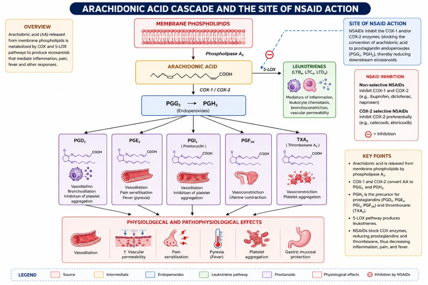
 

Figure 2. The arachidonic acid cascade. Phospholipase A2 liberates arachidonic acid from membrane phospholipids; the cyclooxygenase (COX-1/COX-2) branch yields prostaglandins and thromboxane, while the 5-lipoxygenase (5-LOX) branch yields leukotrienes. NSAIDs act by inhibiting COX-1/COX-2.

Therapeutic Benefits and Limitations

Reduced synthesis of vasodilatory prostaglandins (PGE2, PGI2) underlies the anti-inflammatory and analgesic actions of NSAIDs, while suppression of PGE2-mediated hypothalamic thermoregulatory signalling accounts for their antipyretic effect 120. These benefits are offset by COX-1-dependent loss of gastric cytoprotection, producing gastrointestinal ulceration, bleeding, and perforation risk 121,122. Selective COX-2 inhibitors were developed specifically to preserve efficacy while sparing the gastric mucosa; large trials (CLASS for celecoxib, VIGOR for rofecoxib) confirmed reduced GI complication rates relative to non-selective NSAIDs, although this advantage narrowed in patients co-prescribed low-dose aspirin 123,124.

4.2 Corticosteroids

Corticosteroids are divided into glucocorticoids, used for their anti-inflammatory and immunosuppressive activity, and mineralocorticoids, which govern salt and mineral balance 125. Their anti-inflammatory action begins with induction of lipocortin-1, which suppresses phospholipase A2 and thereby blocks eicosanoid production at its source, while glucocorticoid–receptor complexes additionally inhibit promoter activity of pro-inflammatory genes and reduce cytokine secretion largely through restraint of NF-κB 126,127,128. A single corticosteroid dose produces transient lymphocytopenia within hours through redistribution rather than destruction of circulating lymphocytes, while paradoxically inducing neutrophilia via bone-marrow release and reduced margination 127.

Methylprednisolone and dexamethasone illustrate the class's clinical range. In hospitalised COVID-19 patients, intravenous methylprednisolone pulse therapy reduced mortality and extended survival while lowering CRP and supporting platelet counts 129; low-dose short-course regimens (1–2 mg/kg/day for 5–7 days) shortened hospital stay and reduced respiratory-support needs with a safety profile comparable to standard care 130. Dexamethasone, available in numerous oral, injectable, and topical formulations, remains a reference agent for severe inflammatory and allergic conditions, with reported dose equivalence of roughly 6 mg dexamethasone to 32 mg methylprednisolone, though methylprednisolone's greater pulmonary tissue penetration has been associated with more favourable outcomes in some COVID-19 cohorts 129,131,132.

4.3 Disease-Modifying Anti-Rheumatic Drugs (DMARDs)

Methotrexate (MTX), in clinical use since the 1980s, remains the anchor DMARD for rheumatoid arthritis owing to its efficacy, dosing flexibility, tolerability, and favourable safety-to-efficacy ratio, and current guidelines endorse it as first-line monotherapy or combination therapy in treatment-naïve patients 133,134. A 13-year longitudinal study of 248 patients recorded a five-year continuation rate of 79%, with laboratory abnormalities in only 2.9 per 100 patient-years; of the 19% who discontinued, most did so for gastrointestinal or oral-ulcer adverse effects rather than lack of efficacy 135, and MTX has also demonstrated a survival benefit in rheumatoid arthritis 136.

Sulfasalazine, generally used in combination given its comparatively modest standalone efficacy, causes most of its toxicity — chiefly gastrointestinal and cutaneous effects accounting for over 25% of discontinuations — early in treatment, making long-term use relatively safe once tolerated; rare but serious risks include agranulocytosis (typically within the first six weeks), DRESS syndrome, and hepatotoxicity, warranting monthly monitoring initially and quarterly thereafter 137,138,139. It is considered compatible with pregnancy and lactation, although temporary oligospermia warrants discontinuation in men three months before attempting conception 140,141.

Leflunomide, an oral pyrimidine-synthesis inhibitor with well-documented long-term efficacy, most commonly causes diarrhoea, elevated liver enzymes, alopecia, and rash, most of which are mild, transient, and resolve without discontinuation; long-term safety appears to improve with continued use, and discontinuation rates remain modest even in psoriatic arthritis 142,143.

4.4 Biologic Agents

Etanercept, the first biologic approved for rheumatoid arthritis, is an Fc-fusion soluble TNF receptor construct, distinct in binding behaviour from the chimeric monoclonal antibody infliximab and the fully human antibody adalimumab: infliximab binds both monomeric and trimeric TNF, whereas etanercept preferentially and less stably binds the active trimeric form 144,145,146. These structural differences also translate into differing immunogenicity, summarised in Table 6.

Table 6. Differing risk of anti-drug antibody formation among anti-TNF biologics.

Agent

Structure

Reported antibody risk

Infliximab

Chimeric human–mouse monoclonal antibody

Highest concern; human anti-chimeric antibodies (HACA) can form against the murine variable region

Adalimumab

Fully humanised monoclonal antibody

Lower reported incidence of neutralising antibodies, attributed to fully human structure; human anti-human antibodies (HAHA) still reported in some patients

Etanercept

Fc-fusion soluble TNF receptor

No reported neutralising antibodies; non-neutralising antibody formation reported in ≈ 16% of patients

Adapted from Atzeni & Sarzi-Puttini 147.

IL-6 signals through a membrane receptor (IL-6R) that dimerises with the signal-transducing glycoprotein gp130; unusually, the soluble form of IL-6R facilitates rather than blocks signalling. Siltuximab, a chimeric anti-IL-6 antibody approved in 2014 for multicentric Castleman's disease, has also been investigated in several IL-6-driven malignancies and cachexia syndromes, though its use carries increased risk of upper respiratory infection and other adverse effects including cytopenias and hyperuricaemia 148,149,150.

IL-17 blockade with secukinumab improves outcomes in plaque psoriasis, psoriatic arthritis, and ankylosing spondylitis relative to placebo or etanercept and received FDA approval in 2015; ixekizumab, a second anti-IL-17 antibody, followed for plaque psoriasis in 2016 151,152.

4.5 Janus Kinase (JAK) Inhibitors

First-generation JAK inhibitors — ruxolitinib, tofacitinib, and baricitinib — block multiple JAK isoforms, whereas newer agents achieve greater selectivity: filgotinib, upadacitinib, and solcitinib are JAK1-selective; decernotinib and PF-06651600 target JAK3; and several compounds (BMS-986165, NDI-031232, NDI-031407, PF-06700841, SAR-20347) are TYK2-selective.

Tofacitinib, first studied for prevention of transplant rejection, proved efficacious but was associated with unacceptable over-immunosuppression — including BK viraemia, nephropathy, and post-transplant lymphoproliferative disease — likely reflecting the high doses (10–15 mg twice daily) and concurrent potent immunosuppressants used in those trials 153,154. Baricitinib has shown activity in interferon-signature autoinflammatory conditions but has similarly required comparatively high doses (mean 8.5 mg/day) associated with BK viraemia 155,156. Upadacitinib, engineered for JAK1-selective allosteric binding outside the ATP pocket shared with JAK2, is metabolised via CYP3A but can be co-administered with other CYP3A substrates including statins; in the BALANCE-1 and BALANCE-2 Phase II trials it showed efficacy, in combination with MTX, in rheumatoid arthritis patients who had failed MTX or TNF-inhibitor therapy 157,158.

5. Limitations and Adverse Effects of Conventional Therapies

The efficacy of conventional anti-inflammatory drugs is counterbalanced by organ-specific and systemic toxicity that limits long-term use and motivates the search for safer alternatives.

5.1 Gastrointestinal Toxicity

NSAID-associated mucosal injury ranges from minor petechiae and erosions to ulceration with bleeding, perforation, or obstruction, occasionally fatal 159. Minor epithelial breaks are usually repaired rapidly through "restitution," a division-independent migration of healthy epithelial cells across an intact basement membrane, but this repair capacity can be overwhelmed with continued COX-1 inhibition 160. Risk rises sharply with age, dose, treatment duration, and prior peptic ulcer history, and elderly patients face a three- to six-fold increase in serious upper-GI events 161,162.

5.2 Renal Toxicity

An estimated 1–5% of NSAID users develop renal adverse effects, spanning acute deterioration of renal function, papillary necrosis, acute interstitial nephritis, hyperkalaemia, and sodium/fluid retention 163. Acute effects are generally dose- and duration-dependent and reversible, but a history of acute renal injury increases susceptibility to chronic renal failure 164,165.

5.3 Cardiovascular Risk

Both selective COX-2 inhibitors and traditional non-selective NSAIDs carry increased cardiovascular hazard proportional to the degree of COX-2 inhibition. Placebo-controlled trials of celecoxib, rofecoxib, and valdecoxib recorded increased thrombotic events, and rofecoxib was ultimately withdrawn from the market after its association with myocardial infarction became clear 166,167,168. Population-based studies and meta-analyses subsequently confirmed that this cardiovascular signal extends to some traditional NSAIDs, notably diclofenac, and that risk scales with dose potency 169,170,171.

5.4 Immunogenicity, Infection Risk, and Cost of Biologic Therapy

Anti-TNF biologics can provoke neutralising or non-neutralising anti-drug antibodies, with risk varying by molecular structure: the chimeric antibody infliximab is most prone to antichimeric antibody formation, the fully humanised adalimumab less so, while etanercept has not been reported to generate neutralising antibodies though non-neutralising antibody formation occurs in roughly 16% of patients 147. Rituximab, a B-cell-depleting biologic, is associated with infusion reactions (fever, chills, rash, bronchospasm, hypotension) typically within the first infusion, mitigated by pretreatment with an antihistamine and paracetamol, and requires ongoing surveillance for infection, tuberculosis reactivation, and lymphoma 172. Beyond immunological risk, the high acquisition cost of biologic agents remains a substantial barrier to equitable access and long-term adherence.

6. Herbal and Plant-Derived Anti-Inflammatory Agents

Given the toxicity profile of conventional anti-inflammatory drugs, considerable research attention has turned to plant-derived agents that act through multiple, often complementary, molecular targets while offering a comparatively favourable safety margin. The principal limitations of this class remain poor oral bioavailability, inconsistent standardisation of extracts, and a relative scarcity of large, well-controlled clinical trials. Table 7 summarises the agents discussed below, together with olive leaf extract as a benchmark example of a standardised, mechanistically validated phytomedicine.

Table 7. Selected plant-derived anti-inflammatory agents and their proposed mechanisms.

Plant source

Principal active constituent(s)

Proposed anti-inflammatory mechanism

Curcuma longa (turmeric)

Curcumin

NF-κB inhibition; downstream suppression of cytokines, COX-2, iNOS

Zingiber officinale (ginger)

Gingerols

COX inhibition, reduced prostaglandin synthesis

Ocimum sanctum (holy basil)

Eugenol

Antioxidant activity; cytokine suppression

Aloe vera

Polysaccharides, glycoproteins, aloin

Bradykinin modulation; prostaglandin/cytokine suppression

Camellia sinensis (green tea)

Epigallocatechin gallate (EGCG)

Antioxidant activity; NF-κB and MAPK pathway interference

Portulaca oleracea (purslane)

α-Linolenic acid (omega-3), flavonoids, ascorbic acid

Competitive eicosanoid modulation; antioxidant scavenging

Olea europaea (olive leaf)

Oleuropein, hydroxytyrosol, tyrosol

Reduced TNF-α, IL-1β, COX-2 and NO; RBC/lysosomal membrane stabilisation (Fayez et al., 2023)

6.1 Curcuma longa (Turmeric)

The principal active constituent of turmeric rhizome is curcumin, a diarylheptanoid polyphenol. Its anti-inflammatory activity is attributed largely to inhibition of NF-κB activation, which curtails downstream transcription of pro-inflammatory cytokines, COX-2, and inducible nitric oxide synthase, positioning curcumin as a multi-target modulator of the same central pathway exploited by corticosteroids (see Section 2.4.1).

6.2 Zingiber officinale (Ginger)

Ginger rhizome contains gingerols, pungent phenolic compounds that inhibit cyclooxygenase activity in a manner mechanistically analogous to conventional NSAIDs, thereby reducing prostaglandin synthesis at the site of inflammation while apparently sparing the gastric-protective COX-1 pool to a greater extent than synthetic non-selective NSAIDs.

6.3 Ocimum sanctum (Holy Basil / Tulsi)

Holy basil leaves are a source of eugenol, a phenylpropanoid with combined antioxidant and cytokine-suppressive activity. By scavenging reactive oxygen species and dampening pro-inflammatory cytokine output, eugenol is thought to interrupt the oxidative-stress/cytokine feed-forward loop that sustains chronic low-grade inflammation.

6.4 Aloe vera

Aloe vera gel contains polysaccharides, glycoproteins, and anthraquinones (including aloin) that have been reported to reduce bradykinin-mediated pain signalling and to modulate prostaglandin and cytokine output at the tissue level, supporting its traditional topical and oral use in inflammatory and wound-healing contexts, although rigorous dose–response and mechanistic data remain comparatively limited relative to curcumin or EGCG.

6.5 Camellia sinensis (Green Tea)

Green tea leaves are rich in catechins, of which epigallocatechin gallate (EGCG) is the most extensively studied. EGCG exerts broad antioxidant activity and has been reported to interfere with NF-κB and MAPK signalling, thereby suppressing downstream pro-inflammatory cytokine and COX-2 expression in a manner that overlaps mechanistically with several of the pathways discussed in Section 2.4.

6.6 Portulaca oleracea (Purslane)

Purslane is a succulent, widely distributed leafy vegetable notable among common food plants for its comparatively high omega-3 fatty acid (α-linolenic acid) content, alongside flavonoids, alkaloids, and ascorbic acid. Its anti-inflammatory activity is attributed to a combination of omega-3-mediated eicosanoid modulation — competitively diverting arachidonic-acid metabolism toward less inflammatory lipid mediators — and direct antioxidant scavenging by its phenolic constituents. Preliminary experimental studies support anti-inflammatory and analgesic activity, though clinical evidence in humans remains limited and would benefit from standardised extract characterisation of the kind applied to olive leaf extract and other well-studied phytomedicines.

7. Emerging and Safer Therapeutic Strategies

7.1 Nanotechnology-Based Drug Delivery

Nanocarrier platforms — liposomes, polymeric nanoparticles, and solid lipid nanoparticles — are being explored to overcome the poor aqueous solubility and rapid systemic clearance that limit many plant-derived anti-inflammatory compounds (notably curcumin), while also enabling site-directed delivery of conventional NSAIDs and biologics that could reduce off-target gastrointestinal, renal, and cardiovascular exposure.

7.2 Combination Therapy

Rational combination of a low-dose conventional NSAID with a standardised herbal anti-inflammatory compound, or of a biologic with a conventional DMARD, aims to achieve synergistic efficacy at reduced individual doses, potentially lowering the cumulative toxicity burden associated with monotherapy at full therapeutic dose.

7.3 Personalised Medicine

Biomarker-guided treatment selection — for example, using CRP or cytokine profiling (Section 3.1) — together with pharmacogenomic characterisation of drug-metabolising enzymes, offers a route to matching patients to the anti-inflammatory regimen most likely to be both effective and well tolerated, reducing exposure to agents unlikely to benefit a given individual.

7.4 Molecular Docking and AI-Assisted Drug Discovery

Computational docking against validated inflammatory targets (COX isoforms, NF-κB components, JAK kinases, NLRP3) and machine-learning-assisted screening of natural-product libraries are accelerating identification of candidate anti-inflammatory molecules with predicted target selectivity, potentially shortening the discovery timeline for safer next-generation agents ahead of experimental validation.

8. Future Perspectives

Continued progress in anti-inflammatory therapeutics is likely to depend on several converging trends: precision-medicine approaches that stratify patients by inflammatory phenotype rather than diagnosis alone; multi-target agents capable of modulating more than one node of the NF-κB/MAPK/JAK–STAT/NLRP3 network simultaneously; systems-biology approaches that map how these pathways interact across tissues and disease states; AI-assisted drug design to accelerate lead identification and optimisation; and rigorous standardisation of herbal and plant-derived candidates so that their reported efficacy can be reproduced and validated in controlled clinical trials, following the model already applied to agents such as olive leaf extract.

9. CONCLUSION

Inflammation is executed through a layered system of receptor recognition (PAMPs/DAMPs and PRRs), cellular effectors (macrophages, neutrophils, mast cells, dendritic cells, lymphocytes), chemical mediators (histamine, kinins, prostaglandins, leukotrienes), and a small number of convergent signalling pathways — NF-κB, MAPK, JAK–STAT, and the NLRP3 inflammasome — whose dysregulation underlies a substantial share of global disease burden, as reflected in CRP-linked outcomes across cardiovascular, metabolic, neurodegenerative, renal, and rheumatological disease. Conventional pharmacological control of this system — NSAIDs, corticosteroids, DMARDs, biologics, and JAK inhibitors — remains effective but is constrained by gastrointestinal, renal, cardiovascular, immunological, and economic costs that limit long-term use. Plant-derived agents such as curcumin, gingerols, eugenol, aloe vera constituents, EGCG, and purslane phytochemicals offer multi-targeted, comparatively safer alternatives, but require better standardisation and clinical validation before they can be positioned as substitutes rather than adjuncts to conventional therapy. Nanotechnology-enabled delivery, rational combination regimens, biomarker-guided personalised treatment, and computational drug discovery represent the most promising near-term routes to narrowing the long-standing gap between anti-inflammatory efficacy and safety.

Acknowledgement

The authors received no specific funding for this review and thank the Department of Pharmacy, M.J.P. Rohilkhand University, Bareilly, for institutional support.

Conflicts of Interest

The authors declare no conflicts of interest relevant to the content of this review

REFERENCES

  1. Henson PM: Dampening inflammation. Nature Immunology 2005; 6(12):1179–1181.
  2. Ryan GB and Majno G: Acute inflammation: A review. American Journal of Pathology 1977; 86(1):183–276.
  3. Stankov SV: Definition of inflammation, causes of inflammation and possible anti-inflammatory strategies. The Open Inflammation Journal 2012; 5:1–9.
  4. Rocha e Silva M: A brief survey of the history of inflammation. Agents and actions 1978; 8(1):45-49.
  5. Gruber F: Aulus Cornelius Celsus' De Medicina and His Contributions to Knowledge on Skin Diseases. Acta Dermatovenerologica Croatica 2023; 31(2):92-97.
  6. Scott A, Khan KM, Cook JL and Duronio V: What is 'inflammation'? Are we ready to move beyond Celsus? British Journal of Sports Medicine 2004; 38(3):248-249.
  7. Rather LJ: Disturbance of function (functio laesa): the legendary fifth cardinal sign of inflammation, added by Galen to the four cardinal signs of Celsus. Bulletin of the New York Academy of Medicine 1971; 47(3):303.
  8. Ventura HO: Rudolph Virchow and cellular pathology. Clinical Cardiology 2000; 23(4):295–296.
  9. Schmalstieg Jr FC and Goldman AS: Ilya Ilich Metchnikoff (1845–1915) and Paul Ehrlich (1854–1915): the centennial of the 1908 nobel prize in physiology or medicine. Journal of medical biography 2008; 16(2):96-104.
  10. Henson PM and Johnston RB: Tissue injury in inflammation. Oxidants, proteinases, and cationic proteins. The Journal of clinical investigation 1987; 79(3):669-74.
  11. Vandenabeele P, Galluzzi L, Vanden Berghe T and Kroemer G: Molecular mechanisms of necroptosis: an ordered cellular explosion. Nature reviews Molecular cell biology 2010; 11(10):700-14.
  12. Flower R: The discovery of prostaglandins. In: Nobel Prizes That Changed Medicine. Imperial College Press, London 2011; 171-196.
  13. Serbina NV, Jia T, Hohl TM and Pamer EG: Monocyte-mediated defense against microbial pathogens. Annual Review of Immunology 2008; 26:421–452.
  14. Serbina NV, Salazar-Mather TP, Biron CA, Kuziel WA and Pamer EG: Sequential MyD88-independent and dependent activation of innate immune responses to intracellular bacterial infection. Immunity 2003; 19(6):891–901.
  15. Serbina NV et al: Selective expansion of the monocytic lineage directed by bacterial infection. Journal of Immunology 2009; 183(3):1900–1910.
  16. Stavru F, Archambaud C and Cossart P: Cell biology and immunology of Listeria monocytogenes infections: Novel insights. Immunological Reviews 2011; 240(1):160–184.
  17. Serbina NV and Pamer EG: Monocyte emigration from bone marrow during bacterial infection requires signals mediated by chemokine receptor CCR2. Nature Immunology 2006; 7(3):311–317.
  18. Tsou CL et al: Critical roles for CCR2 and MCP-3 in monocyte mobilization from bone marrow and recruitment to inflammatory sites. Journal of Clinical Investigation 2007; 117(4):902–909.
  19. Tabas I: Consequences and therapeutic implications of macrophage apoptosis in atherosclerosis: The importance of lesion stage and phagocytic efficiency. Arteriosclerosis, Thrombosis, and Vascular Biology 2005; 25(11):2255-2264.
  20. Gautier EL, Jakubzick C and Randolph GJ: Regulation of the migration and survival of monocyte subsets by chemokine receptors and its relevance to atherosclerosis. Arteriosclerosis, Thrombosis, and Vascular Biology 2009; 29(10):1412-1418.
  21. Combadiere C et al: Combined inhibition of CCL2, CX3CR1, and CCR5 abrogates monocytosis and almost abolishes atherosclerosis in hypercholesterolemic mice. Circulation 2008; 117(13):1649–1657.
  22. Swirski FK et al: Ly-6Chi monocytes dominate hypercholesterolemia-associated monocytosis and give rise to macrophages in atheromata. Journal of Clinical Investigation 2007; 117(1):195–205.
  23. Yvan-Charvet L et al: ATP-binding cassette transporters and HDL suppress hematopoietic stem cell proliferation. Science 2010; 328(5986):1689–1693.
  24. Janeway CA: Approaching the asymptote? Evolution and revolution in immunology. Cold Spring Harbor Symposia on Quantitative Biology 1989; 54(Pt. 1):1–13.
  25. Ma M, Jiang W and Zhou R: DAMPs and DAMP-sensing receptors in inflammation and diseases. Immunity 2024; 57(4):752–771.
  26. Cao X: Self-regulation and cross-regulation of pattern-recognition receptor signalling in health and disease. Nature Reviews Immunology 2016; 16(1):35–50.
  27. Eisenhut M and Wallace H: Ion channels in inflammation. Pflügers Archiv 2011; 461(4):401–421.
  28. Hudson BI and Lippman ME: Targeting RAGE signaling in inflammatory disease. Annual Review of Medicine 2018; 69:349–364.
  29. Hancock DB, Eijgelsheim M and Wilk JB et al: Meta-analyses of genome-wide association studies identify multiple loci associated with pulmonary function. Nature Genetics 2010; 42(1):45–52.
  30. Kalea AZ, Schmidt AM and Hudson BI: RAGE: A novel biological and genetic marker for vascular disease. Clinical Science 2009; 116(8):621–637.
  31. Repapi E, Sayers I and Wain LV et al: Genome-wide association study identifies five loci associated with lung function. Nature Genetics 2010; 42(1):36–44.
  32. Falcone C, Emanuele E and D'Angelo A et al: Plasma levels of soluble receptor for advanced glycation end products and coronary artery disease in nondiabetic men. Arteriosclerosis, Thrombosis, and Vascular Biology 2005; 25(5):1032-1037.
  33. Yonekura H, Yamamoto Y and Sakurai S et al: Novel splice variants of the receptor for advanced glycation end-products expressed in human vascular endothelial cells and pericytes, and their putative roles in diabetes-induced vascular injury. Biochemical Journal 2003; 370(3):1097–1109.
  34. Zhang L, Bukulin M and Kojro E et al: Receptor for advanced glycation end products is subjected to protein ectodomain shedding by metalloproteinases. Journal of Biological Chemistry 2008; 283(51):35507–35516.
  35. Al-Robaiy S, Weber B and Simm A et al: The receptor for advanced glycation end-products supports lung tissue biomechanics. American Journal of Physiology-Lung Cellular and Molecular Physiology 2013; 305(7):L491–L500.
  36. Sessa L, Gatti E and Zeni F et al: The receptor for advanced glycation end-products (RAGE) is only present in mammals, and belongs to a family of cell adhesion molecules (CAMs). PLOS ONE 2014; 9(1):e86903.
  37. Wynn TA, Chawla A and Pollard JW: Macrophage biology in development, homeostasis and disease. Nature 2013; 496(7446):445–455.
  38. Laskin DL and Laskin JD: Role of macrophages and inflammatory mediators in chemically induced toxicity. Toxicology 1996; 112(3):183–190.
  39. Laskin DL, Sunil VR, Gardner CR and Laskin JD: Macrophages and tissue injury: agents of defense or destruction ?. Annual review of pharmacology and toxicology 2011; 51(1):267-288.
  40. Mihalache CC and Simon HU: Autophagy regulation in macrophages and neutrophils. Experimental Cell Research 2012; 318(11):1187–1189.
  41. Johnson-Weaver B, Choi HW, Abraham SN and Staats HF: Mast cell activators as novel immune regulators. Current Opinion in Pharmacology 2018; 41:89–95.
  42. Mukai K, Tsai M, Saito H and Galli SJ: Mast cells as sources of cytokines, chemokines, and growth factors. Immunological Reviews 2018; 282(1):121–150.
  43. Piliponsky AM and Romani L: The contribution of mast cells to bacterial and fungal infection immunity. Immunological Reviews 2018; 282(1):188–197.
  44. Metcalfe DD, Kaliner M and Donlon MA: The mast cell. CRC Critical Reviews in Immunology 1981; 3(1):23–74.
  45. Pearce FL: Biological effects of histamine: An overview. Agents and Actions 1991; 33(1–2):4–7.
  46. Bhoola KD, Collier HOJ, Schachter M and Shorley PG: Actions of some peptides on bronchial muscle. British Journal of Pharmacology 1962; 19:190–197.
  47. Keele CA: Clinical and pathological aspects of kinins in man. Proceedings of the Royal Society of Medicine 1969; 173:361–369.
  48. Hamberg M, Svensson J, Wakabayashi T and Samuelsson B: Isolation and structure of two prostaglandin endoperoxides that cause platelet aggregation. Proceedings of the National Academy of Sciences 1974; 71(2):345–349.
  49. Lewis RA, Holgate ST, Roberts 2nd LJ, Oates JA and Austen KF: Preferential generation of prostaglandin D2 by rat and human mast cells. Kroc Foundation series 1981; 14:239-54.
  50. Schwartz LB and Austen KF: Structure and function of the chemical mediators of mast cells. Progress in Allergy 1984; 34:271–321.
  51. Rihoux JP, Masliah J, Bereziat G and König W: G proteins as biological targets for anti-allergic drugs?. International archives of allergy and immunology 1997; 113(1-3):339-41.
  52. Bradding P and Holgate ST: The mast cell as a source of cytokines in asthma. Annals of the New York Academy of Sciences 1996; 796:272–281.
  53. Bradding P, Feather IH, Wilson S, Holgate ST and Howarth PH: Cytokine immunoreactivity in seasonal rhinitis: Regulation by a topical corticosteroid. American Journal of Respiratory and Critical Care Medicine 1995; 151(6):1900–1906.
  54. Benson M, Strannegard IL, Wennergren G and Strannegard O: Cytokines in nasal fluids from school children with seasonal allergic rhinitis. Pediatric Allergy and Immunology 1997; 8(3):143–149.
  55. Han ZN, Boyle DL, Manning AM and Firestein GS: AP-1 and NF-kappa B regulation in rheumatoid arthritis and murine collagen-induced arthritis. Autoimmunity 1998; 28(4):197–208.
  56. Hart LA, Krishnan VL, Adcock IM, Barnes PJ and Chung KF: Activation and localization of transcription factor, nuclear factor-kappaB, in asthma. American Journal of Respiratory and Critical Care Medicine 1998; 158(5):1585–1592.
  57. Neurath MF et al: Cytokine gene transcription by NF-kappa B family members in patients with inflammatory bowel disease. Annals of the New York Academy of Sciences 1998; 859:149–159.
  58. Collins T and Cybulsky MI: NF-κB: pivotal mediator or innocent bystander in atherogenesis?. The Journal of clinical investigation 2001; 107(3):255-64.
  59. Mattson MP and Camandola S: NF-κB in neuronal plasticity and neurodegenerative disorders. Journal of Clinical Investigation 2001; 107(3):247–254.
  60. Cheng L and Karin M: Mammalian MAP kinase signaling cascades. Nature 2001; 410:37–40.
  61. Keshet Y and Seger R: The MAP kinase signaling cascades: A system of hundreds of components regulates a diverse array of physiological functions. Methods in Molecular Biology 2010; 661:3–38.
  62. McKay MM and Morrison DK: Integrating signals from RTKs to ERK/MAPK. Oncogene 2007; 26(22):3113–3121.
  63. Raman M, Chen W and Cobb MH: Differential regulation and properties of MAPKs. Oncogene 2007; 26(22):3100–3112.
  64. Dérijard B, Hibi M and Wu LH et al: JNK1: A protein kinase stimulated by UV light and Ha-Ras that binds and phosphorylates the c-Jun transactivation domain. Cell 1994; 76(6):1025–1037.
  65. Kyriakis JM, Banerjee P and Nikolakaki E et al: The stress-activated protein kinase subfamily of c-Jun kinases. Nature 1994; 369(6476):156–160.
  66. Freshney NW, Rawlinson L and Guesdon F et al: Interleukin-1 activates a novel protein kinase cascade that results in the phosphorylation of Hsp27. Cell 1994; 78(6):1039–1049.
  67. Rouse J, Cohen P and Trigon S et al: A novel kinase cascade triggered by stress and heat shock that stimulates MAPKAP kinase-2 and phosphorylation of the small heat shock proteins. Cell 1994; 78(6):1027–1037.
  68. Ghoreschi K, Laurence A and Shea JJ: Janus kinases in immune cell signaling. Immunological Reviews 2009; 228(1):273–287.
  69. Leonard WJ and Shea JJ: Jaks and STATs: Biological implications. Annual Review of Immunology 1998; 16:293–322.
  70. Seif F, Khoshmirsafa M, Aazami H, Mohsenzadegan M, Sedighi G and Bahar M: The role of JAK-STAT signaling pathway and its regulators in the fate of T helper cells. Cell Communication and Signaling 2017; 15:23.
  71. Gusev E and Zhuravleva Y: Inflammation: A new look at an old problem. International Journal of Molecular Sciences 2022; 23(9):4596.
  72. Gusev EY and Zotova NV: Cellular stress and general pathological processes. Current Pharmaceutical Design 2019; 25(3):251–297.
  73. Franchi L, Eigenbrod T, Muñoz-Planillo R and Nuñez G: The inflammasome: A caspase-1 activation platform regulating immune responses and disease pathogenesis. Nature Immunology 2009; 10(3):241–247.
  74. Lamkanfi M and Dixit VM: Mechanisms and functions of inflammasomes. Cell 2014; 157(5):1013–1022.
  75. Sharma D and Kanneganti T: The cell biology of inflammasomes: Mechanisms of inflammasome activation and regulation. Journal of Cell Biology 2016; 213(6):617–629.
  76. Takeuchi O and Akira S: Pattern recognition receptors and inflammation. Cell 2010; 140(6):805–820.
  77. Elinav E, Strowig T and Kau AL et al: NLRP6 inflammasome regulates colonic microbial ecology and risk for colitis. Cell 2011; 145(5):745–757.
  78. Fernandes-Alnemri T, Wu J and Yu J et al: The pyroptosome: A supramolecular assembly of ASC dimers mediating inflammatory cell death via caspase-1 activation. Cell Death & Differentiation 2007; 14(9):1590–1604.
  79. Dinarello CA: Interleukin-1. In: Thomson AW and Lotze MT (eds), The Cytokine Handbook. Academic Press, San Diego 2003.
  80. Warren JS: Interleukins and tumor necrosis factor in inflammation. Critical Reviews in Clinical Laboratory Sciences 1990; 28(1):37–59.
  81. Dinarello CA: Reduction of inflammation by decreasing production of interleukin-1 or by specific receptor antagonism. International Journal of Tissue Reactions 1992; 14(2):65–75.
  82. Okamura H, Tsutsui H and Komatsu T et al: Cloning of a new cytokine that induces IFN-γ production by T cells. Nature 1995; 378(6552):88–91.
  83. Ushio S, Namba M and Okura T et al: Cloning of the cDNA for human IFN-γ-inducing factor, expression in Escherichia coli, and studies on the biologic activities of the protein. Journal of Immunology 1996; 156(11):4274–4279.
  84. Baumann H and Schendel P: Interleukin-11 regulates the hepatic expression of the same plasma protein genes as interleukin-6. Journal of Biological Chemistry 1991; 266(30):20424–20427.
  85. Paul SR and Schendel P: The cloning and biological characterization of recombinant human interleukin 11. International Journal of Cell Cloning 1992; 10(3):135–143.
  86. Hirano T: Interleukin-6 and its relation to inflammation and disease. Clinical Immunology and Immunopathology 1992; 62(1, Pt 2):S60–S65.
  87. Hirano T, Taga T and Matsuda T et al: Interleukin 6 and its receptor in the immune response and hematopoiesis. International Journal of Cell Cloning 1990; 8(3):155–166.
  88. Border WA and Ruoslahti E: Transforming growth factor-β in disease: The dark side of tissue repair. Journal of Clinical Investigation 1992; 90(1):1–7.
  89. Cooper WO, Fava RA, Gates CA, Cremer MA and Townes AS: Acceleration of onset of collagen-induced arthritis by intra-articular injection of tumour necrosis factor or transforming growth factor-beta. Clinical and Experimental Immunology 1992; 89(2):244–250.
  90. Sporn MB, Roberts AB, Wakefield LM and Assoian RK: Transforming growth factor-β: Biological function and chemical structure. Science 1986; 233(4763):532–534.
  91. Black S, Kushner I and Samols D: C-reactive protein. Journal of Biological Chemistry 2004; 279(47):48487–48490.
  92. Pepys MB and Hirschfield GM: C-reactive protein: A critical update. Journal of Clinical Investigation 2003; 111(12):1805–1812.
  93. Volanakis JE: Human C-reactive protein: Expression, structure, and function. Molecular Immunology 2001; 38(2–3):189–197.
  94. Ridker PM, Rifai N, Rose L, Buring JE and Cook NR: Comparison of C-reactive protein and low-density lipoprotein cholesterol levels in the prediction of first cardiovascular events. New England journal of medicine 2002; 347(20):1557-65.
  95. Engelhart MJ, Geerlings MI, Meijer J, Kiliaan A, Ruitenberg A, Van Swieten JC, Stijnen T, Hofman A, Witteman JC and Breteler MM: Inflammatory proteins in plasma and the risk of dementia: the rotterdam study. Archives of neurology 2004; 61(5):668-72.
  96. Luan YY and Yao YM: The clinical significance and potential role of C-reactive protein in chronic inflammatory diseases. Frontiers in Immunology 2018; 9:1302.
  97. Biasucci LM, Liuzzo G, Colizzi C and Rizzello V: Clinical use of C-reactive protein for the prognostic stratification of patients with ischemic heart disease. Italian Heart Journal 2001; 2:164-71.
  98. Karakas M and Koenig W: CRP in cardiovascular disease. Herz 2009; 34(8):607-13.
  99. Festa A, Hanley AJ, Tracy RP, D'Agostino Jr R and Haffner SM: Inflammation in the prediabetic state is related to increased insulin resistance rather than decreased insulin secretion. Circulation 2003; 108(15):1822-30.
  100. King DE, Mainous III AG, Buchanan TA and Pearson WS: C-reactive protein and glycemic control in adults with diabetes. Diabetes care 2003; 26(5):1535-9.
  101. Andican G, Konukoglu D, Bozluolcay M, Bayülkem K, Firtiına S and Burcak G: Plasma oxidative and inflammatory markers in patients with idiopathic Parkinson's disease. Acta Neurologica Belgica 2012; 112(2):155-9.
  102. Chen H, O'Reilly EJ, Schwarzschild MA and Ascherio A: Peripheral inflammatory biomarkers and risk of Parkinson's disease. American journal of epidemiology 2008; 167(1):90-5.
  103. Kaysen GA and Eiserich JP: Characteristics and effects of inflammation in end-stage renal disease. Seminars in Dialysis 2003; 16(6):438-446.
  104. Bacons PA and Kitas GD: The significance of vascular inflammation in rheumatoid arthritis. Annals of the rheumatic diseases 1994; 53(10):621.
  105. Schett G, McInnes IB and Neurath MF: Reframing immune-mediated inflammatory diseases through signature cytokine hubs. New England Journal of Medicine 2021; 385(7):628–639.
  106. Wu D, Jin Y and Xing Y et al: Global, regional, and national incidence of six major immune-mediated inflammatory diseases. eClinicalMedicine 2023; 64:102193.
  107. Feldmann M, Maini RN and Soriano ER et al: 25 years of biologic DMARDs in rheumatology. Nature Reviews Rheumatology 2023; 19(12):761–766.
  108. Kuek A, Hazleman BL and Ostor AJ: Immune-mediated inflammatory diseases and biologic therapy. Postgraduate Medical Journal 2007; 83(978):251–260.
  109. Zhu X, Yue M and Zhang X et al: Global disease burden of immune-mediated inflammatory diseases (IMIDs), 1990–2021. Med Research 2025; 1:285–296.
  110. GBD 2019 Ageing Collaborators: Global, regional, and national burden of diseases and injuries for adults 70 years and older: systematic analysis for the Global Burden of Disease 2019 Study. bmj 2022; 376.
  111. Felson DT: Safety of nonsteroidal antiinflammatory drugs. New England Journal of Medicine 2016; 375(26):2595-6.
  112. Antman EM, Bennett JS, Daugherty A, Furberg C, Roberts H and Taubert KA: Use of nonsteroidal anti-inflammatory drugs: An update for clinicians. Circulation 2007; 115(12):1634–1642.
  113. Smith WL, DeWitt DL and Garavito RM: Cyclooxygenases: Structural, cellular, and molecular biology. Annual Review of Biochemistry 2000; 69:145–182.
  114. Vane JR: Inhibition of prostaglandin synthesis as a mechanism of action for aspirin-like drugs. Nature New Biology 1971; 231(25):232–235.
  115. Harris RC, McKanna JA, Akai Y, Jacobson HR, Dubois RN and Breyer MD: Cyclooxygenase-2 is associated with the macula densa of rat kidney and increases with salt restriction. Journal of Clinical Investigation 1994; 94(6):2504–2510.
  116. Hoffmann C: COX-2 in brain and spinal cord: Implications for therapeutic use. Current Medicinal Chemistry 2000; 7(11):1113–1120.
  117. Murphy JF and Fitzgerald DJ: Vascular endothelial growth factor induces cyclooxygenase-dependent proliferation of endothelial cells via the VEGF-2 receptor. FASEB Journal 2001; 15(9):1667–1669.
  118. Chandrasekharan NV, Dai H and Roos KL et al: COX-3, a cyclooxygenase-1 variant inhibited by acetaminophen and other analgesic/antipyretic drugs. Proceedings of the National Academy of Sciences 2002; 99(21):13926–13931.
  119. Snipes JA, Kis B, Shelness GS, Hewett JA and Busija DW: Cloning and characterization of cyclooxygenase-1b (putative cyclooxygenase-3) in rat. Journal of Pharmacology and Experimental Therapeutics 2005; 313(2):668–676.
  120. Simmons DL, Botting RM and Hla T: Cyclooxygenase isozymes: The biology of prostaglandin synthesis and inhibition. Pharmacological Reviews 2004; 56(3):387–437.
  121. Hernandez-Diaz S and Rodriguez LA: Association between nonsteroidal anti-inflammatory drugs and upper gastrointestinal tract bleeding/perforation. Archives of Internal Medicine 2000; 160(14):2093–2099.
  122. Jones R: Nonsteroidal anti-inflammatory drug prescribing: Past, present, and future. American Journal of Medicine 2001; 110(1, Suppl. 1):4–7.
  123. Bombardier C, Laine L, Reicin A, Shapiro D, Burgos-Vargas R and Davis B: Comparison of upper gastrointestinal toxicity of rofecoxib and naproxen in patients with rheumatoid arthritis. New England Journal of Medicine 2000; 343(21):1520–1528.
  124. Silverstein FE, Faich G and Goldstein JL et al: Gastrointestinal toxicity with celecoxib vs nonsteroidal anti-inflammatory drugs for osteoarthritis and rheumatoid arthritis: The CLASS study. JAMA 2000; 284(10):1247–1255.
  125. Demoly P and Chung KF: Pharmacology of corticosteroids. Respiratory Medicine 1998; 92(3):385–394.
  126. Coutinho AE and Chapman KE: The anti-inflammatory and immunosuppressive effects of glucocorticoids, recent developments and mechanistic insights. Molecular and Cellular Endocrinology 2011; 335(1):2–13.
  127. Fauci AS: Mechanisms of the immunosuppressive and anti-inflammatory effects of glucocorticosteroids. Journal of Immunopharmacology 1978; 1(1):1–25.
  128. Scheinman RI, Cogswell PC, Lofquist AK and Baldwin AS: Role of transcriptional activation of I kappa B alpha in mediation of immunosuppression by glucocorticoids. Science 1995; 270(5234):283–286.
  129. Ranjbar K, Moghadami M and Mirahmadizadeh A et al: Methylprednisolone or dexamethasone, which one is superior corticosteroid in the treatment of hospitalized COVID-19 patients: A triple-blinded randomized controlled trial. BMC Infectious Diseases 2021; 21:436.
  130. Wang Y, Jiang W and He Q et al: A retrospective cohort study of methylprednisolone therapy in severe patients with COVID-19 pneumonia. Signal Transduction and Targeted Therapy 2020; 5:57.
  131. Hirano T, Homma M, Oka K, Tsushima H, Niitsuma T and Hayashi T: Individual variations in lymphocyte-responses to glucocorticoids in patients with bronchial asthma: Comparison of potencies for five glucocorticoids. Immunopharmacology 1998; 40(1):57–66.
  132. Mager DE, Lin SX, Blum RA, Lates CD and Jusko WJ: Dose equivalency evaluation of major corticosteroids: Pharmacokinetics and cell trafficking and cortisol dynamics. Journal of Clinical Pharmacology 2003; 43(11):1216–1227.
  133. Singh JA, Saag KG and Bridges SL et al: 2015 American College of Rheumatology guideline for the treatment of rheumatoid arthritis. Arthritis Care & Research 2016; 68(1):1–25.
  134. Smolen JS, Landewé R and Bijlsma J et al: EULAR recommendations for the management of rheumatoid arthritis with synthetic and biological disease-modifying antirheumatic drugs: 2016 update. Annals of the Rheumatic Diseases 2017; 76(6):960–977.
  135. Yazici Y, Sokka T and Kautiainen H et al: Long-term safety of methotrexate in routine clinical care: Discontinuation is unusual and rarely due to laboratory abnormalities. Annals of the Rheumatic Diseases 2005; 64(2):207–211.
  136. Wasko MC, Dasgupta A and Hubert H et al: Propensity-adjusted association of methotrexate with overall survival in rheumatoid arthritis. Arthritis & Rheumatism 2013; 65(2):334–342.
  137. Donovan S, Hawley S and MacCarthy J et al: Tolerability of enteric-coated sulphasalazine in rheumatoid arthritis: Results of a co-operating clinics study. British Journal of Rheumatology 1990; 29(3):201–204.
  138. McConkey B: Disease modifying antirheumatic drugs: Gold, penicillamine, antimalarials, and sulfasalazine. Current Opinion in Rheumatology 1991; 3(3):348–354.
  139. Shanoj KC, Joseph S and Shenoy P: Drug reaction with eosinophilia and systemic symptoms syndrome. Indian Journal of Rheumatology 2019; 14(1):59–66.
  140. Aletaha D, Kapral T and Smolen JS: Toxicity profiles of traditional disease-modifying antirheumatic drugs for rheumatoid arthritis. Annals of the Rheumatic Diseases 2003; 62(5):482–486.
  141. Jobanputra P, Amarasena R and Maggs F et al: Hepatotoxicity associated with sulphasalazine in inflammatory arthritis. BMC Musculoskeletal Disorders 2008; 9:48.
  142. Behrens F, Finkenwirth C and Pavelka K et al: Leflunomide in psoriatic arthritis: Results from a large European prospective observational study. Arthritis Care & Research 2013; 65(3):464–470.
  143. Kalden JR, Schattenkirchner M and Sörensen H et al: The efficacy and safety of leflunomide in patients with active rheumatoid arthritis: A five-year follow-up study. Arthritis & Rheumatism 2003; 48(6):1513–1520.
  144. Scallon B, Cai A and Solowski N et al: Binding and functional comparisons of two types of tumor necrosis factor antagonists. Journal of Pharmacology and Experimental Therapeutics 2002; 301(2):418–426.
  145. Seckinger P, Isaaz S and Dayer J: A human inhibitor of tumor necrosis factor α. Journal of Experimental Medicine 1988; 167(4):1511–1516.
  146. Seckinger P, Zhang J, Hauptmann B and Dayer J: Characterization of a tumor necrosis factor α (TNF-α) inhibitor: Evidence of immunological cross-reactivity with the TNF receptor. Proceedings of the National Academy of Sciences 1990; 87(13):5188–5192.
  147. Atzeni F and Sarzi-Puttini P: Autoantibody production in patients treated with anti-TNF-α. Expert Review of Clinical Immunology 2008; 4(2):275–280.
  148. Deisseroth A, Ko C and Nie L et al: FDA approval: Siltuximab for the treatment of patients with multicentric Castleman disease. Clinical Cancer Research 2015; 21(5):950–954.
  149. El-Osta HE and Kurzrock R: Castleman's disease: From basic mechanisms to molecular therapeutics. The Oncologist 2011; 16(4):497–511.
  150. Markham A and Patel T: Siltuximab: First global approval. Drugs 2014; 74(10):1147–1152.
  151. Baeten D, Sieper J and Braun J et al: Secukinumab, an interleukin-17A inhibitor, in ankylosing spondylitis. New England Journal of Medicine 2015; 373(26):2534–2548.
  152. Gottlieb AB, Langley RG and Philipp S et al: Secukinumab improves physical function in subjects with plaque psoriasis and psoriatic arthritis: Results from two randomized, phase 3 trials. Journal of Drugs in Dermatology 2015; 14(8):821–833.
  153. Vincenti F et al: Randomized phase 2b trial of tofacitinib (CP-690,550) in de novo kidney transplant patients: Efficacy, renal function and safety at 1 year. American Journal of Transplantation 2012; 12(9):2446–2456.
  154. Vincenti F et al: Evaluation of the effect of tofacitinib exposure on outcomes in kidney transplant patients. American Journal of Transplantation 2015; 15(6):1644–1653.
  155. Montealegre G et al: Preliminary response to Janus kinase inhibition with baricitinib in chronic atypical neutrophilic dermatosis with lipodystrophy and elevated temperatures (CANDLE). Pediatric Rheumatology 2015; 13:O31.
  156. Montealegre Sanchez GA et al: Lipodystrophy and elevated temperatures (CANDLE): Clinical characterization and initial response to Janus kinase inhibition with baricitinib. Annals of the Rheumatic Diseases 2013; 65:S758–S759.
  157. Genovese MC et al: Efficacy and safety of ABT-494, a selective JAK-1 inhibitor, in a phase IIb study in patients with rheumatoid arthritis and an inadequate response to methotrexate. Arthritis & Rheumatology 2016; 68(12):2857–2866.
  158. Fernández-Ávila DG, Patino-Hernandez D, Kowalskii S, Vargas-Caselles A, Sapag AM, Cachafeiro-Vilar A, Meléndez B, Santiago-Pastelín C, Graf C, Rossetto C and Palleiro D: Rheumatology training in Latin America: a collaborative study by the Pan American League of Associations for Rheumatology. JCR: Journal of Clinical Rheumatology 2022; 28(2):e440-3.
  159. Wallace JL and Chin BC: Inflammatory mediators in gastrointestinal defense and injury. Proceedings of the Society for Experimental Biology and Medicine 1997; 214(3):192–203.
  160. Silen W and Ito S: Mechanisms for rapid re-epithelialization of the gastric mucosal surface. Annual Review of Physiology 1985; 47:217–229.
  161. Day RO, Rowett D and Roughead EE: Towards the safer use of non-steroidal anti-inflammatory drugs. Journal of Quality in Clinical Practice 1999; 19(3):179–184.
  162. Scarpignato C, Lanas A and Blandizzi C et al: Safe prescribing of non-steroidal anti-inflammatory drugs in patients with osteoarthritis: An expert consensus addressing gastrointestinal and cardiovascular risks. BMC Medicine 2015; 13:55.
  163. Harirforoosh S and Jamali F: Renal adverse effects of non-steroidal anti-inflammatory drugs. Expert Opinion on Drug Safety 2009; 8(6):669–681.
  164. Bennett WM, Henrich WL and Stoff JS: The renal effects of nonsteroidal anti-inflammatory drugs: Summary and recommendations. American Journal of Kidney Diseases 1996; 28(1, Suppl. 1):S56–S62.
  165. Breyer MD and Harris RC: Cyclooxygenase 2 and the kidney. Current Opinion in Nephrology and Hypertension 2001; 10(1):89–98.
  166. Bresalier RS, Sandler RS and Quan H et al: Cardiovascular events associated with rofecoxib in a colorectal adenoma chemoprevention trial. New England Journal of Medicine 2005; 352(11):1092–1102.
  167. Grosser T, Fries S and FitzGerald GA: Biological basis for the cardiovascular consequences of COX-2 inhibition. Journal of Clinical Investigation 2006; 116(1):4–15.
  168. Solomon SD, McMurray JJ and Pfeffer MA et al: Cardiovascular risk associated with celecoxib in a clinical trial for colorectal adenoma prevention. New England Journal of Medicine 2005; 352(11):1071–1080.
  169. García Rodríguez LA, Tacconelli S and Patrignani P: Role of dose potency in the prediction of risk of myocardial infarction associated with nonsteroidal anti-inflammatory drugs in the general population. Journal of the American College of Cardiology 2008; 52(20):1628–1636.
  170. Kearney PM, Baigent C and Godwin J et al: Do selective cyclooxygenase-2 inhibitors and traditional nonsteroidal anti-inflammatory drugs increase the risk of atherothrombosis? Meta-analysis of randomised trials. BMJ 2006; 332(7553):1302–1308.
  171. Trelle S, Reichenbach S and Wandel S et al: Cardiovascular safety of non-steroidal anti-inflammatory drugs: Network meta-analysis. BMJ 2011; 342:c7086.
  172. Fonseca JE, Bernardes M and Canhão H et al: Portuguese guidelines for the use of biological agents in rheumatoid arthritis – October 2011 update. Acta Reumatológica Portuguesa 2011; 36:385–388.

Reference

  1. Henson PM: Dampening inflammation. Nature Immunology 2005; 6(12):1179–1181.
  2. Ryan GB and Majno G: Acute inflammation: A review. American Journal of Pathology 1977; 86(1):183–276.
  3. Stankov SV: Definition of inflammation, causes of inflammation and possible anti-inflammatory strategies. The Open Inflammation Journal 2012; 5:1–9.
  4. Rocha e Silva M: A brief survey of the history of inflammation. Agents and actions 1978; 8(1):45-49.
  5. Gruber F: Aulus Cornelius Celsus' De Medicina and His Contributions to Knowledge on Skin Diseases. Acta Dermatovenerologica Croatica 2023; 31(2):92-97.
  6. Scott A, Khan KM, Cook JL and Duronio V: What is 'inflammation'? Are we ready to move beyond Celsus? British Journal of Sports Medicine 2004; 38(3):248-249.
  7. Rather LJ: Disturbance of function (functio laesa): the legendary fifth cardinal sign of inflammation, added by Galen to the four cardinal signs of Celsus. Bulletin of the New York Academy of Medicine 1971; 47(3):303.
  8. Ventura HO: Rudolph Virchow and cellular pathology. Clinical Cardiology 2000; 23(4):295–296.
  9. Schmalstieg Jr FC and Goldman AS: Ilya Ilich Metchnikoff (1845–1915) and Paul Ehrlich (1854–1915): the centennial of the 1908 nobel prize in physiology or medicine. Journal of medical biography 2008; 16(2):96-104.
  10. Henson PM and Johnston RB: Tissue injury in inflammation. Oxidants, proteinases, and cationic proteins. The Journal of clinical investigation 1987; 79(3):669-74.
  11. Vandenabeele P, Galluzzi L, Vanden Berghe T and Kroemer G: Molecular mechanisms of necroptosis: an ordered cellular explosion. Nature reviews Molecular cell biology 2010; 11(10):700-14.
  12. Flower R: The discovery of prostaglandins. In: Nobel Prizes That Changed Medicine. Imperial College Press, London 2011; 171-196.
  13. Serbina NV, Jia T, Hohl TM and Pamer EG: Monocyte-mediated defense against microbial pathogens. Annual Review of Immunology 2008; 26:421–452.
  14. Serbina NV, Salazar-Mather TP, Biron CA, Kuziel WA and Pamer EG: Sequential MyD88-independent and dependent activation of innate immune responses to intracellular bacterial infection. Immunity 2003; 19(6):891–901.
  15. Serbina NV et al: Selective expansion of the monocytic lineage directed by bacterial infection. Journal of Immunology 2009; 183(3):1900–1910.
  16. Stavru F, Archambaud C and Cossart P: Cell biology and immunology of Listeria monocytogenes infections: Novel insights. Immunological Reviews 2011; 240(1):160–184.
  17. Serbina NV and Pamer EG: Monocyte emigration from bone marrow during bacterial infection requires signals mediated by chemokine receptor CCR2. Nature Immunology 2006; 7(3):311–317.
  18. Tsou CL et al: Critical roles for CCR2 and MCP-3 in monocyte mobilization from bone marrow and recruitment to inflammatory sites. Journal of Clinical Investigation 2007; 117(4):902–909.
  19. Tabas I: Consequences and therapeutic implications of macrophage apoptosis in atherosclerosis: The importance of lesion stage and phagocytic efficiency. Arteriosclerosis, Thrombosis, and Vascular Biology 2005; 25(11):2255-2264.
  20. Gautier EL, Jakubzick C and Randolph GJ: Regulation of the migration and survival of monocyte subsets by chemokine receptors and its relevance to atherosclerosis. Arteriosclerosis, Thrombosis, and Vascular Biology 2009; 29(10):1412-1418.
  21. Combadiere C et al: Combined inhibition of CCL2, CX3CR1, and CCR5 abrogates monocytosis and almost abolishes atherosclerosis in hypercholesterolemic mice. Circulation 2008; 117(13):1649–1657.
  22. Swirski FK et al: Ly-6Chi monocytes dominate hypercholesterolemia-associated monocytosis and give rise to macrophages in atheromata. Journal of Clinical Investigation 2007; 117(1):195–205.
  23. Yvan-Charvet L et al: ATP-binding cassette transporters and HDL suppress hematopoietic stem cell proliferation. Science 2010; 328(5986):1689–1693.
  24. Janeway CA: Approaching the asymptote? Evolution and revolution in immunology. Cold Spring Harbor Symposia on Quantitative Biology 1989; 54(Pt. 1):1–13.
  25. Ma M, Jiang W and Zhou R: DAMPs and DAMP-sensing receptors in inflammation and diseases. Immunity 2024; 57(4):752–771.
  26. Cao X: Self-regulation and cross-regulation of pattern-recognition receptor signalling in health and disease. Nature Reviews Immunology 2016; 16(1):35–50.
  27. Eisenhut M and Wallace H: Ion channels in inflammation. Pflügers Archiv 2011; 461(4):401–421.
  28. Hudson BI and Lippman ME: Targeting RAGE signaling in inflammatory disease. Annual Review of Medicine 2018; 69:349–364.
  29. Hancock DB, Eijgelsheim M and Wilk JB et al: Meta-analyses of genome-wide association studies identify multiple loci associated with pulmonary function. Nature Genetics 2010; 42(1):45–52.
  30. Kalea AZ, Schmidt AM and Hudson BI: RAGE: A novel biological and genetic marker for vascular disease. Clinical Science 2009; 116(8):621–637.
  31. Repapi E, Sayers I and Wain LV et al: Genome-wide association study identifies five loci associated with lung function. Nature Genetics 2010; 42(1):36–44.
  32. Falcone C, Emanuele E and D'Angelo A et al: Plasma levels of soluble receptor for advanced glycation end products and coronary artery disease in nondiabetic men. Arteriosclerosis, Thrombosis, and Vascular Biology 2005; 25(5):1032-1037.
  33. Yonekura H, Yamamoto Y and Sakurai S et al: Novel splice variants of the receptor for advanced glycation end-products expressed in human vascular endothelial cells and pericytes, and their putative roles in diabetes-induced vascular injury. Biochemical Journal 2003; 370(3):1097–1109.
  34. Zhang L, Bukulin M and Kojro E et al: Receptor for advanced glycation end products is subjected to protein ectodomain shedding by metalloproteinases. Journal of Biological Chemistry 2008; 283(51):35507–35516.
  35. Al-Robaiy S, Weber B and Simm A et al: The receptor for advanced glycation end-products supports lung tissue biomechanics. American Journal of Physiology-Lung Cellular and Molecular Physiology 2013; 305(7):L491–L500.
  36. Sessa L, Gatti E and Zeni F et al: The receptor for advanced glycation end-products (RAGE) is only present in mammals, and belongs to a family of cell adhesion molecules (CAMs). PLOS ONE 2014; 9(1):e86903.
  37. Wynn TA, Chawla A and Pollard JW: Macrophage biology in development, homeostasis and disease. Nature 2013; 496(7446):445–455.
  38. Laskin DL and Laskin JD: Role of macrophages and inflammatory mediators in chemically induced toxicity. Toxicology 1996; 112(3):183–190.
  39. Laskin DL, Sunil VR, Gardner CR and Laskin JD: Macrophages and tissue injury: agents of defense or destruction ?. Annual review of pharmacology and toxicology 2011; 51(1):267-288.
  40. Mihalache CC and Simon HU: Autophagy regulation in macrophages and neutrophils. Experimental Cell Research 2012; 318(11):1187–1189.
  41. Johnson-Weaver B, Choi HW, Abraham SN and Staats HF: Mast cell activators as novel immune regulators. Current Opinion in Pharmacology 2018; 41:89–95.
  42. Mukai K, Tsai M, Saito H and Galli SJ: Mast cells as sources of cytokines, chemokines, and growth factors. Immunological Reviews 2018; 282(1):121–150.
  43. Piliponsky AM and Romani L: The contribution of mast cells to bacterial and fungal infection immunity. Immunological Reviews 2018; 282(1):188–197.
  44. Metcalfe DD, Kaliner M and Donlon MA: The mast cell. CRC Critical Reviews in Immunology 1981; 3(1):23–74.
  45. Pearce FL: Biological effects of histamine: An overview. Agents and Actions 1991; 33(1–2):4–7.
  46. Bhoola KD, Collier HOJ, Schachter M and Shorley PG: Actions of some peptides on bronchial muscle. British Journal of Pharmacology 1962; 19:190–197.
  47. Keele CA: Clinical and pathological aspects of kinins in man. Proceedings of the Royal Society of Medicine 1969; 173:361–369.
  48. Hamberg M, Svensson J, Wakabayashi T and Samuelsson B: Isolation and structure of two prostaglandin endoperoxides that cause platelet aggregation. Proceedings of the National Academy of Sciences 1974; 71(2):345–349.
  49. Lewis RA, Holgate ST, Roberts 2nd LJ, Oates JA and Austen KF: Preferential generation of prostaglandin D2 by rat and human mast cells. Kroc Foundation series 1981; 14:239-54.
  50. Schwartz LB and Austen KF: Structure and function of the chemical mediators of mast cells. Progress in Allergy 1984; 34:271–321.
  51. Rihoux JP, Masliah J, Bereziat G and König W: G proteins as biological targets for anti-allergic drugs?. International archives of allergy and immunology 1997; 113(1-3):339-41.
  52. Bradding P and Holgate ST: The mast cell as a source of cytokines in asthma. Annals of the New York Academy of Sciences 1996; 796:272–281.
  53. Bradding P, Feather IH, Wilson S, Holgate ST and Howarth PH: Cytokine immunoreactivity in seasonal rhinitis: Regulation by a topical corticosteroid. American Journal of Respiratory and Critical Care Medicine 1995; 151(6):1900–1906.
  54. Benson M, Strannegard IL, Wennergren G and Strannegard O: Cytokines in nasal fluids from school children with seasonal allergic rhinitis. Pediatric Allergy and Immunology 1997; 8(3):143–149.
  55. Han ZN, Boyle DL, Manning AM and Firestein GS: AP-1 and NF-kappa B regulation in rheumatoid arthritis and murine collagen-induced arthritis. Autoimmunity 1998; 28(4):197–208.
  56. Hart LA, Krishnan VL, Adcock IM, Barnes PJ and Chung KF: Activation and localization of transcription factor, nuclear factor-kappaB, in asthma. American Journal of Respiratory and Critical Care Medicine 1998; 158(5):1585–1592.
  57. Neurath MF et al: Cytokine gene transcription by NF-kappa B family members in patients with inflammatory bowel disease. Annals of the New York Academy of Sciences 1998; 859:149–159.
  58. Collins T and Cybulsky MI: NF-κB: pivotal mediator or innocent bystander in atherogenesis?. The Journal of clinical investigation 2001; 107(3):255-64.
  59. Mattson MP and Camandola S: NF-κB in neuronal plasticity and neurodegenerative disorders. Journal of Clinical Investigation 2001; 107(3):247–254.
  60. Cheng L and Karin M: Mammalian MAP kinase signaling cascades. Nature 2001; 410:37–40.
  61. Keshet Y and Seger R: The MAP kinase signaling cascades: A system of hundreds of components regulates a diverse array of physiological functions. Methods in Molecular Biology 2010; 661:3–38.
  62. McKay MM and Morrison DK: Integrating signals from RTKs to ERK/MAPK. Oncogene 2007; 26(22):3113–3121.
  63. Raman M, Chen W and Cobb MH: Differential regulation and properties of MAPKs. Oncogene 2007; 26(22):3100–3112.
  64. Dérijard B, Hibi M and Wu LH et al: JNK1: A protein kinase stimulated by UV light and Ha-Ras that binds and phosphorylates the c-Jun transactivation domain. Cell 1994; 76(6):1025–1037.
  65. Kyriakis JM, Banerjee P and Nikolakaki E et al: The stress-activated protein kinase subfamily of c-Jun kinases. Nature 1994; 369(6476):156–160.
  66. Freshney NW, Rawlinson L and Guesdon F et al: Interleukin-1 activates a novel protein kinase cascade that results in the phosphorylation of Hsp27. Cell 1994; 78(6):1039–1049.
  67. Rouse J, Cohen P and Trigon S et al: A novel kinase cascade triggered by stress and heat shock that stimulates MAPKAP kinase-2 and phosphorylation of the small heat shock proteins. Cell 1994; 78(6):1027–1037.
  68. Ghoreschi K, Laurence A and Shea JJ: Janus kinases in immune cell signaling. Immunological Reviews 2009; 228(1):273–287.
  69. Leonard WJ and Shea JJ: Jaks and STATs: Biological implications. Annual Review of Immunology 1998; 16:293–322.
  70. Seif F, Khoshmirsafa M, Aazami H, Mohsenzadegan M, Sedighi G and Bahar M: The role of JAK-STAT signaling pathway and its regulators in the fate of T helper cells. Cell Communication and Signaling 2017; 15:23.
  71. Gusev E and Zhuravleva Y: Inflammation: A new look at an old problem. International Journal of Molecular Sciences 2022; 23(9):4596.
  72. Gusev EY and Zotova NV: Cellular stress and general pathological processes. Current Pharmaceutical Design 2019; 25(3):251–297.
  73. Franchi L, Eigenbrod T, Muñoz-Planillo R and Nuñez G: The inflammasome: A caspase-1 activation platform regulating immune responses and disease pathogenesis. Nature Immunology 2009; 10(3):241–247.
  74. Lamkanfi M and Dixit VM: Mechanisms and functions of inflammasomes. Cell 2014; 157(5):1013–1022.
  75. Sharma D and Kanneganti T: The cell biology of inflammasomes: Mechanisms of inflammasome activation and regulation. Journal of Cell Biology 2016; 213(6):617–629.
  76. Takeuchi O and Akira S: Pattern recognition receptors and inflammation. Cell 2010; 140(6):805–820.
  77. Elinav E, Strowig T and Kau AL et al: NLRP6 inflammasome regulates colonic microbial ecology and risk for colitis. Cell 2011; 145(5):745–757.
  78. Fernandes-Alnemri T, Wu J and Yu J et al: The pyroptosome: A supramolecular assembly of ASC dimers mediating inflammatory cell death via caspase-1 activation. Cell Death & Differentiation 2007; 14(9):1590–1604.
  79. Dinarello CA: Interleukin-1. In: Thomson AW and Lotze MT (eds), The Cytokine Handbook. Academic Press, San Diego 2003.
  80. Warren JS: Interleukins and tumor necrosis factor in inflammation. Critical Reviews in Clinical Laboratory Sciences 1990; 28(1):37–59.
  81. Dinarello CA: Reduction of inflammation by decreasing production of interleukin-1 or by specific receptor antagonism. International Journal of Tissue Reactions 1992; 14(2):65–75.
  82. Okamura H, Tsutsui H and Komatsu T et al: Cloning of a new cytokine that induces IFN-γ production by T cells. Nature 1995; 378(6552):88–91.
  83. Ushio S, Namba M and Okura T et al: Cloning of the cDNA for human IFN-γ-inducing factor, expression in Escherichia coli, and studies on the biologic activities of the protein. Journal of Immunology 1996; 156(11):4274–4279.
  84. Baumann H and Schendel P: Interleukin-11 regulates the hepatic expression of the same plasma protein genes as interleukin-6. Journal of Biological Chemistry 1991; 266(30):20424–20427.
  85. Paul SR and Schendel P: The cloning and biological characterization of recombinant human interleukin 11. International Journal of Cell Cloning 1992; 10(3):135–143.
  86. Hirano T: Interleukin-6 and its relation to inflammation and disease. Clinical Immunology and Immunopathology 1992; 62(1, Pt 2):S60–S65.
  87. Hirano T, Taga T and Matsuda T et al: Interleukin 6 and its receptor in the immune response and hematopoiesis. International Journal of Cell Cloning 1990; 8(3):155–166.
  88. Border WA and Ruoslahti E: Transforming growth factor-β in disease: The dark side of tissue repair. Journal of Clinical Investigation 1992; 90(1):1–7.
  89. Cooper WO, Fava RA, Gates CA, Cremer MA and Townes AS: Acceleration of onset of collagen-induced arthritis by intra-articular injection of tumour necrosis factor or transforming growth factor-beta. Clinical and Experimental Immunology 1992; 89(2):244–250.
  90. Sporn MB, Roberts AB, Wakefield LM and Assoian RK: Transforming growth factor-β: Biological function and chemical structure. Science 1986; 233(4763):532–534.
  91. Black S, Kushner I and Samols D: C-reactive protein. Journal of Biological Chemistry 2004; 279(47):48487–48490.
  92. Pepys MB and Hirschfield GM: C-reactive protein: A critical update. Journal of Clinical Investigation 2003; 111(12):1805–1812.
  93. Volanakis JE: Human C-reactive protein: Expression, structure, and function. Molecular Immunology 2001; 38(2–3):189–197.
  94. Ridker PM, Rifai N, Rose L, Buring JE and Cook NR: Comparison of C-reactive protein and low-density lipoprotein cholesterol levels in the prediction of first cardiovascular events. New England journal of medicine 2002; 347(20):1557-65.
  95. Engelhart MJ, Geerlings MI, Meijer J, Kiliaan A, Ruitenberg A, Van Swieten JC, Stijnen T, Hofman A, Witteman JC and Breteler MM: Inflammatory proteins in plasma and the risk of dementia: the rotterdam study. Archives of neurology 2004; 61(5):668-72.
  96. Luan YY and Yao YM: The clinical significance and potential role of C-reactive protein in chronic inflammatory diseases. Frontiers in Immunology 2018; 9:1302.
  97. Biasucci LM, Liuzzo G, Colizzi C and Rizzello V: Clinical use of C-reactive protein for the prognostic stratification of patients with ischemic heart disease. Italian Heart Journal 2001; 2:164-71.
  98. Karakas M and Koenig W: CRP in cardiovascular disease. Herz 2009; 34(8):607-13.
  99. Festa A, Hanley AJ, Tracy RP, D'Agostino Jr R and Haffner SM: Inflammation in the prediabetic state is related to increased insulin resistance rather than decreased insulin secretion. Circulation 2003; 108(15):1822-30.
  100. King DE, Mainous III AG, Buchanan TA and Pearson WS: C-reactive protein and glycemic control in adults with diabetes. Diabetes care 2003; 26(5):1535-9.
  101. Andican G, Konukoglu D, Bozluolcay M, Bayülkem K, Firti?na S and Burcak G: Plasma oxidative and inflammatory markers in patients with idiopathic Parkinson's disease. Acta Neurologica Belgica 2012; 112(2):155-9.
  102. Chen H, O'Reilly EJ, Schwarzschild MA and Ascherio A: Peripheral inflammatory biomarkers and risk of Parkinson's disease. American journal of epidemiology 2008; 167(1):90-5.
  103. Kaysen GA and Eiserich JP: Characteristics and effects of inflammation in end-stage renal disease. Seminars in Dialysis 2003; 16(6):438-446.
  104. Bacons PA and Kitas GD: The significance of vascular inflammation in rheumatoid arthritis. Annals of the rheumatic diseases 1994; 53(10):621.
  105. Schett G, McInnes IB and Neurath MF: Reframing immune-mediated inflammatory diseases through signature cytokine hubs. New England Journal of Medicine 2021; 385(7):628–639.
  106. Wu D, Jin Y and Xing Y et al: Global, regional, and national incidence of six major immune-mediated inflammatory diseases. eClinicalMedicine 2023; 64:102193.
  107. Feldmann M, Maini RN and Soriano ER et al: 25 years of biologic DMARDs in rheumatology. Nature Reviews Rheumatology 2023; 19(12):761–766.
  108. Kuek A, Hazleman BL and Ostor AJ: Immune-mediated inflammatory diseases and biologic therapy. Postgraduate Medical Journal 2007; 83(978):251–260.
  109. Zhu X, Yue M and Zhang X et al: Global disease burden of immune-mediated inflammatory diseases (IMIDs), 1990–2021. Med Research 2025; 1:285–296.
  110. GBD 2019 Ageing Collaborators: Global, regional, and national burden of diseases and injuries for adults 70 years and older: systematic analysis for the Global Burden of Disease 2019 Study. bmj 2022; 376.
  111. Felson DT: Safety of nonsteroidal antiinflammatory drugs. New England Journal of Medicine 2016; 375(26):2595-6.
  112. Antman EM, Bennett JS, Daugherty A, Furberg C, Roberts H and Taubert KA: Use of nonsteroidal anti-inflammatory drugs: An update for clinicians. Circulation 2007; 115(12):1634–1642.
  113. Smith WL, DeWitt DL and Garavito RM: Cyclooxygenases: Structural, cellular, and molecular biology. Annual Review of Biochemistry 2000; 69:145–182.
  114. Vane JR: Inhibition of prostaglandin synthesis as a mechanism of action for aspirin-like drugs. Nature New Biology 1971; 231(25):232–235.
  115. Harris RC, McKanna JA, Akai Y, Jacobson HR, Dubois RN and Breyer MD: Cyclooxygenase-2 is associated with the macula densa of rat kidney and increases with salt restriction. Journal of Clinical Investigation 1994; 94(6):2504–2510.
  116. Hoffmann C: COX-2 in brain and spinal cord: Implications for therapeutic use. Current Medicinal Chemistry 2000; 7(11):1113–1120.
  117. Murphy JF and Fitzgerald DJ: Vascular endothelial growth factor induces cyclooxygenase-dependent proliferation of endothelial cells via the VEGF-2 receptor. FASEB Journal 2001; 15(9):1667–1669.
  118. Chandrasekharan NV, Dai H and Roos KL et al: COX-3, a cyclooxygenase-1 variant inhibited by acetaminophen and other analgesic/antipyretic drugs. Proceedings of the National Academy of Sciences 2002; 99(21):13926–13931.
  119. Snipes JA, Kis B, Shelness GS, Hewett JA and Busija DW: Cloning and characterization of cyclooxygenase-1b (putative cyclooxygenase-3) in rat. Journal of Pharmacology and Experimental Therapeutics 2005; 313(2):668–676.
  120. Simmons DL, Botting RM and Hla T: Cyclooxygenase isozymes: The biology of prostaglandin synthesis and inhibition. Pharmacological Reviews 2004; 56(3):387–437.
  121. Hernandez-Diaz S and Rodriguez LA: Association between nonsteroidal anti-inflammatory drugs and upper gastrointestinal tract bleeding/perforation. Archives of Internal Medicine 2000; 160(14):2093–2099.
  122. Jones R: Nonsteroidal anti-inflammatory drug prescribing: Past, present, and future. American Journal of Medicine 2001; 110(1, Suppl. 1):4–7.
  123. Bombardier C, Laine L, Reicin A, Shapiro D, Burgos-Vargas R and Davis B: Comparison of upper gastrointestinal toxicity of rofecoxib and naproxen in patients with rheumatoid arthritis. New England Journal of Medicine 2000; 343(21):1520–1528.
  124. Silverstein FE, Faich G and Goldstein JL et al: Gastrointestinal toxicity with celecoxib vs nonsteroidal anti-inflammatory drugs for osteoarthritis and rheumatoid arthritis: The CLASS study. JAMA 2000; 284(10):1247–1255.
  125. Demoly P and Chung KF: Pharmacology of corticosteroids. Respiratory Medicine 1998; 92(3):385–394.
  126. Coutinho AE and Chapman KE: The anti-inflammatory and immunosuppressive effects of glucocorticoids, recent developments and mechanistic insights. Molecular and Cellular Endocrinology 2011; 335(1):2–13.
  127. Fauci AS: Mechanisms of the immunosuppressive and anti-inflammatory effects of glucocorticosteroids. Journal of Immunopharmacology 1978; 1(1):1–25.
  128. Scheinman RI, Cogswell PC, Lofquist AK and Baldwin AS: Role of transcriptional activation of I kappa B alpha in mediation of immunosuppression by glucocorticoids. Science 1995; 270(5234):283–286.
  129. Ranjbar K, Moghadami M and Mirahmadizadeh A et al: Methylprednisolone or dexamethasone, which one is superior corticosteroid in the treatment of hospitalized COVID-19 patients: A triple-blinded randomized controlled trial. BMC Infectious Diseases 2021; 21:436.
  130. Wang Y, Jiang W and He Q et al: A retrospective cohort study of methylprednisolone therapy in severe patients with COVID-19 pneumonia. Signal Transduction and Targeted Therapy 2020; 5:57.
  131. Hirano T, Homma M, Oka K, Tsushima H, Niitsuma T and Hayashi T: Individual variations in lymphocyte-responses to glucocorticoids in patients with bronchial asthma: Comparison of potencies for five glucocorticoids. Immunopharmacology 1998; 40(1):57–66.
  132. Mager DE, Lin SX, Blum RA, Lates CD and Jusko WJ: Dose equivalency evaluation of major corticosteroids: Pharmacokinetics and cell trafficking and cortisol dynamics. Journal of Clinical Pharmacology 2003; 43(11):1216–1227.
  133. Singh JA, Saag KG and Bridges SL et al: 2015 American College of Rheumatology guideline for the treatment of rheumatoid arthritis. Arthritis Care & Research 2016; 68(1):1–25.
  134. Smolen JS, Landewé R and Bijlsma J et al: EULAR recommendations for the management of rheumatoid arthritis with synthetic and biological disease-modifying antirheumatic drugs: 2016 update. Annals of the Rheumatic Diseases 2017; 76(6):960–977.
  135. Yazici Y, Sokka T and Kautiainen H et al: Long-term safety of methotrexate in routine clinical care: Discontinuation is unusual and rarely due to laboratory abnormalities. Annals of the Rheumatic Diseases 2005; 64(2):207–211.
  136. Wasko MC, Dasgupta A and Hubert H et al: Propensity-adjusted association of methotrexate with overall survival in rheumatoid arthritis. Arthritis & Rheumatism 2013; 65(2):334–342.
  137. Donovan S, Hawley S and MacCarthy J et al: Tolerability of enteric-coated sulphasalazine in rheumatoid arthritis: Results of a co-operating clinics study. British Journal of Rheumatology 1990; 29(3):201–204.
  138. McConkey B: Disease modifying antirheumatic drugs: Gold, penicillamine, antimalarials, and sulfasalazine. Current Opinion in Rheumatology 1991; 3(3):348–354.
  139. Shanoj KC, Joseph S and Shenoy P: Drug reaction with eosinophilia and systemic symptoms syndrome. Indian Journal of Rheumatology 2019; 14(1):59–66.
  140. Aletaha D, Kapral T and Smolen JS: Toxicity profiles of traditional disease-modifying antirheumatic drugs for rheumatoid arthritis. Annals of the Rheumatic Diseases 2003; 62(5):482–486.
  141. Jobanputra P, Amarasena R and Maggs F et al: Hepatotoxicity associated with sulphasalazine in inflammatory arthritis. BMC Musculoskeletal Disorders 2008; 9:48.
  142. Behrens F, Finkenwirth C and Pavelka K et al: Leflunomide in psoriatic arthritis: Results from a large European prospective observational study. Arthritis Care & Research 2013; 65(3):464–470.
  143. Kalden JR, Schattenkirchner M and Sörensen H et al: The efficacy and safety of leflunomide in patients with active rheumatoid arthritis: A five-year follow-up study. Arthritis & Rheumatism 2003; 48(6):1513–1520.
  144. Scallon B, Cai A and Solowski N et al: Binding and functional comparisons of two types of tumor necrosis factor antagonists. Journal of Pharmacology and Experimental Therapeutics 2002; 301(2):418–426.
  145. Seckinger P, Isaaz S and Dayer J: A human inhibitor of tumor necrosis factor α. Journal of Experimental Medicine 1988; 167(4):1511–1516.
  146. Seckinger P, Zhang J, Hauptmann B and Dayer J: Characterization of a tumor necrosis factor α (TNF-α) inhibitor: Evidence of immunological cross-reactivity with the TNF receptor. Proceedings of the National Academy of Sciences 1990; 87(13):5188–5192.
  147. Atzeni F and Sarzi-Puttini P: Autoantibody production in patients treated with anti-TNF-α. Expert Review of Clinical Immunology 2008; 4(2):275–280.
  148. Deisseroth A, Ko C and Nie L et al: FDA approval: Siltuximab for the treatment of patients with multicentric Castleman disease. Clinical Cancer Research 2015; 21(5):950–954.
  149. El-Osta HE and Kurzrock R: Castleman's disease: From basic mechanisms to molecular therapeutics. The Oncologist 2011; 16(4):497–511.
  150. Markham A and Patel T: Siltuximab: First global approval. Drugs 2014; 74(10):1147–1152.
  151. Baeten D, Sieper J and Braun J et al: Secukinumab, an interleukin-17A inhibitor, in ankylosing spondylitis. New England Journal of Medicine 2015; 373(26):2534–2548.
  152. Gottlieb AB, Langley RG and Philipp S et al: Secukinumab improves physical function in subjects with plaque psoriasis and psoriatic arthritis: Results from two randomized, phase 3 trials. Journal of Drugs in Dermatology 2015; 14(8):821–833.
  153. Vincenti F et al: Randomized phase 2b trial of tofacitinib (CP-690,550) in de novo kidney transplant patients: Efficacy, renal function and safety at 1 year. American Journal of Transplantation 2012; 12(9):2446–2456.
  154. Vincenti F et al: Evaluation of the effect of tofacitinib exposure on outcomes in kidney transplant patients. American Journal of Transplantation 2015; 15(6):1644–1653.
  155. Montealegre G et al: Preliminary response to Janus kinase inhibition with baricitinib in chronic atypical neutrophilic dermatosis with lipodystrophy and elevated temperatures (CANDLE). Pediatric Rheumatology 2015; 13:O31.
  156. Montealegre Sanchez GA et al: Lipodystrophy and elevated temperatures (CANDLE): Clinical characterization and initial response to Janus kinase inhibition with baricitinib. Annals of the Rheumatic Diseases 2013; 65:S758–S759.
  157. Genovese MC et al: Efficacy and safety of ABT-494, a selective JAK-1 inhibitor, in a phase IIb study in patients with rheumatoid arthritis and an inadequate response to methotrexate. Arthritis & Rheumatology 2016; 68(12):2857–2866.
  158. Fernández-Ávila DG, Patino-Hernandez D, Kowalskii S, Vargas-Caselles A, Sapag AM, Cachafeiro-Vilar A, Meléndez B, Santiago-Pastelín C, Graf C, Rossetto C and Palleiro D: Rheumatology training in Latin America: a collaborative study by the Pan American League of Associations for Rheumatology. JCR: Journal of Clinical Rheumatology 2022; 28(2):e440-3.
  159. Wallace JL and Chin BC: Inflammatory mediators in gastrointestinal defense and injury. Proceedings of the Society for Experimental Biology and Medicine 1997; 214(3):192–203.
  160. Silen W and Ito S: Mechanisms for rapid re-epithelialization of the gastric mucosal surface. Annual Review of Physiology 1985; 47:217–229.
  161. Day RO, Rowett D and Roughead EE: Towards the safer use of non-steroidal anti-inflammatory drugs. Journal of Quality in Clinical Practice 1999; 19(3):179–184.
  162. Scarpignato C, Lanas A and Blandizzi C et al: Safe prescribing of non-steroidal anti-inflammatory drugs in patients with osteoarthritis: An expert consensus addressing gastrointestinal and cardiovascular risks. BMC Medicine 2015; 13:55.
  163. Harirforoosh S and Jamali F: Renal adverse effects of non-steroidal anti-inflammatory drugs. Expert Opinion on Drug Safety 2009; 8(6):669–681.
  164. Bennett WM, Henrich WL and Stoff JS: The renal effects of nonsteroidal anti-inflammatory drugs: Summary and recommendations. American Journal of Kidney Diseases 1996; 28(1, Suppl. 1):S56–S62.
  165. Breyer MD and Harris RC: Cyclooxygenase 2 and the kidney. Current Opinion in Nephrology and Hypertension 2001; 10(1):89–98.
  166. Bresalier RS, Sandler RS and Quan H et al: Cardiovascular events associated with rofecoxib in a colorectal adenoma chemoprevention trial. New England Journal of Medicine 2005; 352(11):1092–1102.
  167. Grosser T, Fries S and FitzGerald GA: Biological basis for the cardiovascular consequences of COX-2 inhibition. Journal of Clinical Investigation 2006; 116(1):4–15.
  168. Solomon SD, McMurray JJ and Pfeffer MA et al: Cardiovascular risk associated with celecoxib in a clinical trial for colorectal adenoma prevention. New England Journal of Medicine 2005; 352(11):1071–1080.
  169. García Rodríguez LA, Tacconelli S and Patrignani P: Role of dose potency in the prediction of risk of myocardial infarction associated with nonsteroidal anti-inflammatory drugs in the general population. Journal of the American College of Cardiology 2008; 52(20):1628–1636.
  170. Kearney PM, Baigent C and Godwin J et al: Do selective cyclooxygenase-2 inhibitors and traditional nonsteroidal anti-inflammatory drugs increase the risk of atherothrombosis? Meta-analysis of randomised trials. BMJ 2006; 332(7553):1302–1308.
  171. Trelle S, Reichenbach S and Wandel S et al: Cardiovascular safety of non-steroidal anti-inflammatory drugs: Network meta-analysis. BMJ 2011; 342:c7086.
  172. Fonseca JE, Bernardes M and Canhão H et al: Portuguese guidelines for the use of biological agents in rheumatoid arthritis – October 2011 update. Acta Reumatológica Portuguesa 2011; 36:385–388.

Photo
Mohd Ahmad
Corresponding author

M.Pharm Department of Pharmacy, M.J.P. Rohilkhand University, Bareilly (243006), Uttar Pradesh, India

Photo
Kamal Kishore Mahashwari
Co-author

M.Pharm Department of Pharmacy, M.J.P. Rohilkhand University, Bareilly (243006), Uttar Pradesh, India

Photo
Zafar Akbar
Co-author

M.Pharm Department of Pharmacy, M.J.P. Rohilkhand University, Bareilly (243006), Uttar Pradesh, India

Mohd Ahmad*, Kamal Kishore Mahashwari, Zafar Akbar, Inflammation And Anti-Inflammatory Therapeutics: Molecular Mechanisms, Clinical Burden, Therapeutic Limitations, And Emerging Safer Alternatives, Int. J. of Pharm. Sci., 2026, Vol 4, Issue 8, 1039-1066. https://doi.org/10.5281/zenodo.21825753

More related articles
Synthesis, Characterization, Molecular Docking Stu...
Dr. Shobhit Srivastava, Soban Khan, Sandeep Kumar...
Review: Influence of Dietary Antinutrients on Rheu...
Deepak Venkataraman N, Tejasri V.S, Aishwarya M.R, Girija P, Dine...
Pharmacosomes As Amphiphilic Vesicular Carriers: A...
Praveena V A, Shabnam Shibu Sekumeeran, Krishna Haridas, Gayathri...
Glimepiride : A Comprehensive Clinical and Pharmacological Review of Its Spe...
R. V. Sivaprakash, D. Rajalingam , G. Nanasekar, S. Mahalakshmi, S. Arunagiri, M. Gowtham...
Related Articles
Biofilm-Targeted Gastroretentive Drug Delivery Systems For Helicobacter Pylori E...
Ankush Maurya, Manshi Saxena , Mohd. Zubair Arshad, Shreya Chaurasiya , Dr. Saurabh Bhargava...
Formulation and Evaluation of Herbal Face Wash Tablets Using Terminalia Arjuna B...
Samruddhi Gaikwad, Nikita Mali, Akanksha Putale, Baburao Chandakavathe...
Review on Caffeine, Loaded NLC Gel for Periorbital Skin Care, Anti-Cellulite and...
Tejas S., Anju K. P., Supriya R., Simran Sagar, Supriya Upadhaya...
A Comprehensive Review of Citrus sinensis (L.) Osbeck: Phytochemical Composition...
Vageesh Revadigar, Deeksha G, Chandana K R, Ranjitha N D, Priya mariswamy...
More related articles
Review: Influence of Dietary Antinutrients on Rheumatoid Arthritis...
Deepak Venkataraman N, Tejasri V.S, Aishwarya M.R, Girija P, Dinesh M, Aswini R.P, Aazath D, Gokul D...
Pharmacosomes As Amphiphilic Vesicular Carriers: A Comprehensive Review...
Praveena V A, Shabnam Shibu Sekumeeran, Krishna Haridas, Gayathri P M, Aswathy Mohan, Divyamol A K, ...
Review: Influence of Dietary Antinutrients on Rheumatoid Arthritis...
Deepak Venkataraman N, Tejasri V.S, Aishwarya M.R, Girija P, Dinesh M, Aswini R.P, Aazath D, Gokul D...
Pharmacosomes As Amphiphilic Vesicular Carriers: A Comprehensive Review...
Praveena V A, Shabnam Shibu Sekumeeran, Krishna Haridas, Gayathri P M, Aswathy Mohan, Divyamol A K, ...
