
We use cookies to ensure our website works properly and to personalise your experience. Cookies policy
Department of Pharmaceutical Chemistry, College of Pharmaceutical Sciences, Government Medical College, Thiruvananthapuram
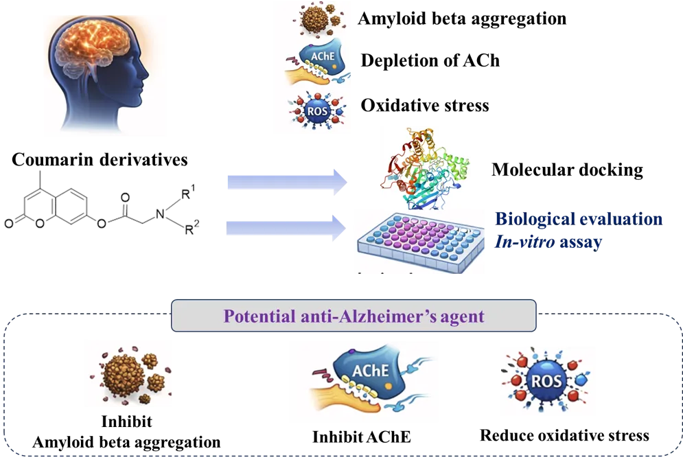
Alzheimer’s disease is a progressive neurodegenerative disorder marked by dementia, motor incoordination, cognitive decline, and respiratory depression. Its complex pathophysiology—characterized by hippocampal neuronal death, amyloid-? plaques, and neurofibrillary tangles—limits the development of disease-modifying therapies. This study explores 7-hydroxy-4-methylcoumarin derivatives as multitarget agents acting on acetylcholinesterase (AChE), ?-secretase (BACE), and glycogen synthase kinase-3? (GSK-3?). Fifteen derivatives were designed via literature review and in-silico modeling. Docking studies identified MU-4, MU-5, MU-7, and MU-9 as promising candidates based on binding affinity and synthetic feasibility. FTIR confirmed successful synthesis. In-vitro AChE inhibition assays revealed MU-7 as a potent inhibitor, outperforming the standard. MU-4 and MU-7 showed antioxidant activity, though less than ascorbic acid. MU-9 demonstrated strong BACE selectivity and amyloid-inhibitory potential, increasing the viability of amyloid-?-induced cell lines to 83.58% at 25??g/ml. MU-7 (secondary amine) is a potent AChE inhibitor, MU-4 (tertiary amine) exhibits antioxidant properties, and MU-9 (nitro-substituted) shows neuroprotective effects. These findings suggest that coumarin-based derivatives may offer both symptomatic relief and disease-modifying potential in Alzheimer’s therapy. Further studies are warranted to validate these results and advance drug development.
Alzheimer’s is a complex neurodegenerative illness that impacts around 45 million people worldwide and is a major socioeconomic burden.1 It is the sixth leading cause of death in the United States. The disease manifests progressive and irreversible memory loss, behavioural changes, cognitive impairment, motor incoordination, and finally death.
The pathophysiology of Alzheimer’s disease is characterized by progressive neurodegeneration. The precise aetiology is still unknown but known to be multifaceted, complex, and interrelated.3 There is a formation of amyloid plaques and neurofibrillary tangles (NFT) in the brain.4 The alterations in genes coding amyloid precursor protein (APP), presenilin-1 (PS1), and presenilin (PS2) are also responsible for the deposition of senile plaques in extracellular regions and the intracellular formation of NFTs.5 This causes damage to nerve cells in the cortex region, basal region of the forebrain, and loss of cholinergic neurons.6 Various hypotheses aid in the therapeutic formulation against Alzheimer’s disease.
All of the existing treatments used today aim to counterbalance the neurotransmitter imbalance associated with the disease. As of now, two classes of drugs are widely used for the management of AD, including cholinesterase inhibitors (AChEIs) and N-methyl-d-aspartate (NMDA) receptor antagonists.7 Donepezil remains a drug of choice for Alzheimer's disease. Despite ongoing research in this field, few drugs can be efficiently used for the management of Alzheimer's. Most of the candidate drugs failed during the clinical trials. It is reported that the reason for the failure of the candidate drug is the wrong selection of the treatment target and incomplete knowledge about the complex pathophysiology of Alzheimer’s disease.8 This can be overcome to an extent by designing multitargeted drug ligands. The study aims to design, synthesize, and evaluate 7-Hydroxy-4-methylcoumarin derivatives as multitargeted ligands for the development of anti-Alzheimer’s drugs.
MATERIALS AND METHODS
In-silico designing of 7-hydroxy-4-methylcoumarin derivatives
The study started with a thorough literature review to identify potential lead molecules for showing anti Alzheimer’s property and coumarin was found to be a better ring moiety for interaction with Acetylcholinesterase (AChE) and beta-secretase (BACE) enzymes.
Fifteen derivatives were designed based on an intense literature review (Table 1). The derivatives contain a coumarin core linked with various substituted amines. The designed derivatives are screened for various properties like drug likeness and physicochemical properties using software and in-silico methods. Among the fifteen derivatives, four derivatives having better properties and blood-brain barrier (BBB) permeability are selected for synthesis
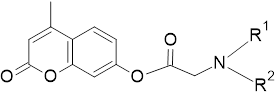
Table 1: 7-Hydroxy-4-methylcoumarin derivatives designed for study.
|
Compound code |
R1 |
R2 |
|
MU-1 |
Phenyl |
H |
|
MU-2 |
Phenyl |
Methyl |
|
MU-3 |
Phenyl |
Ethyl |
|
MU-4 |
Phenyl |
Phenyl |
|
MU-5 |
p-chlorophenyl |
H |
|
MU-6 |
p-bromophenyl |
H |
|
MU-7 |
p-fluorophenyl |
H |
|
MU-8 |
p-nitrophenyl |
H |
|
MU-9 |
m-nitrophenyl |
H |
|
MU-10 |
o-nitrophenyl |
H |
|
MU-11 |
o-aminophenyl |
H |
|
MU-12 |
Benzyl |
H |
|
MU-13 |
Benzyl |
Methyl |
|
MU-14 |
Ethyl |
Ethyl |
|
MU-15 |
Methyl |
Methyl |
Molecular docking studies
Molecular docking studies are carried out to evaluate the binding affinity of the selected derivatives towards AChE (1EVE), BACE (1W51) and glycogen synthase kinase-3b (4PTC) receptors. The software PyRx9 and biovia discovery studio10 are used for docking and visualisation.
Synthesis of 7-hydroxy-4-methyl coumarin derivatives
Synthesis of 7-hydroxy-4-methyl coumarin using Pechman condensation
7.5 ml of con. sulphuric acid cooled to 10℃ and stirred with a solution of resorcinol (15 mmol) and ethyl acetoacetate (18 mmol) for about 30-45 minutes. Pour the mixture into crushed ice. Filter and collect the precipitate after washing, drying, and recrystallization using ethanol.11 (figure 1)
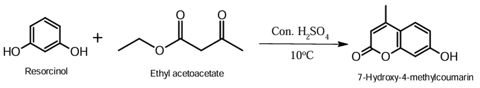
Figure 1: Chemical reaction involved in the synthesis of 7-hydroxy-4-methylcoumarin.
Synthesis of intermediate 1
A mixture of 7-Hydroxy-4-methylcoumarin (10 mmol), and triethyl amine (10 mmol) in anhydrous dichloromethane (25 ml) was cooled to 0 to 5℃. To this reaction mixture, add chloroacetyl chloride (10 mmol) and stir for 1 hr. Then, stir for 5 hr at room temperature. Wash with 5% HCl, 5% NaOH, and 5% brine solution. Once collected, the organic layer was dried over sodium sulfate. Filtered, the solvent was eliminated using less pressure. (Figure 2)

Figure 2: chemical reaction involved in the synthesis of intermediate 1.
Synthesis of derivatives using primary amines. (MU-5, MU-7, MU-9)
Conventional method: 2.5 mmol of substituted primary amine mixed with 10 ml anhydrous DCM. Intermediate 2 (0.5 mmol) was added to this mixture. Refluxed for 7 hours in the presence of anhydrous K2CO3 at 35℃. Evaporate the solvent DCM under reduced pressure.
Microwave-assisted synthesis: Intermediate (0.5 mmol) dissolved in 4ml anhydrous acetonitrile and suspended under magnetic stirring with K2CO3(0.5 mmol) & catalytic amount of KI. Substituted amine (2.5mmol) was added. Irradiated with microwave 520 nm for 30 min. The solvent DCM evaporated under reduced pressure. (figure 3).
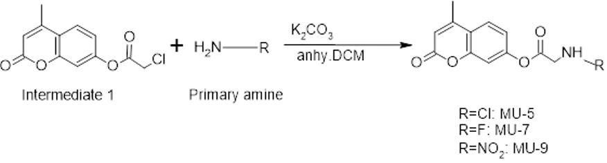
Figure 3: Reaction involved in the synthesis of primary amine derivatives.
Synthesis of derivative using Diphenylamine (MU-4)
Diphenylamine (0.04 M) was dissolved in toluene (200ml) & intermediate 2 (0.04 M) was added to it. The reaction mixture was refluxed at 80℃ for 4 hours. The precipitate of compound MU-4 was then poured into crushed ice and kept overnight for precipitation. It was washed with cold water, filtered, and dried. (Figure 4)
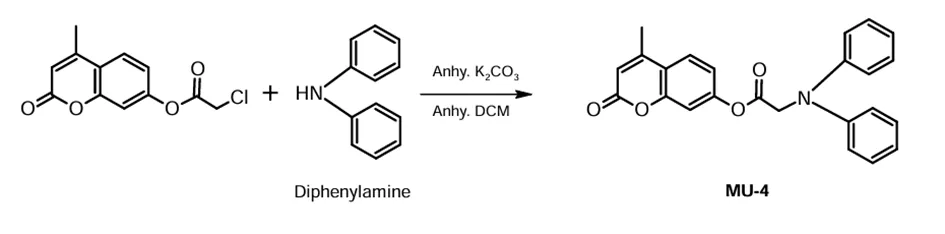
Figure 4: synthesis of MU-4
In-vitro biological evaluation
Evaluation of in-vitro antioxidant activity by DPPH assay
Antioxidant property is an additional benefit for an anti-Alzheimer’s drug. Antioxidant property of the synthesised derivatives was evaluated using DPPH assay.
The samples were made into different concentrations ranging from 12.5µg/mL to 200µg/mL from the stock solution. Samples were made up to a final volume of 20ml with DMSO, and added 1.48 ml of DPPH (0.1 mM) solution. A control with an equivalent amount of distilled water instead of the test compound was also taken. The reaction mixture was incubated at 25℃ in the dark for twenty minutes. SHIMADZU(UV-1900i) UV-VIS spectrophotometer measures absorbance at 517 nm. The following formula calculates the percentage inhibition of the sample and the standard.12
Evaluation of AChE inhibitory activity by Ellman’s assay
AChE (acetylcholinesterase) activity was measured using a modified 96-well microplate assay based on Ellman’s method.
The AChE used in this assay was sourced from Electrophorus electricus (electric eel) in the form of Type VI-S lyophilized powder, with specific activities of 222 U/mg solid and 268 U/mg protein. The enzyme stock solution was prepared at a concentration of 222 U/mL and stored at -80˚C. Further dilutions of the enzyme were made in a solution containing 0.1% BSA (bovine serum albumin) in the buffer. DTNB was dissolved in the buffer, which also contained 0.1 M NaCl and 0.02 M MgCl2. Acetylthiocholine (ATCI) was dissolved in deionized water. 100 µL of 3 mM DTNB, 20 µL of a 0.26 U/mL AChE solution, and 40 µL of the Tris buffer (pH 8.0) were added to each well of the 96-well plate, along with 20 µL of each extract at various concentrations (25, 50, and 100 µg/mL) dissolved in the buffer. After mixing, the plate was incubated for 15 minutes at 25˚C, and the absorbance was measured at 412 nm. The reaction was initiated by the addition of 20 µL of ATCI. Following a second mixing step, the absorbance was measured at two, time intervals (5 minutes and 20 minutes).13
In-vitro neuroprotective effect determination by neutral red assay
Cell lines SHSY-5Y (human neuroblastoma) was selected. Cell lines (Purchased from NCCS Pune) maintained in DMEM (from NCCS Pune), India. Cultured using medium -DMEM in a tissue culture flask (25cm2). Supplement with L-glutamine, 10% FBS, NaHCO3, and an antibiotic solution (100U/ml Penicillin, 100mg/ml streptomycin, and 2.5 mg/ml amphotericin B). Incubated in a 5% humidified CO2 incubator at 37℃. Cell vitality was estimated using an inverted phase contrast microscope, under MTT assay. Two days old, a single layer of confluent cells was trypsinized. Suspended in 10% growth medium and 100 ml of suspension was seeded in a 96-well tissue culture plate Incubated in 5% humidity in a CO2 incubator at 37℃.
To induce toxicity beta-amyloid (10µM) was added and incubated for one hour. 25 µg/ml, 12.5µg/ml, 6.25 µg/ml, 3.1 µg/ml, and 1.5 µg/ml of freshly prepared compounds were then added. Each concentration was added three times to the wells and incubated at 37°C at 5% humidity in a CO2 incubator for 24 hours. Untreated cells (control) and Beta-amyloid-treated cells were also maintained.
Culture plates containing the sample were treated with 10 microliters of the neutral red solution, and incubated for three hours at 37 ℃ in a CO2 incubator. Following a PBS wash, 200µL of fixing solution (consisting of 50% ethanol and 1% acetic acid) was used to fix the cells. Plates were left at room temperature for 20 minutes after the fixation solution was removed and 200µl of extraction buffer was added and thoroughly mixed. A microplate reader was used to measure the absorbance at 540 nm, and the % viability was computed.14,15
RESULTS
Molecular docking studies
The fifteen designed derivatives were docked in selected receptors. Donepezil and a nanomolar non- peptidic inhibitor (L01), are used as standards for AChE and b-secretase respectively. No standard drug available in the market inhibits GSK-3b except lithium. So, the activity of derivatives to inhibit GSK is evaluated among themselves. (Table 2)
Table 2: Docking scores of proposed 7-hydroxy-4-methylcoumarin derivatives
|
Compound |
Docking score |
||
|
1EVE |
1W51 |
4PTC |
|
|
MU-1 |
-10.4 |
-8 |
-7 |
|
MU-2 |
-10.4 |
-8 |
-7 |
|
MU-3 |
-9.8 |
-7.2 |
-6 |
|
MU-4 |
-10.4 |
-9.8 |
-8 |
|
MU-5 |
-9.8 |
-8.4 |
-6.6 |
|
MU-6 |
-9.2 |
8 |
-6.9 |
|
MU-7 |
-10.9 |
-7.7 |
-6.9 |
|
MU-8 |
-10.5 |
-8.8 |
-7.1 |
|
MU-9 |
-9.6 |
-9.8 |
-7.6 |
|
MU-10 |
-9.7 |
-7.8 |
-6.8 |
|
MU-11 |
-8.5 |
-8.5 |
-6.7 |
|
MU-12 |
-10.9 |
-7.4 |
-6.6 |
|
MU-13 |
-9.4 |
-7.4 |
-6.5 |
|
MU-14 |
-9.3 |
-6.9 |
-6.6 |
|
MU-15 |
-8.2 |
-7.2 |
-6.7 |
|
Donepezil |
-11.1 |
|
|
|
L01 |
|
-9.6 |
|
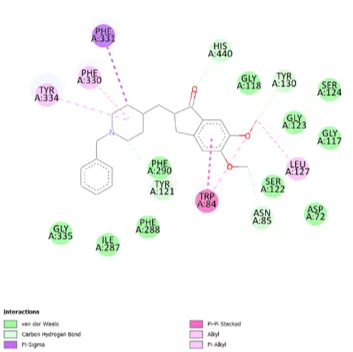
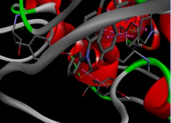
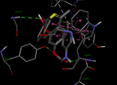
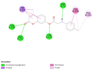
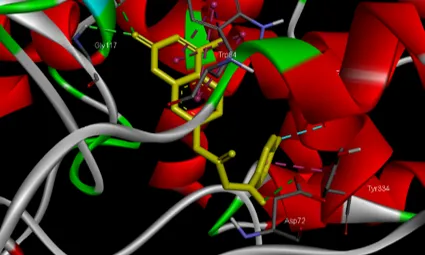
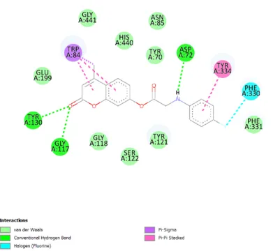
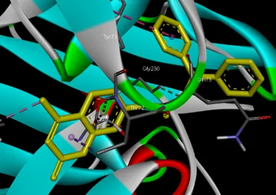
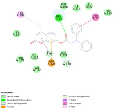
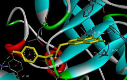
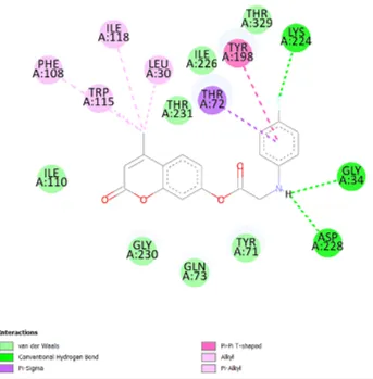
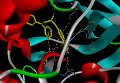
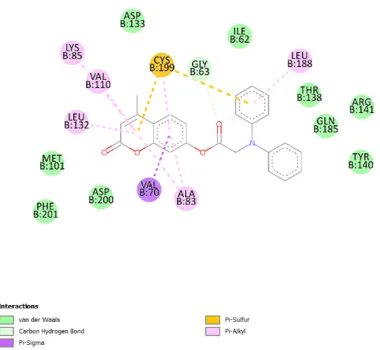
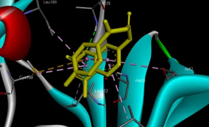
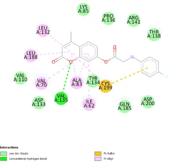
The active sites of the AChE enzyme can be identified as Catalytically Active Sites (CAS) & Peripheral Anionic Sites (PAS). The main amino acids present in PAS include ASP 72, TYR 334, TYR 70, TYR 121, and TRP 279 and amino acids in CAS are PHE 330, TRP 84, TYR 130, and GLY 118. All of these selected compounds show comparable interactions with active site amino acids. (Figure 5 : Docking images of donepezil and selected compound with AChE active site (PDB ID: 1EVE)
Donepezil

MU-4
MU-5
MU-7
MU-9
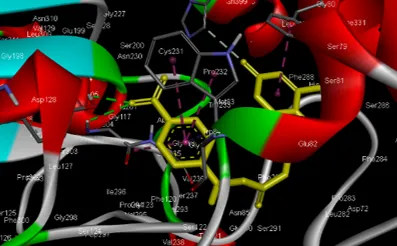
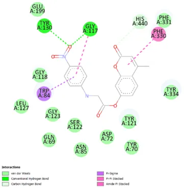
Figure 5: Docking images of donepezil and selected compound with AChE active site (PDB ID: 1EVE).
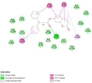
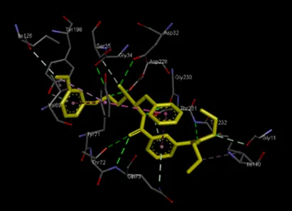
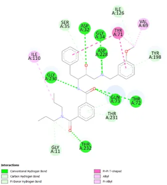
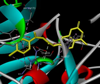
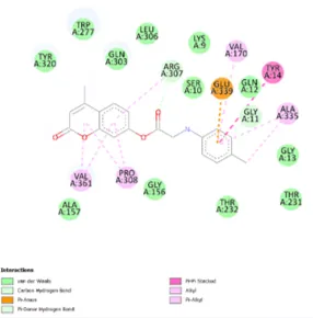
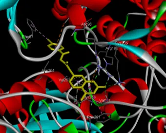
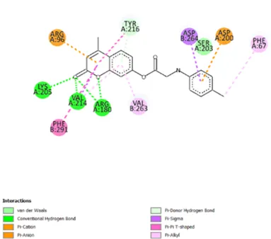
In this study, we use the high-resolution apo-structure of BACE and the structure of BACE in complex with an inhibitor. The selected compound exhibits the same interactions with the active site of BACE as the standard compound. (Figure 6: Docking images of standard compound L01 and selected compounds with BACE (PDB ID: 1W51).
L01 (nanomolar non-peptidic inhibitor)
MU-4
MU-5
MU-7
MU-9
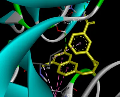
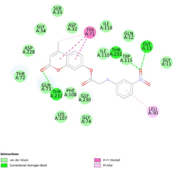
Figure 6: Docking images of standard compound L01 and selected compounds with BACE (PDB ID: 1W51)
No standard is available for GSK-3b. The interactions are compared among the designed derivatives.
MU-4
MU-5
MU-7
MU-9
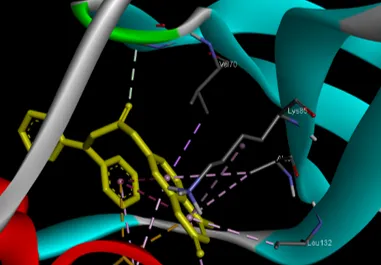
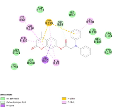
Figure 7: Docking images of selected compounds with GSK-3b (PDB ID: 4PTC)
The in-silico studies suggested that compounds MU-4, MU-5, MU-6, and MU-7 show affinity towards all three selected targets. All these compounds show increased affinity towards AChE except MU-9. MU-9 shows the highest activity at BACE. So, the compounds MU-4, MU-5, MU-7 and MU-9 were selected for synthesis.
CHEMISTRY
7-hydroxy-4-methylcoumarin
Thin layer chromatography (TLC) was carried out to ensure reaction completion using n-hexane: ethyl acetate (3:2) and visualised using UV short wavelength. The Rf value = 0.6. white crystalline powder. M.P. 187℃. IR spectra peaks: O-H stretching:3159.79 cm-1, =C-H stretching 3075.9 cm-1, -C-H stretching 2924.52 cm-1, Conjugated C=O 1677.77 cm-1, C=C stretching 1598.7 cm-1, O-H bending 1448.28 cm-1, C=C bending 842.74 cm-1, C-O-C ester 1066.44 cm-1.
H1 NMR spectra peaks at 2.391 ppm, 6.133 ppm, 6.795 ppm, 6.863 ppm, 7.488 ppm.
Figure 8: IR spectrum of 7-Hydroxy-4-methyl coumarin.
Figure 9: H1NMR spectrum of 7-hydroxy-4-methylcoumarin.
Intermediate 2
TLC using n-hexane:ethyl acetate (2:1). The visualisation was carried out under UV long wavelength—The Rf value =0.4. cream colour crystalline solid, M.P. 162℃ - 164℃.
IR spectra peaks: =C-H stretching 3080.73 cm-1 -C-H stretching 2918.73 cm-1, C=O ester 1761.65 cm-1, C=O lactone 1730.8 cm-1, C=C aromatic 1618.95 cm-1, C-O stretching 1165.79 cm-1, C-O-C ester 1063.55 cm-1, C-Cl stretching 867.81 cm-1.
H1 NMR peaks: 2.437 ppm, 4.334 ppm, 6.288 ppm, 7.106 ppm, 7.161 ppm, 7.640 ppm.
Figure 10: IR spectrum of intermediate 1
Figure 11: H1 NMR spectrum of intermediate 1
Compound MU-4
TLC uses petroleum ether:ethyl acetate (3:1), Rf value = 0.33. Brownish white crystalline powder with M.P. 114℃ to116 ℃.
IR spectra peaks: =C-H stretching 3380.83 cm-1, -C-H stretching 3040.41 cm-1, Ester C=O 1720.53 cm-1, C=C stretching 1492.84 cm-1, C-H Bending 1492.84 cm-1, Ester C-O stretching 1210 cm-1, C=C bending 809.33 cm-1.
H1 NMR peaks: 2.488 ppm, 4.334 ppm, 6.133 ppm, 6.353 ppm, 6.857 ppm
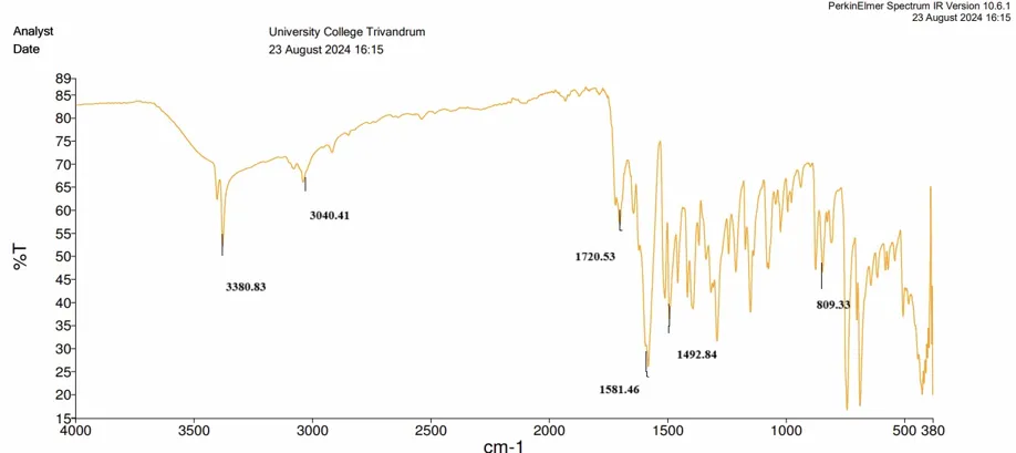
Figure 12: IR spectrum of compound MU-4
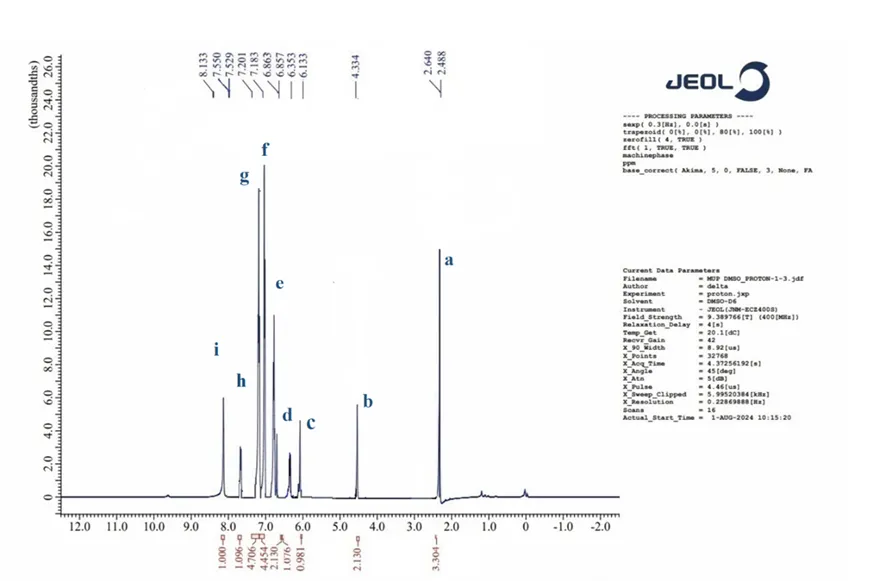
Figure 13: H1NMR spectrum of compound MU-4
Compound MU-5
TLC using n-hexane: ethyl acetate (2:1) as a solvent system. Rf value: 0.91. creamy white crystalline powder. M.P. 108℃ to 110 ℃.
IR spectra peaks: =C-H stretching 3325.12 cm-1, -C-H stretching 3080 cm-1, N-H stretching 3050 cm-1, C=O ester 1761.65 cm-1, C=O lactone 1733.16 cm-1, C=C aromatic 1618.50 cm-1, N-H bending 1510.01 cm-1, C-Cl stretching 850.05 cm-1.
H1NMR peaks: 2.428 ppm, 3.630 ppm, 5.876 ppm, 6.640 ppm,6.833 ppm, 7.017 ppm, 7.401 ppm, 8.133ppm.
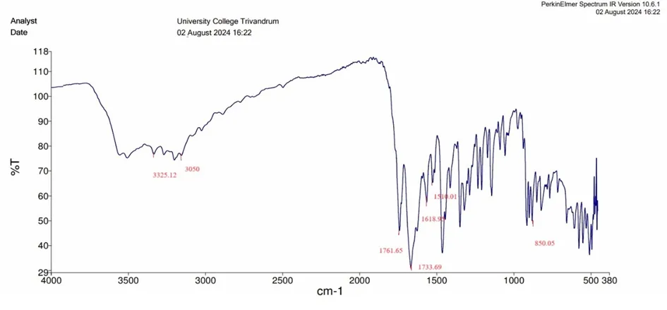
Figure 14: IR spectrum of compound MU-5
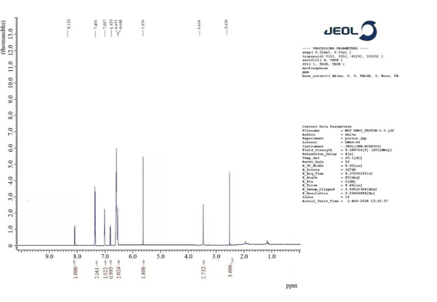
Figure 15: H1NMR Spectrum of compound MU-5
Compound MU-7
TLC: Rf value 0.833 using solvent system n-hexane: ethyl acetate (3:1). Creamy white solid. M.P. 121℃ to 123 ℃.
IR spectra: =C-H stretching 3435.59 cm-1, -C-H stretching 3225.5 cm-1 , N-H stretching 3128.67 cm-1, Ester C=O stretching 1667 cm-1, Aromatic C=C stretching 1594 cm-1, C-F stretching 1389.11 cm-1, C-H bending 810 cm-1.
H1NMR Peaks: 2.528 ppm, 3.430 ppm, 5.676 ppm, 6.540 ppm, 6.738 ppm, 6.911 ppm, 7.106 ppm, 8.133 ppm.
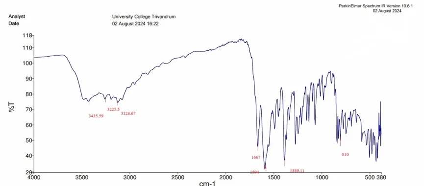
Figure 16: IR spectrum of compound MU-7
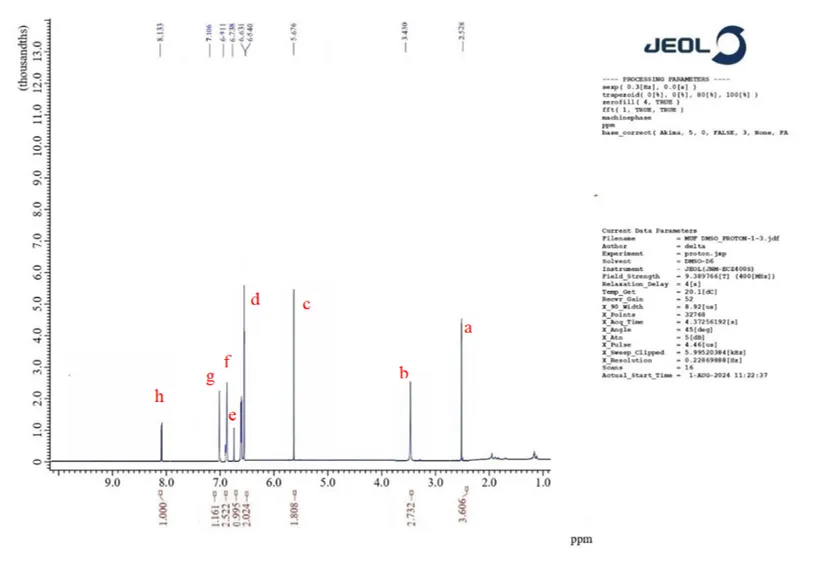
Figure 17: H1NMR Spectrum of compound U-7
Compound MU-9
TLC was done using Pet ether: ethyl acetate (2:1) as a solvent system. Rf value = 0.629. yellow crystalline powder. M.P. 96℃ to 98 ℃.
IR spectra peaks: =C-H stretching 3429.19 cm-1, N-H stretching 3333.95 cm-1, -C-H stretching 3077.69 cm-1, C=O ester 1761.65 cm-1, C=O lactone 1733.16 cm-1, C=C aromatic 1618 cm-1, N-O stretching 1495.65 cm-1.
H1NMR peaks: 2.426 ppm,3.530 ppm, 5.676 ppm, 6.540 ppm, 7.017 ppm, 7.201ppm, 7.629 ppm, 7.650ppm, 7.850 ppm, 8.130 ppm.
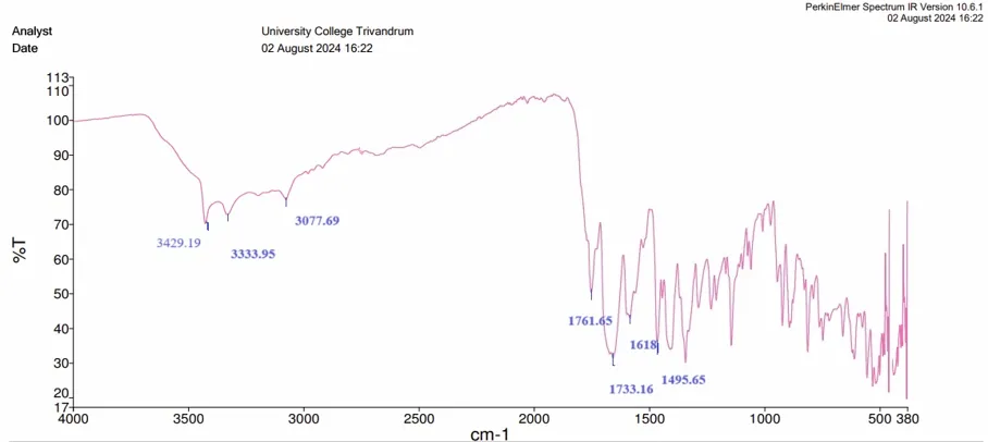
Figure 18: IR spectrum of compound MU-9
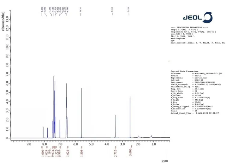
Figure 19: H1NMR Spectrum of compound MU-7
Biological activity
Evaluation of in-vitro acetylcholinesterase inhibitory activity
Among the four derivatives, the compounds MU-4 and MU-7 show the highest AChE inhibitory activity in in-silico studies. These are selected for the in-vitro acetylcholinesterase inhibition assay. The evaluation is carried out by Ellman’s assay method. The activity of MU-4 and MU-7 is compared with that of standard donepezil.
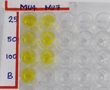
Figure 20: 96-well microplate assay for AChE inhibition.
Table 3: Percentage inhibition of the acetylcholinesterase inhibitory activity of compounds MU-4 and MU-7.
|
Compound |
Concentration (mg/ml) |
DOD |
Percentage inhibition |
|
|
Enzyme control (E) = 0.9758 |
||
|
Donepezil |
25 |
0.5388 |
53.88 |
|
50 |
0.6439 |
64.39 |
|
|
100 |
0.8034 |
80.35 |
|
|
MU-4 |
25 |
0.6171 |
36.75 |
|
50 |
0.4818 |
50.62 |
|
|
100 |
0.2608 |
73.27 |
|
|
MU-7 |
25 |
0.4622 |
52.63 |
|
50 |
0.2586 |
73.49 |
|
|
100 |
0.1912 |
80.40 |
|
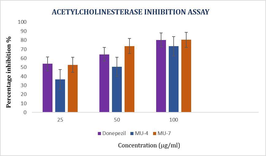
Figure 21: Graphical representation of the concentration v/s percentage inhibition of samples MU-4, MU-7 and standard donepezil.
Table 4: IC50 values of compounds (Calculated using ED50 PLUS V 1.0 Software) .
|
Compounds |
IC50 (mg/ml) |
|
Donepezil |
23.19 |
|
MU-4 |
48.88 |
|
MU-7 |
21.84 |
Antioxidant activity by DPPH assay
The synthesized compounds MU-4 and MU-7 are selected for evaluating antioxidant activity by DPPH assay and compared with ascorbic acid.
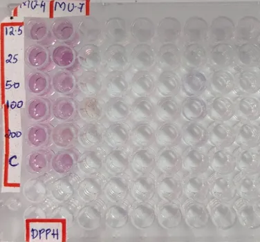
Figure 22: DPPH assay of MU-4 And MU-7.
Table 5: Percentage inhibition of different concentrations of compounds MU-4 and MU-7 by DPPH assay.
|
Concentration (mg/ml) |
Absorbance |
Percentage Inhibition (%) |
||||
|
MU-4 |
MU-7 |
Ascorbic acid |
MU-4 |
MU-7 |
Ascorbic acid |
|
|
12.5 |
0.2432 |
0.2501 |
0.2075 |
17.47 |
15.13 |
28.79 |
|
25 |
0.2261 |
0.2318 |
0.1765 |
23.28 |
21.34 |
39.43 |
|
50 |
0.1935 |
0.1882 |
0.1273 |
34.34 |
36.13 |
56.31 |
|
100 |
0.1508 |
0.153 |
0.0726 |
48.82 |
48.08 |
75.08 |
|
200 |
0.1371 |
0.1347 |
0.285 |
53.47 |
54.29 |
90.21 |
Table 6: IC50 values of MU-4 and MU-7 in DPPH assay
|
Compounds |
IC50 (mg/ml) |
|
MU-4 |
125.18 |
|
MU-7 |
130.87 |
|
Ascorbic acid |
40.65 |
Amyloid-b inhibitory activity by neutral red assay
The amyloid-b inhibitory activity of compound MU-9 was evaluated by a cell viability assay on human neuroblastoma cell lines (SHSY-5Y) using neutral red as a stain. Untreated cells are kept as a control and all other cell lines are treated with amyloid beta. Then different concentrations of sample solution were added to each test well to evaluate percentage viability.
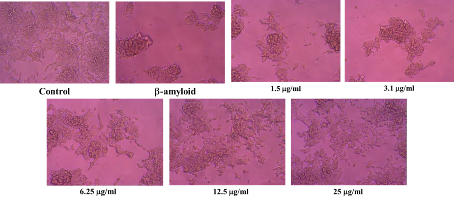
Figure 23: Cell lines treated with various concentrations of sample MU-9.
Table 7: Percentage viability of cell lines treated with MU-9
|
Sample Concentration (µg/ml) |
OD I |
OD II |
OD III |
Average Absorbance @ 540nm |
Percentage Viability |
|
CONTROL |
0.4139 |
0.4216 |
0.4189 |
0.4181 |
100.00 |
|
Beta-amyloid |
0.2089 |
0.1987 |
0.1995 |
0.2024 |
48.40 |
|
SAMPLE CODE: MU-9 |
|
||||
|
1.5 |
0.2256 |
0.2345 |
0.2364 |
0.2322 |
55.52 |
|
3.1 |
0.2459 |
0.2548 |
0.2539 |
0.2515 |
60.15 |
|
6.25 |
0.2728 |
0.2739 |
0.2731 |
0.2733 |
65.35 |
|
12.5 |
0.3026 |
0.3128 |
0.3105 |
0.3086 |
73.81 |
|
25 |
0.3521 |
0.3485 |
0.3476 |
0.3494 |
83.56 |
DISCUSSION
From a thorough literature review, fifteen derivatives of 7-hydroxy-4-methylcoumarin were designed and evaluated for anti-Alzheimer’s property in-silico. From the in-silico studies, based on the lipophilicity, BBB permeability and docking scores on all the three selected receptors, four compounds were selected for synthesis. The synthesised compounds MU-4, MU-5, MU-7 & MU-9 were confirmed using appearance, melting point, IR spectra and NMR spectra. Of the four synthesized compounds, MU-4 and MU-7 were chosen for in-vitro testing for AChE inhibitory activity due to their high docking scores. The results from Ellman’s assay for AChE inhibitory activity showed that both compounds were active, with compound MU-7 exhibiting better inhibitory activity than the standard drug donepezil. The compounds MU-4 and MU-7 underwent in-vitro anti-oxidant activity assessment using the DPPH assay. The results indicate that both compounds exhibit anti-oxidant potency, though lower than the standard compound ascorbic acid. The neuroprotective ability of compound MU-9 was evaluated by a neutral red assay using neuroblastoma cell lines. The percentage viability of cells on treatment with 25mg/ml solution was 83.56%.
The designed 4-methyl umbelliferone derivatives, particularly MU-7, show promise as potential anti-Alzheimer’s agents. These compounds warrant further investigation and development due to their ability to inhibit key enzymes involved in Alzheimer's disease and their antioxidant properties.
LIMITATIONS OF THE STUDY
The synthesized derivatives are esters, which could function as a prodrug in the body. Consequently, understanding the drug’s fate requires comprehensive in vivo studies. The compounds showed lower antioxidant activity than the standard ascorbic acid, indicating the need for further optimization to enhance their antioxidant properties.
The study does not offer insights into the in-vivo pharmacokinetic and pharmacodynamic properties.
CONCLUSION
In conclusion, this study has successfully designed, synthesized, and evaluated novel 7-Hydroxy-4-methylcoumarin derivatives with potential anti-Alzheimer's properties. The results, especially for compound MU-7, are encouraging and suggest that these derivatives could be valuable candidates for further research and development in the field of Alzheimer's disease treatment.
REFERENCES

Chithra P, Arul K, 7-Hydroxy-4-Methylcoumarin Derivatives: A Multitargeted Ligand for the Management of Alzheimer's Disease, Int. J. of Pharm. Sci., 2026, Vol 4, Issue 6, 857-876. https://doi.org/10.5281/zenodo.20529277
 10.5281/zenodo.20529277