

We use cookies to ensure our website works properly and to personalise your experience. Cookies policy
Shree Krishna College of Pharmacy, Sitapur.
Topical drug delivery has emerged as an attractive alternative to oral and parenteral administration owing to its ability to provide localized therapeutic action while minimizing systemic adverse effects. However, the highly organized structure of the stratum corneum significantly restricts the penetration of many therapeutic agents, particularly hydrophilic and high molecular weight compounds. Conventional topical formulations such as creams, ointments, and gels often exhibit limited skin permeation and poor bioavailability, resulting in suboptimal therapeutic outcomes. Nanovesicular carrier systems, especially ethosomes, have gained considerable attention because of their superior ability to enhance transdermal and dermal drug delivery through improved skin penetration.Ethosomes are soft, malleable phospholipid vesicles containing high concentrations of ethanol, which fluidizes both vesicular lipids and the stratum corneum lipid bilayer, thereby facilitating deeper penetration into the skin. Incorporation of ethosomes into hydrogel matrices further improves formulation stability, spreadability, residence time, patient compliance, and controlled drug release. The combination of bioactive compounds such as Vitamin A, Vitamin E, and ginger extract within an ethosomal hydrogel offers a multifunctional therapeutic strategy owing to their antioxidant, anti-inflammatory, antimicrobial, collagen-stimulating, and wound-healing properties.Vitamin A regulates epidermal differentiation and collagen synthesis, making it highly effective in skin rejuvenation and treatment of photoaging. Vitamin E protects cellular membranes against oxidative damage through its potent free radical scavenging activity while improving skin hydration and barrier function. Ginger extract contains several bioactive phytoconstituents including 6-gingerol, 6-shogaol, and zingerone that exhibit remarkable anti-inflammatory, antioxidant, antimicrobial, and wound-healing activities. The synergistic combination of these bioactives within an ethosomal hydrogel has the potential to enhance dermal penetration, improve therapeutic efficacy, and provide sustained drug release.This review comprehensively discusses the anatomical structure of the skin, challenges associated with dermal drug delivery, principles of ethosomal drug delivery systems, hydrogel-based formulations, pharmacological importance of Vitamin A, Vitamin E, and ginger extract, formulation strategies, evaluation parameters, recent research advances, and future prospects. The review also identifies current research gaps and highlights the potential of multifunctional ethosomal hydrogels as promising nanocarrier systems for advanced topical drug delivery.
The skin is the largest organ of the human body, accounting for approximately 15–16% of total body weight and covering an average surface area of nearly 2 m² in adults. Besides serving as a physical barrier against environmental insults, pathogens, ultraviolet radiation, and chemical agents, the skin also plays a critical role in thermoregulation, immune surveillance, sensory perception, and maintenance of fluid homeostasis [1,2]. Owing to its accessibility and extensive vascular network, the skin has become an attractive route for localized as well as systemic drug delivery.
Topical drug delivery offers several advantages over conventional oral and injectable routes, including avoidance of hepatic first-pass metabolism, improved patient compliance, reduced gastrointestinal irritation, prolonged drug residence at the target site, and lower systemic toxicity [3]. Despite these advantages, effective dermal drug delivery remains challenging due to the barrier properties of the stratum corneum, which restricts the penetration of most therapeutic molecules. The "brick-and-mortar" organization of corneocytes embedded within a lipid matrix acts as the primary obstacle to drug permeation [4].
Several strategies have been investigated to overcome the skin barrier, including chemical penetration enhancers, microneedles, iontophoresis, sonophoresis, lipid nanoparticles, liposomes, transfersomes, niosomes, and ethosomes [5]. Among these advanced nanocarrier systems, ethosomes have demonstrated remarkable potential because of their unique composition consisting of phospholipids, ethanol, and water. High ethanol concentration increases membrane fluidity and disrupts the highly ordered lipid arrangement of the stratum corneum, thereby facilitating enhanced penetration of therapeutic molecules into deeper skin layers [6].
Hydrogels are three-dimensional hydrophilic polymeric networks capable of absorbing large amounts of water while maintaining structural integrity. Owing to their excellent biocompatibility, non-greasy nature, ease of application, and controlled drug release properties, hydrogels have become one of the most widely used vehicles for topical drug delivery [7]. The incorporation of ethosomal vesicles into hydrogel systems combines the penetration-enhancing capability of ethosomes with the sustained-release characteristics and patient-friendly properties of hydrogels, resulting in improved therapeutic performance.
Among various dermatological bioactive compounds, Vitamins A and E have received considerable attention because of their essential roles in maintaining skin integrity and preventing oxidative damage. Vitamin A stimulates epidermal renewal, regulates keratinocyte differentiation, enhances collagen production, and reduces photoaging [8]. Vitamin E (α-tocopherol) is one of the most effective lipid-soluble antioxidants, protecting skin cells against oxidative stress induced by ultraviolet radiation and environmental pollutants while improving skin hydration and elasticity [9].
Natural herbal extracts have also gained increasing interest owing to their excellent safety profile and multiple pharmacological activities. Ginger (Zingiber officinale Roscoe), an important medicinal plant belonging to the family Zingiberaceae, contains numerous bioactive constituents such as 6-gingerol, 6-shogaol, paradols, and zingerone. These phytochemicals exhibit potent antioxidant, anti-inflammatory, antimicrobial, analgesic, and wound-healing activities, making ginger extract a promising candidate for topical therapeutic applications [10].
The combination of Vitamin A, Vitamin E, and ginger extract within an ethosomal hydrogel provides a multifunctional nanocarrier capable of addressing multiple pathological pathways simultaneously. Vitamin A promotes skin regeneration, Vitamin E protects against oxidative stress, whereas ginger extract suppresses inflammatory mediators and microbial growth. Encapsulation of these bioactive compounds into ethosomal vesicles further enhances their stability, skin penetration, and therapeutic efficacy while minimizing degradation and irritation.
Recent advances in nanotechnology have further strengthened the application of ethosomal hydrogels for treatment of inflammatory skin diseases, wound healing, anti-aging therapy, psoriasis, acne vulgaris, fungal infections, and cosmetic dermatology. Nevertheless, challenges related to formulation stability, large-scale manufacturing, regulatory approval, and long-term safety still require extensive investigation before successful clinical translation.
Therefore, this review summarizes the current understanding of ethosomal hydrogel systems containing Vitamin A, Vitamin E, and ginger extract, with particular emphasis on formulation strategies, characterization techniques, dermal delivery mechanisms, recent research progress, and future perspectives for advanced topical drug delivery.
2. Skin Anatomy and Barrier Function
The skin is a complex multilayered organ composed of three principal layers: epidermis, dermis, and hypodermis (subcutaneous tissue). Each layer possesses unique structural and physiological characteristics that collectively contribute to protection, thermoregulation, immune defense, and maintenance of internal homeostasis [11].
The epidermis is the outermost layer and consists primarily of keratinocytes arranged into five distinct strata: stratum basale, stratum spinosum, stratum granulosum, stratum lucidum (present only in thick skin), and stratum corneum. Among these, the stratum corneum is the principal barrier to percutaneous drug absorption. It is composed of dead, flattened corneocytes embedded within an extracellular lipid matrix containing ceramides, cholesterol, and free fatty acids, often described as the "brick-and-mortar" model [12].
The dermis, located beneath the epidermis, is composed of collagen fibers, elastin, fibroblasts, blood vessels, lymphatics, nerves, and skin appendages such as hair follicles and sweat glands. This layer provides mechanical strength, elasticity, nutrient supply, and immune support. Drug molecules reaching the dermis may enter systemic circulation through the extensive capillary network [13].
The hypodermis consists mainly of adipose tissue and connective tissue that serve as thermal insulation, mechanical cushioning, and energy storage. It also acts as a depot for lipophilic drugs and contributes to prolonged drug residence in certain formulations [14].
Drug permeation across the skin occurs primarily through three pathways:
Among these, the intercellular pathway is the predominant route for passive diffusion of lipophilic molecules. Ethosomal vesicles enhance drug penetration by increasing lipid fluidity within the stratum corneum and facilitating deeper transport through these pathways [15]
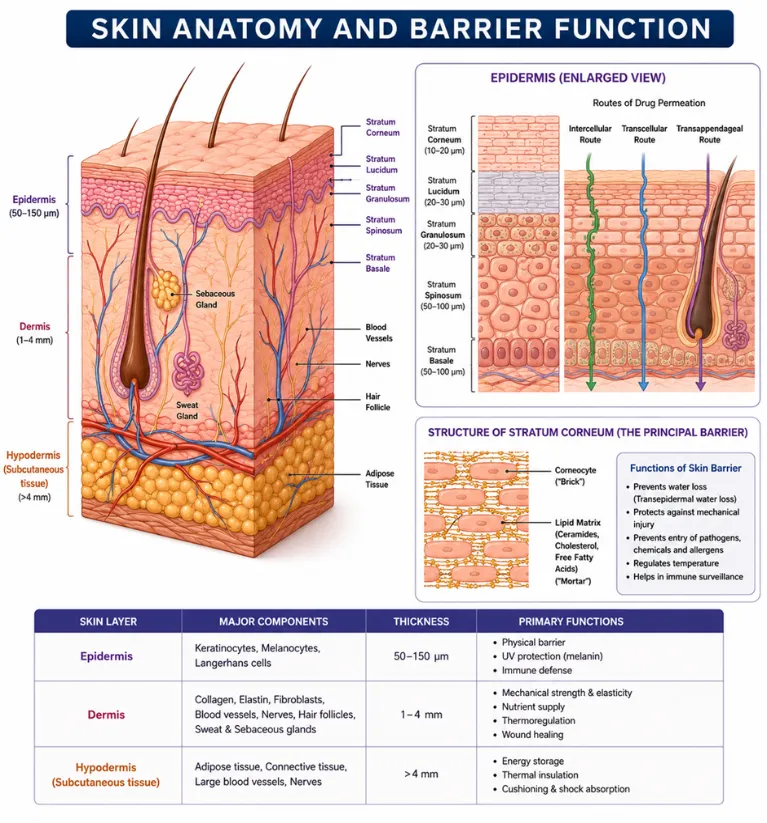
Figure 1 Skin Anatomy and Barrier Function
Table 1. Major Layers of Human Skin and Their Functions
|
Skin Layer |
Major Components |
Primary Functions |
|
Epidermis |
Keratinocytes, melanocytes, Langerhans cells |
Physical barrier, UV protection, immune defense |
|
Stratum Corneum |
Corneocytes, ceramides, cholesterol |
Principal barrier to drug permeation |
|
Dermis |
Collagen, elastin, fibroblasts, blood vessels |
Mechanical support, nutrient supply, wound healing |
|
Hypodermis |
Adipose tissue, connective tissue |
Thermal insulation, energy storage, cushioning |
Table 2. Advantages and Limitations of Topical Drug Delivery
|
S.No. |
Advantages |
Limitations |
|
|
Avoids first-pass metabolism |
Limited penetration through stratum corneum |
|
|
Localized drug delivery |
Low bioavailability of hydrophilic drugs |
|
|
Reduced systemic toxicity |
Skin irritation from some excipients |
|
|
Improved patient compliance |
Variable permeability among individuals |
|
|
Sustained therapeutic effect |
Enzymatic degradation of certain drugs |
3. Ethosomes: A Novel Vesicular Drug Delivery System
Ethosomes are advanced phospholipid-based nanovesicular carriers specifically designed to enhance the dermal and transdermal delivery of therapeutic agents. Introduced by Touitou and co-workers in 2000, ethosomes consist primarily of phospholipids, a relatively high concentration of ethanol (20–45%), water, and optional additives such as glycols or cholesterol [16]. Unlike conventional liposomes, the high ethanol content imparts remarkable flexibility and deformability to the vesicular membrane, allowing ethosomes to penetrate the compact lipid architecture of the stratum corneum more effectively.
The unique combination of ethanol and phospholipids enables ethosomes to transport both hydrophilic and lipophilic drugs into deeper skin layers without causing irreversible damage to the skin barrier. Ethosomes have demonstrated significant success in delivering corticosteroids, antifungal agents, antibiotics, antioxidants, anti-inflammatory drugs, herbal extracts, peptides, and cosmetic ingredients [17].
The increasing interest in ethosomal drug delivery is attributed to their high encapsulation efficiency, improved drug stability, controlled release behavior, and enhanced patient compliance. Recent developments have focused on integrating ethosomes into semisolid dosage forms such as hydrogels, creams, and emulgels to further improve topical retention and therapeutic efficacy [18].
3.1 Composition of Ethosomes
Ethosomes are composed of four major components, each contributing a distinct function to the vesicular system.
Phospholipids
Phospholipids constitute the structural framework of ethosomal vesicles. Commonly used phospholipids include: Soy phosphatidylcholine, Egg phosphatidylcholine, Hydrogenated phosphatidylcholine & Phosphatidylserine.
These amphiphilic molecules self-assemble into bilayer vesicles capable of encapsulating both hydrophilic and lipophilic therapeutic agents.
Ethanol
Ethanol is the most important component of ethosomes and is generally present at concentrations ranging from 20–45%. Ethanol performs multiple functions:
Water
Purified water forms the continuous phase of the vesicular dispersion and hydrates phospholipid molecules.
Polyols
Propylene glycol or Transcutol® may be incorporated to improve vesicle stability, enhance skin hydration, and further increase drug permeation
Table 3. Major Components of Ethosomes and Their Functions
|
Component |
Typical Concentration |
Function |
|
Phospholipid |
1–5% |
Vesicle formation |
|
Ethanol |
20–45% |
Penetration enhancement |
|
Water |
q.s. |
Hydration medium |
|
Propylene glycol |
5–20% |
Co-solvent and humectant |
|
Cholesterol (optional) |
0.5–2% |
Membrane stabilization |
3.2 Preparation Methods of Ethosomes
Several preparation methods have been reported depending upon drug characteristics and formulation requirements.
3.2.1 Cold Method
The cold method is the most widely employed technique for laboratory-scale preparation.
Procedure
3.2.2 Hot Method
3.2.3 Thin Film Hydration Method
Table 4. Comparison of Ethosome Preparation Methods
|
Method |
Advantages |
Limitations |
|
Cold Method |
Simple, reproducible |
Requires sonication |
|
Hot Method |
Suitable for some lipids |
Not ideal for thermolabile drugs |
|
Thin Film Hydration |
Uniform vesicles |
Time-consuming |
|
Reverse Phase Evaporation |
High encapsulation |
Organic solvent exposure |
3.3 Mechanism of Skin Penetration
The superior skin penetration ability of ethosomes is primarily attributed to the synergistic action of ethanol and phospholipid vesicles.
Step 1: Ethanol Interaction
Ethanol penetrates the stratum corneum and disrupts the highly ordered lipid arrangement by increasing lipid fluidity.
Step 2: Vesicle Softening
The presence of ethanol makes phospholipid bilayers highly flexible and deformable.
Step 3: Enhanced Vesicle Penetration
Soft vesicles squeeze through the disrupted lipid domains and migrate into deeper epidermal layers.
Step 4: Drug Release
Once inside viable skin layers, the vesicles gradually release encapsulated drug molecules, producing prolonged therapeutic action.
This mechanism results in significantly greater drug deposition compared with conventional liposomes.
3.4 Factors Affecting Ethosomal Characteristics
Several formulation variables influence the physicochemical properties and performance of ethosomes.
Ethanol Concentration
Increasing ethanol concentration decreases vesicle size and increases flexibility. However, concentrations above approximately 45% may destabilize vesicles.
Phospholipid Concentration
Higher phospholipid concentration generally increases vesicle size and entrapment efficiency.
Cholesterol Content
Cholesterol enhances membrane rigidity but excessive amounts may reduce drug release
Sonication Time
Longer sonication reduces particle size but excessive sonication may result in drug leakage.
Storage Temperature
Refrigerated storage (4–8°C) generally improves vesicle stability
3.5 Advantages of Ethosomes
Ethosomes possess several advantages over conventional topical formulations:
3.6 Limitations of Ethosomes
Despite numerous advantages, ethosomes also exhibit certain limitations:
Table 5. Comparison of Liposomal Nanocarriers
|
Property |
Liposomes |
Transfersomes |
Ethosomes |
Transethosomes |
|
Ethanol |
No |
No |
High |
High |
|
Edge Activator |
No |
Yes |
No |
Yes |
|
Vesicle Flexibility |
Moderate |
High |
Very High |
Extremely High |
|
Skin Penetration |
Moderate |
High |
Excellent |
Excellent |
|
Drug Entrapment |
Moderate |
High |
High |
High |
|
Dermal Delivery |
Good |
Better |
Excellent |
Excellent |
4. Hydrogel-Based Drug Delivery Systems
Hydrogels are three-dimensional crosslinked polymeric networks capable of absorbing large quantities of water while maintaining their structural integrity. Owing to their hydrophilic nature, excellent biocompatibility, softness, and controlled drug release behavior, hydrogels have become one of the most widely investigated semisolid dosage forms for topical drug delivery [19].
Hydrogels provide a moist environment at the site of application, improving skin hydration and facilitating enhanced drug permeation. Their non-greasy texture, cooling sensation, ease of washing, and patient acceptability make them superior to conventional ointments and creams for many dermatological applications.
When ethosomal dispersions are incorporated into hydrogel matrices, the resulting ethosomal hydrogel combines the penetration-enhancing capability of ethosomes with the prolonged residence time and controlled release properties of hydrogels. Such hybrid systems improve formulation stability, reduce vesicle aggregation, enhance skin retention, and increase therapeutic efficacy [20].
4.1 Ideal Characteristics of Hydrogels
An ideal hydrogel intended for topical application should possess the following characteristics:
Common polymers include: Carbopol 934, Carbopol 940, Carbopol 971, Hydroxypropyl methylcellulose (HPMC), Sodium carboxymethyl cellulose (NaCMC),Poloxamer 407, Chitosan, Xanthan gum, Sodium alginate and Polyvinyl alcohol (PVA).
Among these, Carbopol 934 and Carbopol 940 are the most extensively used owing to their excellent gelling ability, transparency, bioadhesion, and compatibility with ethosomal dispersions.
5. Vitamin A in Dermal Drug Delivery
Vitamin A is a fat-soluble vitamin that plays an indispensable role in maintaining skin integrity, cellular differentiation, epithelial growth, immune regulation, and collagen synthesis. It belongs to the retinoid family, which includes retinol, retinaldehyde, retinoic acid, and retinyl esters. Among these, retinol and retinoic acid are the most extensively utilized in dermatological and cosmetic formulations due to their proven efficacy in improving skin texture, reducing wrinkles, stimulating collagen production, and treating various hyperproliferative skin disorders [21].
Topically applied Vitamin A promotes epidermal renewal by regulating keratinocyte proliferation and differentiation through interaction with nuclear retinoic acid receptors (RARs) and retinoid X receptors (RXRs). Activation of these receptors modulates the expression of genes responsible for collagen synthesis, epidermal turnover, and extracellular matrix remodeling [22].
Vitamin A also inhibits matrix metalloproteinases (MMPs), enzymes responsible for collagen degradation during aging and photoaging. Consequently, topical Vitamin A formulations improve skin elasticity, reduce fine wrinkles, accelerate wound healing, and restore epidermal barrier function [23].
Despite its remarkable therapeutic potential, Vitamin A exhibits several pharmaceutical limitations including poor aqueous solubility, sensitivity to light and oxygen, rapid degradation, and skin irritation at higher concentrations. Nano-vesicular carriers such as ethosomes provide protection against oxidative degradation while significantly improving dermal penetration and sustained release [24].
6. Vitamin E in Dermal Drug Delivery
Vitamin E represents a family of lipid-soluble antioxidants consisting of tocopherols and tocotrienols, among which α-tocopherol exhibits the highest biological activity. Vitamin E is naturally present within the epidermis and sebaceous glands where it protects cellular membranes against oxidative damage induced by ultraviolet radiation and environmental pollutants [25].
Oxidative stress generated by reactive oxygen species (ROS) accelerates lipid peroxidation, collagen degradation, inflammation, and premature skin aging. Vitamin E interrupts these oxidative chain reactions by donating hydrogen atoms to lipid radicals, thereby preventing membrane damage and preserving cellular integrity [26].
Topical Vitamin E also enhances epidermal hydration by stabilizing intercellular lipids within the stratum corneum. Moreover, it exhibits anti-inflammatory activity through inhibition of prostaglandin synthesis and suppression of inflammatory cytokines
Nanoencapsulation of Vitamin E within ethosomal vesicles protects it against oxidation and enhances penetration into deeper skin layers, providing prolonged antioxidant protection.
7. Ginger Extract in Dermal Drug Delivery
Ginger (Zingiber officinale Roscoe) is one of the oldest medicinal plants belonging to the family Zingiberaceae. Its rhizome contains numerous pharmacologically active phytoconstituents including: 6-Gingerol, 8-Gingerol, 10-Gingerol, 6-Shogaol, Zingerone, Paradols, Essential oils, Flavonoids and Terpenoids
These compounds exhibit broad-spectrum biological activities including antioxidant, anti-inflammatory, antimicrobial, analgesic, anticancer, and wound-healing effects [28].
Among these constituents, 6-gingerol is considered the principal bioactive compound responsible for most pharmacological actions
8. Synergistic Therapeutic Effects of Vitamin A, Vitamin E and Ginger Extract
Table 6. Comparative Pharmacological Activities of Vitamin A, Vitamin E and Ginger Extract
|
Parameter |
Vitamin A |
Vitamin E |
Ginger Extract |
|
Antioxidant activity |
Moderate |
Excellent |
Excellent |
|
Anti-inflammatory activity |
Moderate |
High |
Very High |
|
Collagen stimulation |
Excellent |
Moderate |
Moderate |
|
Wound healing |
Excellent |
High |
High |
|
Antimicrobial activity |
Limited |
Mild |
Excellent |
|
Anti-aging effect |
Excellent |
Excellent |
Moderate |
|
Skin hydration |
Moderate |
Excellent |
Moderate |
|
Photo-protection |
Moderate |
Excellent |
Moderate |
|
Stability |
Low |
Moderate |
Moderate |
Table 7. Major Bioactive Components and Their Pharmacological Activities
|
Bioactive Compound |
Source |
Major Pharmacological Activities |
|
Retinol |
Vitamin A |
Cell differentiation, collagen synthesis |
|
Retinoic acid |
Vitamin A |
Anti-aging, acne treatment |
|
α-Tocopherol |
Vitamin E |
Antioxidant, membrane stabilization |
|
γ-Tocopherol |
Vitamin E |
Anti-inflammatory |
|
6-Gingerol |
Ginger |
Anti-inflammatory, antioxidant |
|
6-Shogaol |
Ginger |
Antioxidant, antimicrobial |
|
Zingerone |
Ginger |
Wound healing, anti-inflammatory |
|
Paradols |
Ginger |
Anti-inflammatory |
9. Rationale for Developing an Ethosomal Hydrogel Containing Vitamin A, Vitamin E and Ginger Extract
Despite the established therapeutic benefits of these bioactive agents, their clinical application is often limited by poor aqueous solubility, instability, susceptibility to oxidation, and inadequate penetration across the stratum corneum. Incorporating these compounds into an ethosomal hydrogel addresses these limitations by combining the penetration-enhancing properties of ethosomes with the sustained-release and patient-friendly characteristics of hydrogels. Such a formulation has the potential to improve drug stability, increase dermal retention, reduce dosing frequency, and enhance patient compliance. This integrated strategy may therefore offer a promising approach for the topical management of inflammatory skin disorders, wound healing, photoaging, and other dermatological conditions
10. Formulation Development of Ethosomal Hydrogel
The development of an ethosomal hydrogel containing Vitamin A, Vitamin E, and ginger extract requires careful optimization of formulation variables to ensure high encapsulation efficiency, desirable vesicle size, stability, controlled drug release, and enhanced skin permeation. A systematic approach involving pre-formulation studies, vesicle preparation, hydrogel incorporation, and physicochemical evaluation is essential for obtaining a stable and effective topical formulation.
10.1 Pre-formulation Studies
Pre-formulation studies provide essential information regarding the physicochemical properties of the active ingredients and excipients. Parameters commonly evaluated include:
These studies help identify potential incompatibilities and guide the selection of suitable formulation components.
10.2 Preparation of Drug-Loaded Ethosomes
The cold method is generally preferred for preparing ethosomes containing heat-sensitive vitamins and herbal extracts.
Typical formulation composition
|
Ingredient |
Function |
|
Soy phosphatidylcholine |
Vesicle former |
|
Ethanol |
Penetration enhancer |
|
Propylene glycol |
Co-solvent |
|
Vitamin A |
Skin regeneration |
|
Vitamin E |
Antioxidant |
|
Ginger extract |
Anti-inflammatory |
|
Purified water |
Vehicle |
After preparation, the ethosomal suspension is incorporated into a pre-neutralized Carbopol hydrogel under gentle stirring to obtain a homogeneous ethosomal hydrogel.
11. Evaluation of Ethosomal Hydrogel
Comprehensive characterization is essential to ensure formulation quality, stability, and therapeutic performance
11.1 Physical Appearance
The hydrogel should be evaluated for:
A smooth, homogeneous, translucent gel without phase separation is generally considered acceptable.
11.2 pH Determination
Skin-compatible pH is essential to minimize irritation.
Maintaining physiological pH helps preserve skin barrier integrity and enhances patient acceptability
11.3 Vesicle Size and Polydispersity Index (PDI)
Particle size significantly influences skin penetration.
Measurement:
Ideal values:
Smaller and uniformly distributed vesicles exhibit superior dermal penetration.
11.4 Zeta Potential
Zeta potential indicates colloidal stability.
Ideal value:
High absolute zeta potential prevents vesicle aggregation during storage.
11.5 Entrapment Efficiency
Entrapment efficiency determines the proportion of active compounds encapsulated within the ethosomes.
Entrapment Efficiency (%)=Total Drug - Free DrugTotal Drug×100

Higher entrapment efficiency generally correlates with improved sustained release and therapeutic efficacy.
11.6 Drug Content
Drug content is determined using UV-visible spectrophotometry or High-Performance Liquid Chromatography (HPLC) to ensure uniform distribution of active ingredients within the formulation
11.7 Viscosity
Viscosity affects spreadability, patient compliance, and residence time.
Instrument:
An optimized viscosity ensures easy application without runoff.
11.8 Spreadability
Spreadability determines the ease of application over the skin surface.
S=M×LT

Where:
Good spreadability enhances patient compliance and uniform drug distribution.
11.9 Extrudability
Extrudability evaluates the ease with which gel can be expelled from a collapsible tube.
Excellent extrudability indicates convenient patient use.
11.10 In Vitro Drug Release
Drug release studies are generally performed using:
Membrane:
Release media:
Drug release kinetics may be analyzed using:
11.11 Ex Vivo Skin Permeation Study
Animal skin (rat, porcine, or goat skin) mounted on a Franz diffusion cell is commonly used to assess:
These studies simulate topical drug delivery under physiological conditions.
11.12 Stability Studies
Stability studies should follow ICH Q1A(R2) guidelines to evaluate formulation robustness under different storage conditions.
Parameters monitored include: Appearance, pH, Particle size, Entrapment efficiency,Drug content & Viscosity.
Table 8. Evaluation Parameters of Ethosomal Hydrogel
|
Parameter |
Instrument/Method |
Acceptance Criteria |
|
Appearance |
Visual inspection |
Smooth, homogeneous |
|
Ph |
Digital pH meter |
5.0–6.5 |
|
Particle size |
Dynamic Light Scattering |
100–300 nm |
|
Polydispersity Index |
DLS |
<0.30 |
|
Zeta potential |
Zeta analyzer |
> ±30 mV |
|
Drug content |
UV/HPLC |
95–105% |
|
Entrapment efficiency |
Centrifugation |
>80% |
|
Viscosity |
Brookfield Viscometer |
Optimized consistency |
|
Spreadability |
Glass slide method |
Good |
|
Drug release |
Franz Diffusion Cell |
Sustained release |
|
Stability |
ICH guidelines |
No significant changes |
12. Recent Advances in Ethosomal Hydrogel Research
Recent investigations have highlighted several innovations in ethosomal technology for dermal drug delivery:
These advances have broadened the potential of ethosomal hydrogels for treating inflammatory skin diseases, wound healing, fungal infections, acne, psoriasis, and photoaging,
13. CHALLENGES AND FUTURE PERSPECTIVES
Although ethosomal hydrogels show significant promise, several challenges remain:
Future research should focus on optimizing formulation stability, incorporating advanced nanocarrier technologies, conducting well-designed clinical studies, and developing scalable manufacturing processes. The application of artificial intelligence and machine learning for formulation optimization may further accelerate the translation of ethosomal hydrogels from laboratory research to clinical practice.
CONCLUSION
Ethosomal hydrogels represent a promising platform for enhanced dermal drug delivery by combining the penetration-enhancing properties of ethosomes with the sustained-release and patient-friendly characteristics of hydrogels. Incorporation of Vitamin A, Vitamin E, and ginger extract into an ethosomal hydrogel provides a multifunctional therapeutic approach that targets multiple aspects of skin health, including oxidative stress, inflammation, impaired collagen synthesis, and microbial contamination.
Vitamin A promotes epidermal renewal and collagen production, Vitamin E provides potent antioxidant protection and improves barrier function, while ginger extract contributes anti-inflammatory, antimicrobial, and wound-healing activities. Encapsulation within ethosomal vesicles enhances the stability and skin permeation of these bioactive agents, resulting in improved local bioavailability and therapeutic performance.
Current evidence supports the potential of ethosomal hydrogels as advanced topical delivery systems for applications in wound healing, photoaging, inflammatory skin disorders, acne, and cosmetic dermatology. However, further optimization, comprehensive stability studies, and well-controlled clinical trials are necessary to establish their long-term safety, efficacy, and commercial feasibility. With continued advances in nanotechnology and formulation science, ethosomal hydrogels are expected to play an increasingly important role in the development of next-generation dermal drug delivery systems.
REFERENCES



Mohd. Faizan Khan, Vinod Kumar, Dr Anurag Kumar, Formulation and Evaluation of Ethosomal Hydrogel Containing Vitamin A, Vitamin E and Ginger Extract for Enhanced Dermal Delivery: A Comprehensive Review, Int. J. of Pharm. Sci., 2026, Vol 4, Issue 8, 298-313, https://doi.org/10.5281/zenodo.21772811
 10.5281/zenodo.21772811