
We use cookies to ensure our website works properly and to personalise your experience. Cookies policy
Institute of Pharmacy & Research, Badnera-Amravati
Microbeads have emerged as highly versatile carriers in modern drug delivery systems due to their controlled drug release behavior, biocompatibility, and ability to enhance therapeutic effectiveness. These spherical, polymer-based particles can be formulated using natural, semi-synthetic, or synthetic polymers, allowing precise control over drug release, stability, and targeting performance. This review provides a comprehensive overview of microbead-based drug delivery systems, covering formulation techniques, polymer selection, molecular interactions, drug release mechanisms, characterization methods, therapeutic applications, regulatory considerations, and future research directions. The advantages of microbeads in oral, topical, intranasal, transdermal, and targeted drug delivery are critically discussed. In addition, key challenges such as biocompatibility concerns, environmental impact, regulatory constraints, and scale-up difficulties are highlighted. Overall, microbeads represent a promising and continuously evolving platform for advanced and efficient drug delivery applications.
Microbeads diameter ranges from 0.5 micrometer to 1000 micrometer, and they are clearly spherical in shape. Spherical polymeric matrices are used to encapsulate drugs, which aid in their gradual and prolonged release. Drugs can exist as crystals or as solutions. With a variety of uniformly distributed active agents, MB offers multiple release and sustained release profiles of treatment with no significant adverse effects. In order to maintain optimal therapeutic levels, the beads can also deliver a high concentration of incorporated drug at the target site.MBs are polymeric matrices made up of several polymers that cause the active pharmaceutical ingredient (API) to be released continuously. One of the most popular methods for obtaining a controlled release formulation is the use of MBs. The preparation uses a variety of polymers, including cationic chitosan, anionic sodium alginate, and binding agents such gelatin, chondroitin sulphate, and avidin.[1]
Microbeads have a number of benefits. Low variations within the therapeutic range, reduced dose frequency and adverse effects, improved bioavailability and patient compliance, and sustained release qualities are a few of them.[2]
Formulation Techniques for Microbeads
Microbeads can be prepared using a variety of techniques, including ionotropic gelation, emulsion gelation, polyelectrolyte complexation, and many more. Ionotropic gelation and emulsion gelation are the two most popular techniques.
Ionotropic gelation technique
One of the most promising methods for creating microbeads is ionotropic gelation, which makes it simple to encapsulate numerous naturally occurring polysaccharides (which serve as biopolymers) and numerous micro and macro therapeutic molecules in their hydrogel meshwork structure. Alginates, gellan gum, chitosan, pectin, and carboxymethyl cellulose are examples of natural or semi-synthetic polymers that are frequently utilized for drug encapsulation using this method. [3]
The method is further separated into two categories: internal and external gelation methods. The cross-linking ion source varies between the two techniques. As the name implies, the cross-linker ion was added to the polymeric solution in the internal gelation approach, while it is placed externally in the external gelation technique.[4]
In order to create microbeads using the external ionotropic gelation approach, sodium alginate must be dissolved in distilled water at 800 rpm for 30 minutes in order to produce a bubble-free solution. The medication is added to this solution, and the resulting dispersion is then added dropwise to the calcium chloride (CaCl2) solution while being continuously stirred using a 21-gauge needle. The ionically cross-linked moiety of three-dimensional lattices is created when the cations permeate into the drug-loaded polymeric matrix. After being prepared, the MBs are collected by filtering and then cleaned to get rid of the calcium that is sticking to the surface. Finally, these are allowed to dry at ambient temperature. [5]
The concentration of CaCl2 is one of the key variables that determines the production of MBs. A thermo-stable gel forms as a result of Ca2+ ions' ability to attach to alginate's carboxylic group. The diameter of the nozzle, the concentration of the alginate solution, and the distance between the hardening bath and the output all have an impact on the size of microbeads. [6,7]
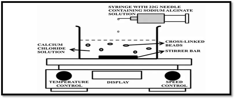
Figure No. 1 schematic representation of Ionotropic gelation technique
Emulsion gelation technique
Microbeads are made using the Emulsion Gelatin Method, which involves preparing a pre-gelation liquid using sodium alginate solution. The process entails adding light liquid paraffin, coconut oil, and olive oil to the polymer solution and homogenizing it for 20 minutes at 1000 rpm in order to stabilize the emulsion before adding an emulsifier. After the emulsion is formed, the medication is added. The room-temperature, gently stirred CaCl2 solution is filled with the bubble-free emulsion using a 21-gauge syringe needle. The emulsification gel beads are left in the solution for forty minutes before being separated and cleaned with distilled water.[8,9]
The schematic procedure is shown in Figure
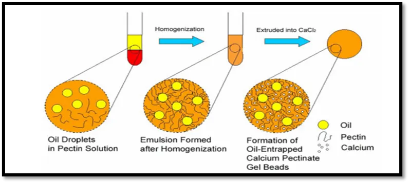
Figure No. 2 schematic representation of Emulsion gelation technique
Polyelectrolyte Complexation Technique
The complicated coacervation of polyelectrolytes with opposing charges, polycation and polyanion materials, and biocompatible and biodegradable alginate-chitosan microcapsules is another method for producing microbeads. These microcapsules are suitable for use in biomedical fields since they can be made under mild or even physiological conditions. In recent years, there has been increased interest in the use of alginate–chitosan microcapsules as drug-delivery vehicles for proteins and polypeptides. Depending on the pH, ionic strength, and polyion content, the liquid will separate with this method into a dense coacertive phase that contains the microbeads and a diluted equilibrium phase. For example, complex coacervation between alginic acid and chitosan was achieved by spraying the sodium alginate solution into the chitosan solution, producing strong microbeads that remained stable over a broad pH range.[10]
Polymers Used in Microbead Formulation
Since microbeads are composed of polymeric matrices, choosing the right polymers is essential to the drug delivery process in order to improve bioavailability. There are two types of polymers: natural and manmade. Proteins (gelatin, collagen, lectins) and polysaccharides (alginate, pectin’s, and chitosan) are examples of natural polymers. Polylactic acid (PLA), polycaprolactone (PCL), polyglycolic acid (PGA), and polylactic-glycolic acid (PLGA) are examples of biodegradable synthetic polymers; silicones and ethyl vinyl acetate (EVA) are examples of non-biodegradable polymers. [11]
The following benefits make natural polymers more popular than synthetic ones: a) natural abundance; b) inexpensive cost; c) renewable resource; d) less toxic; e) biocompatible and biodegradable. Artificial polymers are biocompatible and safe, but their main drawback is that the body breaks them down hydrolytically. They also have a propensity to migrate away from the injection site, which can cause embolism or other organ injury. Consequently, it has been discovered that natural polymers are a perfect option for drug delivery systems. [12]
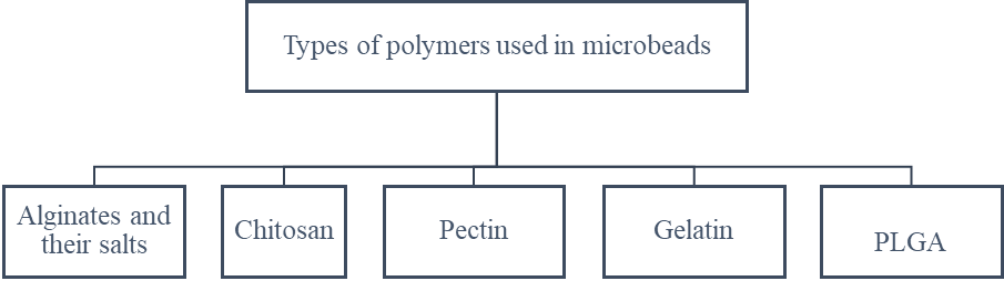
Figure No. 3 Types of polymers used in microbeads
Alginates and their salts
These are separated from brown sea wood by dissolving the alginic acid found in alginates in a diluted alkaline solution. The linear polymer of D-mannuronic acid and L-guluronic acid, which are organized as blocks in the polymeric chain, makes up alginic acid. When alginic acid and sodium hydroxide react, sodium alginate is produced. Naturally occurring alginate polymers have a number of advantageous characteristics, including their accessibility, capacity to produce pH-sensitive hydrogels, and lack of toxicity. Alginic acid's calcium and sodium salts are regarded as safe and biocompatible. There are more than 200 commercial grades available. Because alginates are naturally obtained, they contained a variety of contaminants, such as proteins, endotoxins, and heavy metals. These contaminants should be eliminated for parenteral use. Because of their low pyrogenicity, ultrapure grades of alginates can be utilized as implants in combination with other medications. [13,14]
Sodium alginate:
The ionic polymer polysaccharide sodium alginate is made up of hundreds of oxidized sugar "units" that have been bonded together. The six-membered rings that make up the repeating units include negatively charged CO2 groups. An oxygen atom connects the C-1 carbon atom of one ring in the polymer chain to the C-4 carbon atom of the subsequent ring. The combination of ionic CO2, side chains, and several-OH groups makes sodium alginate very hydrophilic. It is utilized in numerous processed foods as a thickening agent. [15,16]
Chitosan
A possible biopolymer for applications where bioactivity is desired is chitosan, a biodegradable and biocompatible amino-polysaccharide that is created by alkaline deacetylation of chitin, a naturally occurring component of shrimp or crab shells. 7 Cell encapsulation is extremely difficult because chitosan may only dissolve in an acidic solution. This problem can be resolved by combining a weak base, like beta-glycerophosphate, with the thermoresponsive properties of chitosan (BGP). This produces a physiologically pH solution at room temperature (RT) that undergoes the sol-gel transition when heated to body temperature. By making minor adjustments to the emulsification stage and employing the acidification process to replace internal gelation with thermal gelation, the emulsification method designed for the production of alginate microbeads was adapted to chitosan. [17]
Pectin
Pectin’s are fascinating since they are used in many different industries. For example, pectin’s are utilized as thickening and gelling agents in food. Because of their exceptional biocompatibility and customized pH sensitivity, which allow for targeted distribution, such as colon-specific, pectin’s are employed in pharmaceutical drug delivery applications. Pectin’s are safe for humans to consume on a daily basis. Additionally, pectin’s are believed to have anticancer properties, divalent metals with urine, facilitate the removal of dangerous substances, slow down the absorption of glucose, and lower blood cholesterol. Pectin gel microbeads have the potential to serve as drug delivery vehicles. The temporal profile of substance release from the beads can be changed by regulating the rate of gel disintegration.[18]
Gelatin
Microbeads made of biodegradable polymers like gelatin may be able to create the best controlled release systems. Microchannel emulsification can be used to encapsulate proteins or peptides because it requires very little energy to create the emulsion droplets. The possibility for creating monodisperse microbeads is demonstrated via microchannel emulsification. The average particle diameter decreased from 40.7 to 15.6 m while the gelatin bead preparation went on, while the relative standard deviations varied from 5.1% to 7.3%. Even though the gelatin beads significantly reduced after drying, the relative standard deviations stayed almost the same at 5.1% to 5.9%. [19]
PLGA
PLGA, a synthetic polymer that can contain proteins, viral or bacterial DNA, and anticancer drugs, is one of the most widely used drug delivery systems. PLGA is made by randomly polymerizing lactic acid and glycolic acid, which have high biodegradability and biocompatibility. PLGA is broken down in living things into glycolic and lactic acids, which are then broken down into carbon dioxide and water via the tricarboxylic acid cycle before being exhaled through the lungs. PLGA polymers with a high MW degrade more quickly because of their enhanced structural stability.[20]
Drug Release Mechanisms
Characterization of Microbeads
The characterization of any formulation plays a vital role in the drug development process.
Swelling Index
Microbeads with a known weight are suspended in 0.1N HCl at pH 1.2 (stimulated gastrointestinal fluids). Liquid globules adhered to the outside of microspheres are gathered using blotting paper and subsequently weighed using a microbalance. The enlarged microspheres are next dried in an oven set at 60 degrees Celsius for five hours, or until a consistent weight is shown, in order to eliminate any remaining water. [22]
Flow Properties of Microbeads
The angle of repose is measured using the fixed funnel method to determine the flow characteristics of produced microbeads. [23]
Surface Morphology
Scanning electron microscopy is used to detect the particle size distribution, surface topography, texture, and morphology of fractured or sectioned surfaces. Due to its ease of use and simplicity in sample preparation, SEM is the most popular technique for characterizing drug delivery systems. [24]
FT-IR Spectroscopy Measurements
Fourier Transform-Infrared spectroscopy is used to determine the degree of degradation of the polymeric matrix of the transport system. The surface of the microbeads is analyzed using alternating total reflectance (ATR) measurements. Using the ATR cell, the IR beam was repeatedly reflected to create the IR spectra of the sample's surface components. The ATR-FTIR provides information about the microspheres' surface proportions, depending on the manufacturing methods. [25]
Entrapment Efficiency
MBs are suspended in 0.1N HCl as part of the process. After filtering and diluting the solution to 10 microgram/mL, the absorbance of the mixture is measured using a UV-visible spectrophotometer against distilled water, which serves as a blank. [26]
Drying Rate of Microbeads
The drying rate is the amount of time MBs take to eliminate water in an incubator. Weighed beads are placed in open glass vials and kept in an incubator at 37°C. The beads are first weighed at short intervals-5, 10, and 15 minutes-up to 100 minutes, and then at larger intervals-200, 300, and 550 minutes. Until balance is reached, the beads are weighed. [27]
Loose Surface Crystal Study (LSC)
LSC, which exhibits immediate release in dissolving fluid, predicts the amount of medication on the outside of the microbeads. In phosphate buffer (pH 7.4), which mimics the dissolving medium, 100 mg of MBs are added and forcefully agitated.
Spectrophotometric analysis is used to determine how much medication has leached from the surface. [28]
In-Vitro Drug Release Studies
Drug-loaded microbeads are subjected to in-vitro drug release experiments utilizing USP type I equipment in various media. The surface of 900 millilitres of dissolving media is evenly covered with 100 milligrams of microbeads. At 37±0.5C and 50 rpm, the content is rotating. The drug dis solution study period is kept at ideal sink conditions. In order to simulate gastrointestinal transit, the pH of the dissolving medium is changed at various intervals. For two hours, the dissolving medium's pH of 1.2 is maintained using 0.01N HCl.
After a few hours, the medium is filtered via a membrane filter (0.45 mm). As soon as the sample is removed, the residue on the filter paper is put to the next medium, which is phosphate buffer pH 7.4. Additionally, samples (10 ml) are removed from another medium at specific intervals of 15 minutes. A UV-visible spectrophotometer is used to evaluate the samples and determine the medication release. After every withdrawal, the volume of dissolution medium is kept constant by substituting an equal volume of medium.[29]
Mucoadhesive Test
The mucoadhesive property of MBs is assessed using an in-vitro adhesion testing method. Cotton thread is used to gather freshly removed chicken intestinal mucosa fragments on glass slides. About 20 MBs are applied to each prepared glass slide, and the slides are then immediately suspended for the USP II tablet disintegration test. When the test apparatus is triggered, the sample is moved slowly up and down in the test fluid at 37C inside a vessel. At intervals of 30 minutes to 8 hours, the machine is halted, and the number of beads that remain on the mucosal surface is determined. [30]
Applications
Microbeads offer several advantages:
FUTURE PROSPECTIVE
Due to its unique characteristics, such as enhanced thermal, chemical, and physical properties, chemical stability, reduced irritation, improved drug release characteristics, prolonged drug launch, enhanced product sophistication and efficiency, the microbeads medication transport approach offers significant promise for numerous pharmaceutical applications in the foreseeable future. Another promising future direction is the use of microbeads in targeted and personalized medicine. By modifying the surface of microbeads with specific ligands, antibodies, peptides, or aptamers, drugs can be directed more accurately to diseased tissues or cells. This targeted approach helps deliver the medicine exactly where it is needed, improving treatment effectiveness while reducing unwanted effects on healthy tissues.
In addition, microbead formulations can be designed to suit individual patient needs. By considering patient-specific factors such as drug absorption, metabolism, and response, personalized microbead s can provide more consistent and effective therapy. With the growing emphasis on precision medicine, such customized drug delivery systems are expected to play an important role in achieving safer, more efficient, and patient-centered treatments.
CONCLUSION
Microbead-based drug delivery systems have gained considerable attention as a reliable and flexible strategy for achieving controlled and sustained drug release. Their spherical polymeric structure, along with the use of natural, semi-synthetic, and synthetic polymers, makes it possible to fine-tune drug release behavior, improve stability, and enhance overall therapeutic effectiveness. The availability of multiple formulation techniques—such as ionotropic gelation, emulsion gelation, and polyelectrolyte complexation—allows researchers to design microbeads with properties tailored to specific drugs and therapeutic needs. The choice of polymer is especially important, as it directly influences biocompatibility, biodegradability, and release characteristics. Natural polymers like alginate, chitosan, pectin, and gelatin are widely preferred because they are safe, biodegradable, and well accepted by the body, whereas synthetic polymers such as PLGA offer greater control over degradation and mechanical strength. Drug release from microbeads occurs through a combination of mechanisms, including diffusion, degradation, erosion, and swelling, which together ensure a more consistent and predictable release of the drug over time. Thorough characterization of microbeads—covering parameters such as swelling behavior, flow properties, surface morphology, entrapment efficiency, and in-vitro drug release—is essential to confirm formulation quality and performance. Owing to these well-defined properties, microbeads have found applications in a wide range of drug delivery routes, including oral, topical, mucoadhesive, vaccine, and targeted delivery systems, particularly for drugs that are sensitive or require prolonged action. Although microbeads offer many advantages, certain challenges remain, including issues related to polymer purity, long-term biocompatibility, environmental concerns, regulatory requirements, and large-scale manufacturing. Even so, continuous progress in polymer science, formulation methods, and targeted delivery approaches is steadily overcoming these limitations. Overall, microbeads represent a promising and evolving drug delivery platform with strong potential for improving therapeutic outcomes and patient care in the future.
REFERENCES


Ankita Jaypurkar, S. Bonde, P. Keche, Dr. S. Dighade, A Comprehensive Review on Microbeads as Advanced Drug Delivery: Preparation Techniques, Release Mechanisms, Applications, And Future Prospects, Int. J. of Pharm. Sci., 2026, Vol 4, Issue 6, 7026-7035, https://doi.org/10.5281/zenodo.20960862
 10.5281/zenodo.20960862