

We use cookies to ensure our website works properly and to personalise your experience. Cookies policy
Department of Pharmaceutics, IVM'S Krishnarao Bhegade Institute of Pharmaceutical Education and Research, Talegaon Dabhade, Pune 410506.
Nanoparticles have emerged as a promising platform in pharmaceutical sciences due to their unique physicochemical properties and ability to enhance the delivery of therapeutic agents. Their nanoscale size, large surface area, and tunable surface characteristics enable improved drug solubility, bioavailability, stability, and targeted delivery. Nanoparticle-based drug delivery systems have gained significant attention for overcoming the limitations of conventional dosage forms, including poor pharmacokinetics, non-specific distribution, and undesirable side effects. Various types of nanoparticles, such as polymeric nanoparticles, lipid-based nanoparticles, metallic nanoparticles, magnetic nanoparticles, dendrimers, and mesoporous silica nanoparticles, have been extensively investigated for pharmaceutical applications. The synthesis of nanoparticles can be achieved through physical, chemical, and biological approaches, while advanced characterization techniques are essential for evaluating their size, morphology, surface charge, crystallinity, and stability. Nanoparticles have demonstrated remarkable potential in targeted drug delivery, cancer therapy, gene delivery, vaccine development, antimicrobial therapy, diagnostic imaging, and theranostic applications. Despite these advantages, challenges related to large-scale manufacturing, regulatory approval, long-term safety, and biological interactions continue to limit their widespread clinical translation. Recent advancements in artificial intelligence, personalized nanomedicine, biomimetic nanocarriers, and next-generation lipid nanoparticles are opening new avenues for the development of more effective and safer nanotherapeutics. This review provides a comprehensive overview of nanoparticle synthesis methods, characterization techniques, pharmaceutical applications, regulatory considerations, current challenges, and future perspectives. Continued progress in nanotechnology and pharmaceutical engineering is expected to accelerate the successful integration of nanoparticle-based systems into modern healthcare and precision medicine.
1.1 Definition and Fundamental Concepts
Nanoparticles are defined as particulate materials with at least one dimension in the nanoscale range of 1 to 1000 nanometers (nm). In the context of pharmaceutical sciences, nanoparticles typically refer to colloidal systems with sizes ranging from 10 to 1000 nm that can serve as carriers for therapeutic agents including small molecule drugs, proteins, peptides, nucleic acids, and imaging agents. The term "nanoparticle" encompasses a broad spectrum of carrier systems including polymeric nanoparticles, lipid nanoparticles, metallic nanoparticles, dendrimers, micelles, and hybrid systems combining multiple materials.[1] The unique properties of nanoparticles arise from their nanoscale dimensions, which confer distinct physicochemical characteristics compared to their bulk counterparts. The extremely high surface area-to-volume ratio of nanoparticles enables enhanced interaction with biological membranes and cellular components. Quantum effects become apparent at the nanoscale, particularly for metallic nanoparticles, leading to unique optical, electronic, and magnetic properties. The small size allows nanoparticles to penetrate biological barriers that are impermeable to larger particles, including the blood-brain barrier, tight junctions of the intestinal epithelium, and tumor vasculature through the enhanced permeability and retention (EPR) effect.[2]
1.2 Historical Evolution and Milestones
The concept of using colloidal particles for drug delivery dates back to the early 20th century, but the modern era of pharmaceutical nanoparticles began in the 1970s with the development of liposomes by Gregoriadis and colleagues. The first FDA-approved nanomedicine, Doxil (doxorubicin liposomes), was introduced in 1995, marking a pivotal milestone in clinical translation. Since then, the field has experienced exponential growth, with over 100 nanomedicine products receiving regulatory approval worldwide by 2025. The evolution of pharmaceutical nanoparticles can be categorized into several generations:
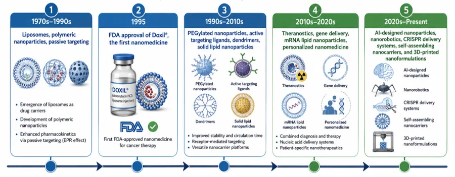
Figure 1. Historical evolution of nanoparticle-based drug delivery systems from conventional carrier systems to advanced intelligent nanomedicines.
1.3 Significance and Advantages in Pharmaceutical Sciences
The incorporation of nanoparticles into pharmaceutical formulations offers numerous compelling advantages that address critical limitations of conventional drug delivery approaches:
1.4 Classification of Pharmaceutical Nanoparticles
Pharmaceutical nanoparticles can be classified based on their composition, structure, and origin:
A. Organic Nanoparticles:
B. Inorganic Nanoparticles:
C. Hybrid Nanoparticles:
2. SYNTHESIS OF NANOPARTICLES
2.1 Top-Down Approaches
2.1.1 Ball Milling
Ball milling is a mechanical attrition technique where bulk material is ground in a rotating cylindrical container filled with milling media (balls) under controlled conditions. The impact and shear forces generated during milling break down the material into nanoparticles. Parameters such as milling speed, duration, ball-to-powder ratio, and use of surfactants or stabilizers significantly influence the particle size and morphology. High-energy ball milling can produce nanoparticles in the range of 10-100 nm, though the size distribution is often broad. The technique is scalable and cost-effective but may introduce contamination from the milling media and lacks precise control over particle characteristics.[8]
2.1.2 High-Pressure Homogenization
High-pressure homogenization forces a coarse suspension through a small orifice under high pressure (typically 100-2000 bar), creating intense shear, cavitation, and impact forces that reduce particle size. This technique is widely used for producing nanosuspensions of poorly water-soluble drugs. Multiple homogenization cycles are typically required to achieve the desired particle size. The method is suitable for thermolabile compounds as it operates at ambient or slightly elevated temperatures. However, the process can be energy-intensive and may require high drug concentrations.[9]
2.1.3 Media Milling (Nanocrystal Technology)
Media milling involves grinding drug particles in a liquid medium using milling beads (typically 0.2-0.5 mm) in a stirred media mill. Stabilizers such as surfactants or polymers are added to prevent agglomeration. This technique has been successfully commercialized for producing nanocrystal formulations (e.g., Rapamune, Emend). The process can achieve particle sizes below 400 nm and is scalable for industrial production. Challenges include potential contamination from milling media and the need for extensive washing steps.[10]
2.2 Bottom-Up Approaches
2.2.3 Supercritical Fluid Technology
2.2.4 Coacervation and Ionic Gelation Methods
Coacervation Method: Coacervation involves the phase separation of a polymer solution into a polymer-rich phase (coacervate) and a polymer-poor phase. In simple coacervation, a non-solvent or change in temperature causes the polymer to separate from solution. In complex coacervation, two oppositely charged polymers interact to form a complex that separates as a coacervate phase. The drug is dispersed in the polymer solution, and coacervation is induced, causing the polymer to encapsulate the drug particles. The coacervate droplets are then hardened by cross-linking, temperature change, or solvent removal. This method is particularly useful for natural polymers like gelatin, alginate, and chitosan. Ionic Gelation Method: Ionic gelation is a mild, aqueous-based method suitable for encapsulating sensitive biomolecules. In this technique, a polyelectrolyte solution (such as sodium alginate or chitosan) containing the drug is added dropwise to a solution containing multivalent counterions (such as calcium chloride for alginate or tripolyphosphate for chitosan). The electrostatic interaction between the polyelectrolyte and counterions causes gelation and formation of nanoparticles. The method is simple, does not require organic solvents or high temperatures, and is biocompatible. However, the particle size is relatively large (typically 200-1000 nm) and the drug loading may be limited.[16]
2.3 Green Synthesis Approaches (Emerging Trend 2023-2026)
2.3.1 Plant-Mediated Green Synthesis
Plant-mediated synthesis utilizes aqueous extracts of plant parts (leaves, roots, fruits, seeds, bark) as reducing and stabilizing agents for the production of metallic nanoparticles. The plant extracts contain a rich diversity of phytochemicals including polyphenols, flavonoids, terpenoids, alkaloids, and proteins that serve as both reducing agents (converting metal ions to metal nanoparticles) and capping agents (stabilizing the formed nanoparticles and preventing aggregation).
Recent studies have demonstrated the successful green synthesis of gold and silver nanoparticles using crude extracts of Aconitum violaceum, a medicinal plant from the Himalayan region. The synthesized AuNPs and AgNPs exhibited spherical and triangular morphologies with average sizes below 100 nm. The phytochemical screening revealed the presence of alkaloids, flavonoids, terpenoids, saponins, and anthraquinones in the extract, with alkaloids and flavonoids identified as the primary reducing and capping agents. The green-synthesized nanoparticles demonstrated effective antibacterial activities with minimum inhibitory concentrations (MICs) of 95 and 70 microg/mL against Lactobacillus acidophilus and 90 and 65 microg/mL against Escherichia coli for AuNPs and AgNPs, respectively. The enhanced antibacterial activity compared to the crude extract was attributed to the synergistic effect of phytochemicals and the nanoparticles themselves.
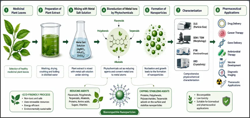
Figure 2. Green Synthesis of Nanoparticles Using Plant Extracts
2.3.2 Microbial-Mediated Green Synthesis
Microorganisms including bacteria, fungi, actinomycetes, and algae have been exploited for the biosynthesis of nanoparticles. The mechanism involves enzymatic reduction of metal ions by microbial cellular components or secreted biomolecules. Intracellular synthesis occurs within the microbial cell, while extracellular synthesis utilizes enzymes or metabolites secreted into the culture medium.
Bacteria such as Pseudomonas, Bacillus, and Escherichia coli can synthesize nanoparticles through enzymatic reduction of metal ions. This method offers rapid growth, easy cultivation, and scalability, although purification is required to remove endotoxins.
Fungi including Aspergillus, Fusarium, and Penicillium species are widely used for nanoparticle synthesis due to their high enzyme production and metal tolerance. Fungal synthesis often provides higher yields and facilitates easier downstream processing.
Marine and freshwater algae can produce nanoparticles using naturally occurring polysaccharides, proteins, and pigments as reducing agents. This eco-friendly approach generates biocompatible nanoparticles suitable for pharmaceutical and biomedical applications.
2.3.3 Biopolymer-Mediated Synthesis
Biopolymers such as chitosan, alginate, starch, and cellulose derivatives can serve as both reducing and stabilizing agents for nanoparticle synthesis. Chitosan, a cationic polysaccharide derived from chitin, has been extensively studied for silver and gold nanoparticle synthesis due to its biocompatibility, antimicrobial properties, and ability to form complexes with metal ions. The amino and hydroxyl groups of chitosan participate in the reduction of metal ions and stabilization of nanoparticles through coordination and electrostatic interactions.[19,20]
2.4 Comparison of Synthesis Methods
The selection of an appropriate synthesis method depends on various factors including the nature of the drug, desired particle characteristics, scale of production, regulatory requirements, and cost considerations. Table 1 presents a comprehensive comparison of the major synthesis methods for pharmaceutical nanoparticles.[21]
Table No. 1: Comparison of all Synthesis Methods
|
Method |
Particle Size Range |
Advantages |
Limitations |
|
Ball Milling |
10-1000 nm |
Scalable, cost-effective |
Broad size distribution, contamination |
|
High-Pressure Homogenization |
100-1000 nm |
Suitable for thermolabile compounds |
Energy-intensive, high drug concentration needed |
|
Media Milling (Nanocrystal) |
<400 nm |
Commercially proven (Rapamune) |
Media contamination, extensive washing |
|
Solvent Evaporation |
100-1000 nm |
Versatile, high encapsulation |
Organic solvent residue, complex process |
|
Nanoprecipitation |
100-300 nm |
Simple, low energy, small particles |
Limited to water-miscible solvents |
|
Single Emulsion (O/W) |
100-1000 nm |
Versatile, widely used |
Organic solvents, limited to hydrophobic drugs |
|
Double Emulsion (W/O/W) |
200-1000 nm |
Both hydrophilic and hydrophobic drugs |
Lower efficiency, broader size distribution |
|
Supercritical Fluid Technology |
100-500 nm |
Solvent-free, environmentally friendly |
High equipment cost, limited solubility |
|
Coacervation |
200-1000 nm |
Mild conditions, natural polymers |
Complex process control |
|
Ionic Gelation |
200-1000 nm |
Simple, aqueous, biocompatible |
Large particles, limited loading |
|
Green Synthesis (Plant/Microbial) |
10-100 nm |
Sustainable, biocompatible, cost-effective |
Batch variability, limited mechanistic understanding |
2.5 Recent Advances in Synthesis (2023-2026)
2.5.1 Microfluidic Synthesis
Microfluidic technology has emerged as a powerful platform for controlled nanoparticle synthesis, offering precise control over mixing, reaction conditions, and residence time. The laminar flow regime in microchannels enables rapid and uniform mixing of reagents, leading to narrow particle size distributions. Microfluidic devices can be designed with various geometries (T-junction, Y-junction, flow-focusing) to control the mixing pattern and nanoparticle formation. This technology is particularly valuable for the production of lipid nanoparticles for mRNA delivery, where precise control over lipid composition and particle size is critical for vaccine efficacy. Recent advances include the development of 3D-printed microfluidic devices and continuous flow systems for scalable production.
2.5.2 3D Printing of Nanoparticles
Three-dimensional printing technologies have been adapted for nanoparticle fabrication, enabling the creation of complex nanostructures with precise spatial control. Techniques such as two-photon polymerization and direct laser writing can produce nanoparticles and nanostructures with resolutions below 100 nm. 3D printing allows for the fabrication of personalized nanoparticle-based drug delivery systems with tailored release profiles and geometries optimized for specific administration routes.
2.5.3 AI-Driven Nanoparticle Design
Artificial intelligence and machine learning algorithms are being increasingly applied to predict and optimize nanoparticle properties. Machine learning models trained on large datasets of nanoparticle formulations can predict the optimal synthesis parameters for desired particle characteristics, significantly reducing the time and cost of formulation development. AI-driven design is particularly promising for lipid nanoparticles, where the vast chemical space of lipid combinations makes experimental screening impractical.[22,23]
3. CHARACTERIZATION OF NANOPARTICLES
Characterization is a crucial step in the development of nanoparticle-based drug delivery systems because the physicochemical properties of nanoparticles directly influence their biological performance, therapeutic efficacy, stability, and safety. Comprehensive characterization helps in understanding particle size, morphology, surface charge, crystallinity, chemical composition, thermal behavior, drug loading capacity, and release characteristics. Regulatory agencies also require detailed characterization data to ensure product quality, reproducibility, and clinical safety.
3.1 Particle Size Analysis
Particle size is one of the most important parameters influencing nanoparticle behavior. It affects cellular uptake, biodistribution, circulation time, drug release kinetics, and targeting efficiency. Nanoparticles with smaller particle sizes generally exhibit improved cellular internalization and enhanced permeability through biological barriers.
Particle size distribution is commonly expressed in terms of mean particle diameter and polydispersity index (PDI). A narrow size distribution is desirable because it ensures formulation uniformity and reproducibility.
Dynamic Light Scattering is the most widely used technique for measuring nanoparticle size in suspension. The method is based on the analysis of fluctuations in scattered light caused by the Brownian motion of particles. The diffusion coefficient obtained from these fluctuations is used to calculate the hydrodynamic diameter of nanoparticles.
Advantages:
3.2 Polydispersity Index (PDI)
PDI is a measure of particle size distribution uniformity. It indicates the degree of heterogeneity within a nanoparticle formulation.Lower PDI values indicate better formulation quality and stability.[25]
Table no.2: Polydispersity Index (PDI)
|
PDI Value |
Interpretation |
|
< 0.1 |
Highly monodisperse system |
|
0.1-0.3 |
Acceptable size distribution |
|
> 0.3 |
Broad size distribution |
|
> 0.7 |
Highly polydisperse system |
3.3 Zeta Potential Analysis
Zeta potential is a measure of the electrical charge present on the nanoparticle surface. It is an important indicator of colloidal stability because surface charges generate electrostatic repulsion between particles, preventing aggregation.The zeta potential also influences interactions with biological membranes, protein adsorption, cellular uptake, and biodistribution.[26]
Table no.3: Polydispersity Index (PDI)
|
Zeta Potential (mV) |
Stability |
|
Above +30 |
Highly stable |
|
Between +20 and +30 |
Moderately stable |
|
Between -20 and +20 |
Limited stability |
|
Below -30 |
Highly stable |
3.4 Morphological Characterization
Morphological analysis provides information regarding nanoparticle shape, surface texture, aggregation state, and structural organization. Morphology significantly affects drug loading, release behavior, and biological interactions.
SEM uses a focused electron beam to scan the sample surface and generate high-resolution images.
Applications:
Advantages:
TEM involves transmission of electrons through a thin sample to obtain highly detailed images.
Applications:
Advantages:
AFM employs a nanoscale probe that scans the surface of nanoparticles to generate three-dimensional topographical images.
Applications:
Advantages:
3.5 Structural Characterization
Structural characterization determines crystalline structure, phase composition, and molecular organization of nanoparticles.
XRD is used to identify crystalline phases and evaluate crystal structure. The technique measures diffraction patterns generated when X-rays interact with crystalline materials.The Debye-Scherrer equation is commonly used to estimate crystallite size from XRD peak broadening.
Applications:
Advantages:
3.6 Chemical Characterization
Chemical characterization identifies functional groups, molecular interactions, and surface chemistry.
FTIR detects absorption of infrared radiation by molecular bonds, generating characteristic spectra for specific functional groups.
Applications:
Advantages:
Raman spectroscopy provides molecular information based on inelastic scattering of light.
Applications:
Advantages:
3.7 Surface Area Analysis
Surface area is a critical parameter influencing drug loading, dissolution rate, and adsorption capacity.
BET analysis determines specific surface area by measuring gas adsorption onto the nanoparticle surface. Higher surface area generally results in improved drug loading capacity and enhanced dissolution.
Applications:
4. PHARMACEUTICAL APPLICATIONS OF NANOPARTICLES
Nanoparticles have revolutionized modern pharmaceutical sciences by providing innovative solutions to challenges associated with conventional drug delivery systems. Their unique physicochemical properties, including small particle size, large surface area-to-volume ratio, tunable surface characteristics, and ability to encapsulate a wide range of therapeutic agents, make them highly effective carriers for pharmaceutical applications. Nanoparticles can improve drug solubility, enhance bioavailability, prolong circulation time, facilitate controlled release, and enable site-specific drug delivery. As a result, nanoparticle-based systems have found widespread applications in drug delivery, cancer therapy, gene therapy, vaccine development, antimicrobial treatment, diagnostic imaging, and theranostics.[30]
4.1 Targeted Drug Delivery
Targeted drug delivery is a major application of nanoparticles that improves therapeutic efficacy while reducing systemic toxicity. Drug-loaded nanoparticles circulate in the bloodstream and accumulate at diseased tissues or tumors through passive or active targeting mechanisms. After binding to target cells, they are internalized by endocytosis and release the drug in a controlled manner, resulting in enhanced treatment outcomes and reduced damage to healthy tissues.
Passive targeting relies on the Enhanced Permeability and Retention (EPR) effect observed in tumor tissues and inflamed sites. Tumors possess abnormal blood vessels with large fenestrations and poor lymphatic drainage, allowing nanoparticles to preferentially accumulate within tumor tissues.
Advantages of passive targeting include:
Active targeting involves surface modification of nanoparticles with ligands such as antibodies, peptides, folic acid, aptamers, or transferrin. These ligands specifically recognize receptors overexpressed on target cells, facilitating receptor-mediated uptake.
Examples include:
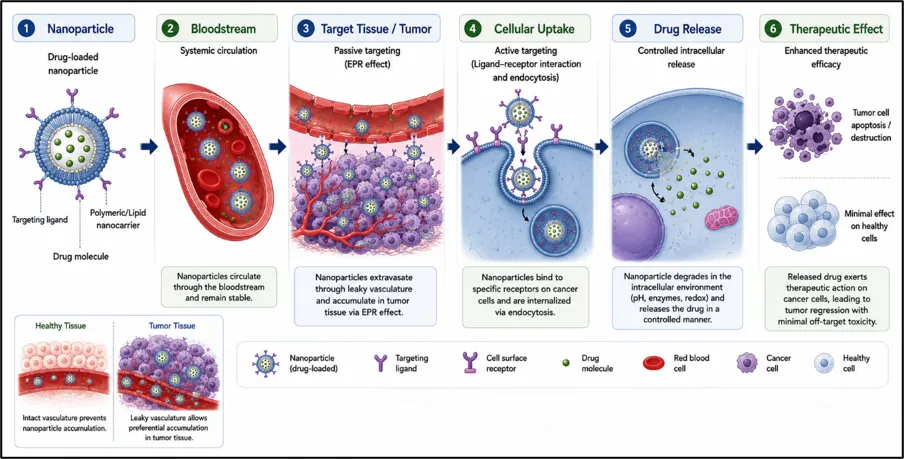
Figure 3. Schematic of Nanoparticle-Mediated Targeted Drug Delivery.
4.2 Cancer Therapy
Cancer remains one of the leading causes of mortality worldwide. Conventional chemotherapy is often associated with severe adverse effects due to non-specific distribution of anticancer drugs. Nanoparticle-based drug delivery systems have emerged as effective strategies for improving cancer treatment.
Nanoparticles can encapsulate chemotherapeutic agents and deliver them selectively to tumor tissues. This approach improves drug accumulation at the tumor site while reducing systemic exposure.
Polymeric nanoparticles composed of PLGA, PLA, and chitosan have been extensively investigated for delivering anticancer drugs such as doxorubicin, paclitaxel, and docetaxel.
Benefits include:
Liposomes and lipid nanoparticles have demonstrated considerable success in cancer treatment.
Examples:
4.3 Gene Delivery
Gene therapy involves the delivery of genetic material to modify or regulate gene expression for therapeutic purposes. Efficient gene delivery remains a major challenge because nucleic acids are susceptible to enzymatic degradation and exhibit poor cellular uptake. Nanoparticles serve as non-viral vectors capable of protecting genetic material and facilitating intracellular delivery.
Therapeutic agents delivered include:
Advantages of nanoparticle-mediated gene delivery include:
4.4 Vaccine Delivery
Nanoparticles have transformed vaccine development by enhancing antigen stability, immune response, and targeted delivery to immune cells. Traditional vaccines often require adjuvants to stimulate adequate immune responses. Nanoparticles can function as both carriers and adjuvants, improving vaccine efficacy.The recent success of mRNA vaccines against COVID-19 demonstrated the tremendous potential of lipid nanoparticles in vaccine delivery.
Advantages of nanoparticle-based vaccines include:
Types of nanoparticle vaccine carriers include:
4.5 Antimicrobial Applications
The increasing prevalence of antimicrobial resistance has created an urgent need for novel therapeutic approaches. Nanoparticles possess intrinsic antimicrobial activity and can enhance the effectiveness of existing antimicrobial agents.
Silver nanoparticles are among the most extensively studied antimicrobial nanomaterials.
Mechanisms of action include:
Silver nanoparticles exhibit activity against:
Nanoparticles can improve antibiotic therapy by:
5. REGULATORY ASPECTS AND SAFETY CONSIDERATIONS
The rapid growth of nanotechnology has led to the development of numerous nanoparticle-based pharmaceutical products. Despite their therapeutic advantages, nanoparticles pose unique regulatory and safety challenges due to their small size, high surface reactivity, and complex biological interactions.[40] Therefore, thorough evaluation of quality, safety, efficacy, and manufacturing consistency is essential before clinical use. Regulatory agencies such as the FDA and EMA assess nanomedicines under existing drug and biologic regulations using a case-by-case approach. Comprehensive characterization, including particle size, surface charge, morphology, drug loading, and stability, is required to ensure product quality and reproducibility. Manufacturing processes must also comply with Good Manufacturing Practices (GMP). Safety assessment is a critical aspect of nanoparticle development. Extensive preclinical studies are conducted to evaluate cytotoxicity, genotoxicity, immunotoxicity, biodistribution, and long-term safety. Since nanoparticles may accumulate in organs such as the liver, spleen, lungs, and kidneys, their potential to induce oxidative stress, inflammation, or tissue damage must be carefully examined.[41]
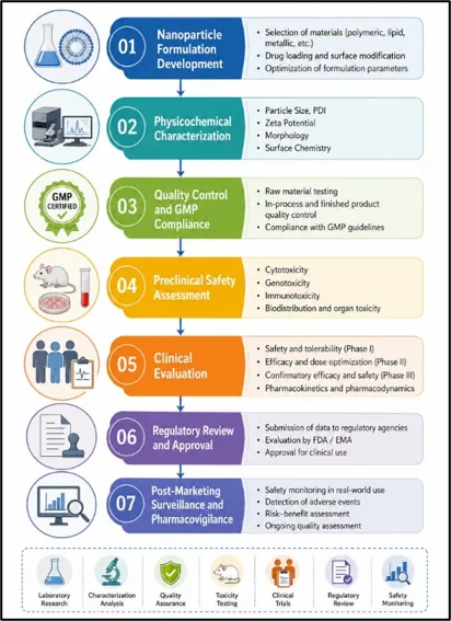
Figure 4. Regulatory and Safety Assessment Framework for Nanoparticle-Based Drug Delivery Systems
Clinical studies further assess pharmacokinetics, therapeutic efficacy, and adverse effects, while post-marketing surveillance monitors long-term safety. Although several nanomedicines have gained regulatory approval, challenges related to standardization, toxicity evaluation, and regulatory harmonization remain. Continued collaboration among researchers, industry, and regulatory authorities is essential for the safe and effective advancement of nanoparticle-based therapeutics.[42]
6. CHALLENGES AND FUTURE PERSPECTIVES
7. CONCLUSION
Nanoparticles have emerged as a transformative platform in pharmaceutical sciences, offering significant advantages over conventional drug delivery systems through improved solubility, bioavailability, stability, controlled release, and targeted delivery. A wide range of synthesis approaches, including physical, chemical, and green synthesis methods, enable the development of nanoparticles with tailored physicochemical properties for specific therapeutic applications. Comprehensive characterization techniques are essential for ensuring product quality, safety, and reproducibility. The application of nanoparticles in targeted drug delivery, cancer therapy, gene delivery, vaccine development, antimicrobial treatment, diagnostic imaging, and theranostic systems highlights their immense potential in modern healthcare. Recent advances in lipid nanoparticles, particularly for nucleic acid delivery, have further accelerated the clinical translation of nanomedicine and demonstrated the versatility of nanoparticle-based platforms. Green synthesis approaches have also gained considerable attention due to their sustainability, cost-effectiveness, and improved biocompatibility. Despite these promising developments, challenges related to large-scale manufacturing, regulatory approval, long-term safety, and targeting efficiency continue to limit broader clinical implementation. Addressing these limitations through improved characterization methods, standardized regulatory frameworks, and advanced manufacturing technologies will be crucial for future success. Emerging innovations such as artificial intelligence-driven nanoparticle design, personalized nanomedicine, biomimetic nanocarriers, and nanoparticle-mediated gene editing are expected to further expand the scope of nanotechnology in healthcare. Overall, nanoparticles represent a rapidly evolving field with substantial potential to advance precision medicine and improve therapeutic outcomes across a wide range of diseases.
REFERENCES

Daiwashala Pawar, Amol Ratke, Sneha Wadu, Ruchira Medankar, A Review on Nanoparticles: Synthesis, Characterisation, and Applications in Pharmaceutical Sciences, Int. J. of Pharm. Sci., 2026, Vol 4, Issue 7, 1184-1203. https://doi.org/10.5281/zenodo.21222368
 10.5281/zenodo.21222368