

We use cookies to ensure our website works properly and to personalise your experience. Cookies policy
Karnataka College of Pharmacy.
Solid lipid nanoparticles have gained considerable attention as a promising drug delivery system due to their ability to improve the bioavailability, stability, and therapeutic efficacy of poorly soluble drugs. Composed of biocompatible and biodegradable lipids, solid lipid nanoparticles provide controlled and sustained drug release while minimizing toxicity. This review highlights the structure, classification, preparation techniques, characterization methods, and evaluation parameters of slns. The advantages, limitations, and diverse pharmaceutical applications of slns across various routes of administration are also discussed. Overall, slns represent a versatile and efficient nanocarrier platform with significant potential for targeted and controlled drug delivery in modern pharmaceutical development.
Ulcerative colitis, a type of inflammatory bowel disease, primarily targets the mucous membrane layer of the colon, leading to various distressing symptoms such as abdominal discomfort, diarrhoea, rectal bleeding, diminished appetite, and weight loss [1]. The impact and prevalence of ulcerative colitis are considerable, affecting millions of individuals globally [2]. The presence of the intestinal epithelial barrier presents a challenge for the bidirectional communication between gut microbes and the host [3].
The progression of uc may evolve through a sequence of inflammation, typical hyperplasia, and subsequent cancerization, culminating in the development of colitis- associated cancer [4]. In recent years, the incidence of uc has been rising, but its specific pathogenesis, likely influenced by genetic factors, alterations in intestinal microbiota, and inflammatory damage, remains unclear [5].
It has been shown that the intestinal microbiota is one of the important biological indicators of the development of uc [6]. Pathogenic bacteria are considered to negatively impact the status of patients with uc in multiple ways [7]. Recent evidence revealed that the disease severity in patients with uc is closely related to the pathogenic bacteria [8]. Conversely, probiotic microbes alleviate uc symptoms by modulating the host inflammatory response [9]. Probiotic microbes can regulate immune homeostasis against uc patients through their own or secreted bioactive substances [10].
The presence of the intestinal epithelial barrier presents a challenge for the bidirectional communication between gut microbes and the host [11]. Therefore, most microbe-host interactions likely occur via characteristic derivatives secreted by bacteria. Extracellular vesicles are derivatives of bacteria that consist of a membrane bilayer nanostructure and that are released during growth. Extracellular vesicles contain a range of parental bacterial components [12].
Recent work has highlighted the versatility of probiotic-derived Extracellular vesicles in regulating intestinal homeostasis [13]. A significant challenge in treating uc is identifying patients at higher risk of disease progression who may benefit from early intensive treatment while avoiding overtreatment of low-risk patients. Current treatment decisions rely on cross-sectional disease activity, which may not accurately reflect disease progression or complications over time.
The international inflammatory bowel disease research organization’s severity index does not fully overcome these issues, suggesting that clinical characteristics alone are insufficient for accurate risk stratification. Therefore, objective biomarkers are essential for advancing precision medicine In uc. Multi-omics studies integrating clinical data, biopsies, and blood biomarkers show great potential in identifying novel diagnostic and prognostic markers [14].
Additionally, personalized therapies that utilize biomarkers to predict treatment response and adverse drug events are urgently needed. Although the concept of histologic healing as a marker of remission is widely accepted, the stride-ii consensus does not consider it a therapeutic goal [15]. Ongoing trials, such as verdict (nct04259138), are investigating the role of corticosteroid-free symptomatic remission, endoscopic remission, and histologic healing in treatment algorithms [16].
Various environmental factors can cause or prevent uc, and cigarette smoking is one of the most consistent. A meta-analysis found that smoking is related to a decreased risk of developing ulcerative colitis than not smoking [17,18]. The diagnosis of uc is established on clinical symptoms supported by objective results from endoscopic or histological testing. On October 12, 2023, the us food and drug administration approved velsipity, a revolutionary ulcerative colitis treatment derived from Etrasimod that works by binding to the body’s receptors, particularly the s1p receptors 1, 4, and 5.
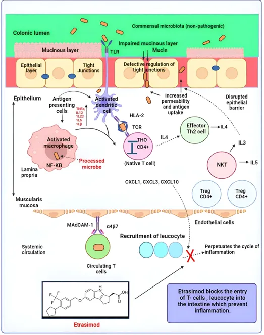
Figure No. 1: Pathophysiology of Ulcerative Colitis
This interaction is critical in restricting the movement of particular immune cells, mainly lymphocytes, which reduces their presence in the bloodstream. In the frame work of controlling ulcerative colitis, Etrasimod successfully inhibits the escape of these immune cells into the colon. The article details the newly approved drug for treating uc, including its structure, synthetic scheme, mechanism of action, pharmacokinetic and pharmacodynamic data, and adverse reactions, providing comprehensive information on the drug’s effectiveness.
Pathophysiology:
The main pathway for ulcerative colitis pathophysiology is described in the following parts:
Epithelial Barrier
The epithelial barrier acts as the first line of Défense in the mucosal immune system by protecting host cells from pathogens and producing antibacterial peptides. In ulcerative colitis, impaired synthesis and sulfation of the colonic mucin muc2 weakens this barrier, leading to tight-junction dysfunction and increased intestinal permeability, which allows bacteria to more easily penetrate the mucosa [19,20].
Commensal Microflora
In a healthy state, the intestinal immune system strikes a balance between tolerating beneficial flora and food-related antigens while appropriately responding to harmful gut pathogens. Uc appears to arise from a disturbance in this balanced connection between the body’s mucosal immunity and the gastrointestinal flora. This disturbance creates an abnormal immune reaction directed against typically harmless, non-pathogenic bacteria in the gut [21,22].
Antigen Recognition
Antigens trigger the immune system’s natural response by interacting with both macrophages and dendritic cells. Dendritic cells extend their dendrites across the epithelial cell layer, connecting between intestinal epithelial cells to collect bacteria and other material from the intestinal lumen [23]. In individuals affected by uc, there’s an increase in both the quantity and activation of dendritic cells, with a heightened ability to stimulate immune reactions. These elevated circulating dendritic cell levels correspond to the disease’s activity, indicating their crucial role in initiating and sustaining inflammation [24].
Dysregulation Of Immune Responses
The delicate balance involving regulator and effector t-cells, particularly t-helper (th)1, th2, and th17 cells, is altered in the mucosal lining of ulcerative colitis patients20. This disturbance con tributes to the disease process. Interleukin 13 is capable of triggering a positive feedback loop affecting natural killer t-cells, thereby intensifying tissue damage. Both interleukin 13 and natural killer t-cells appear to play pivotal roles in the development of uc [25].
Leucocyte Recruitment
The secretion of chemo attractors like cxcl8, which is elevated in individuals with ulcerative colitis, It plays a crucial role in attracting circulating white blood cells from bloodstream to inflamed mucosal tissue. This recruitment is vital in intensifying the inflammatory response [26].
Anatomy And Physiology of The Colon:
Anatomy Of the Colon:
The colon is the major part of the large intestine and extends from the ileocecal junction to the rectum, with a total length of about 1.5 meters. It is anatomically divided into four main segments: the ascending colon, transverse colon, descending colon, and sigmoid colon.
the colon ends at the rectum, which stores feces before elimination. Structurally, the colon wall consists of four layers: the mucosa, submucosa, muscularis externa, and serosa.
The mucosa of the colon is lined by simple columnar epithelium rich in goblet cells, which secrete mucus to lubricate the intestinal contents and protect the epithelial surface. Unlike the small intestine, the colon lacks villi but contains numerous intestinal glands (crypts of Lieberkühn). The muscularis externa is arranged into an inner circular layer and an outer longitudinal layer, which is concentrated into three distinct bands called teniae coli. These structural features give the colon its characteristic haustrated appearance [27].
Physiology Of The Colon:
The primary physiological functions of the colon include absorption, secretion, fermentation, and faecal formation. The colon absorbs water and electrolytes, particularly sodium and chloride, thereby concentrating the intestinal contents into semi-solid feces. It also absorbs short-chain fatty acids produced by bacterial fermentation, which serve as an important energy source for colonic epithelial cells.
The colon houses a dense and diverse population of microorganisms that play a vital role in digestion and immune regulation. These bacteria ferment undigested carbohydrates, synthesize certain vitamins such as vitamin k and some b vitamins, and help maintain mucosal immunity. Mucus secretion by goblet cells protects the epithelium from mechanical damage and bacterial invasion [28]. Colonic motility is characterized by slow segmental contractions that facilitate absorption and periodic mass movements that propel fecal matter toward the rectum. Neural control is mediated by the enteric nervous system, with modulation from the autonomic nervous system. Overall, the colon plays a crucial role in maintaining fluid balance, supporting gut microbiota, and ensuring proper elimination of waste from the body [29].
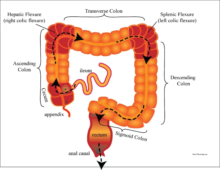
Figure No. 2: Anatomy And Physiology of The Colon
Factors Affecting Ulcerative Colitis: [30,31,32].
Ulcerative colitis is a chronic inflammatory bowel disease influenced by multiple interacting factors. The main factors affecting uc are:
1. Genetic Factors
A family history of ulcerative colitis or other inflammatory bowel diseases increases the risk, indicating a genetic predisposition.
2. Immune System Dysregulation
An abnormal immune response leads to excessive inflammation of the colonic mucosa, where the immune system mistakenly attacks healthy intestinal tissue.
3. Environmental Factors
Urban living, pollution, dietary habits, stress, and exposure to infections may trigger or worsen the disease in genetically susceptible individuals.
4. Gut Microbiota Imbalance (Dysbiosis)
Alterations in normal intestinal flora can disrupt the mucosal barrier and promote inflammation in the colon.
5. Dietary Factors
Diets high in fat, refined sugars, and low in Fiber may aggravate symptoms, while certain foods can act as triggers during active disease.
Histamine And Histamine Receptors:
Histamine, a structurally simple chemical messenger, is a natural body constituent synthesized from the amino acid histidine by l-histidine decarboxylase, an enzyme expressed in many different cell types. Histamine plays an important physiologic role in human health, exerting its diverse effects through 4 subtypes of receptors [33,34].
Through the h1-receptor, it contributes to regulation of cell proliferation and differentiation, haematopoiesis, embryonic development, regeneration and wound healing and plays an important role in neurotransmission in the central nervous system.
It is produced in neurons with cell bodies in the tuberomammillary nucleus of the posterior hypothalamus that send their axons throughout the cerebrum, cerebellum, posterior pituitary, and spinal cord.
It has anticonvulsant activity and contributes to regulation of vigilance (alertness and attention), cognition, learning, memory and the circadian sleep-wake cycle, as well as to energy and endocrine homeostasis [35,36].
Antihistamines
The first-generation antihistamines were discovered approximately 60 years ago and have been available for at least the last 40 years. These agents are highly potent competitive inhibitors for histamine, acting at the histamine h1-receptor site on most target cells in the respiratory mucosa. However, these agents are characterized by ethylamine moieties, which make them highly lipophilic and hence easily able to penetrate the blood brain barrier and occupy h1-receptor sites in the brain, a large number of which are located on the frontal lobes and in the deep structures of the brain. In fact, positron emission tomography studies conducted by Yanai et al3 have demonstrated that the first-generation agents occupy approximately 75% of the h1-receptor sites in the brain [37].
Table No. 1. First and second-generation antihistamines
|
First-generation antihistamines |
|||
|
Drug (brand name) |
Onset of action (h) |
Sedative side effects |
Possible cardiac side effects |
|
Brompheniramine (dimetapp) |
1 |
Yes |
No |
|
Chlorpheniramine maleate (chlor-trimeton) |
1 |
Yes |
No |
|
Diphenhydramine hydrochloride (benadryl) |
1 |
Yes |
No |
|
Second-generation antihistamines |
|||
|
Drug (brand name) |
Onset of action (h) |
Sedative side effects |
Possible cardiac side effects |
|
Fexofenadine (allegra) |
1–2 |
No |
No |
|
Cetirizine (zyrtec) |
1–2 |
Yes |
No |
|
Loratadine (claritin) |
1–2 |
No |
No |
Antihistamine Drugs And Mechanism Of Action:
Nobel prize winner Daniel Bovet, a Swiss-born Italian pharmacologist, is best known for his synthesis and testing of antihistamines in 1937. Antihistamines were once considered histamine receptor antagonists; however, they have been reclassified as inverse agonists that have an affinity for g-protein-coupled histamine receptors, to which they bind, returning equilibrium to the cell and reducing the effects of an allergic response [38,39]. 1-antihistamines, thereby, inhibit respiratory, vascular and gastrointestinal smooth muscle constriction and decrease histamine-activated salivary and lacrimal gland secretions. H (1)-antihistamines are generally categorised as old or first-generation or new, second-generation h (1) antihistamines. The first generation of h (1)-antihistamines have poor receptor selectivity for the h (1)-receptor, occupying muscarinic cholinergic, α-adrenergic, serotonin receptors and ion channels [40].
Studies looking at the binding to h (1)-receptors in the brain in adults have shown between 50% and 90% occupancy by first-generation h (1)-antihistamines, compared with 30% cetirizine and a negligible amount for fexofenadine using positron emission tomography. These older h (1)-antihistamines may therefore be used for nausea (promethazine), migraine (pizotifen) and as preoperative medication, but the multiple receptors binding also means potential for related adverse effects [41].
In the 1980s, new h(1)-antihistamines were developed to be minimally sedating or non-sedating, with limited blood-brain barrier penetration by addition of a carboxylic moiety with a protonated amine, reducing the drug’s blood-brain barrier penetration capacity and increasing h(1)-selectivity.11 consensus on the use of these second-generation h(1)-antihistamines was published in 2003.12 some texts refer to the active metabolites derived from second-h(1)-generation antihistamines as ‘third generation’ h(1)-antihistamines (desloratadine, levoce- tirizine and fexofenadine), but they are more commonly classified under the second-generation h(1)-antihistamine category [42,43].
Fexofenadine
Fexofenadine hydrochloride is an antihistamine used to treat allergic symptoms. It is considered a second-generation antihistamine agent due to its less potential to penetrate the blood-brain boundary. Also, fexofenadine is therapeutically used to treat chronic urticaria and prevent allergic rhinitis [44,45]. Fexofenadine is chemically 2- [4- [1-hydroxy-4- [4-hydroxy (diphenyl) methyl] piperidin-1-yl] butyl] phenyl]-2-methylpropanoic acid. Fexofenadine hydrochloride mainly competes with the endogenous histamine receptor in the human body, exerts a temporary relief on histaminic effects, and does not exert anticholinergic and anti-dopaminergic effects. The side effects reported are cough, otitis media, fever, fatigue, respiratory tract infection, allergy and insomnia. Fexofenadine produces enhanced action in relieving symptoms of nasal congestion and itchy, watery eyes than cetirizine [46,47]. The half-life of fexofenadine hydrochloride is 1 to 2 h, and it is an active metabolite of terfenadine. Both the enantiomeric forms of fexofenadine have equal antihistaminic action [48,49].
Fexofenadine is administered orally for the treatment of allergic conditions such as allergic rhinitis and chronic idiopathic urticaria. In adults and adolescents aged 12 years and above, the usual recommended dose is 120 mg or 180 mg once daily. In children aged 6-11 years, fexofenadine is given at a dose of 30 mg twice daily, while in children aged 2-5 years, the recommended dose is 30 mg once daily. It should be taken with water, fruit juices may reduce its absorption, dose adjustment may be necessary in patients with renal impairment [50,51].
Mechanism of Action of Fexofenadine:
Fexofenadine is a second-generation h1 antihistamine that acts as a selective peripheral h1-receptor inverse agonist. It blocks the binding of histamine to h1 receptors on target cells, thereby inhibiting histamine-mediated effects such as vasodilation, increased capillary permeability, itching, sneezing and wheal-and-flare reactions. Due to its polar structure and low lipophilicity, fexofenadine shows minimal penetration across the blood-brain barrier, resulting in negligible sedation. It has high h1-receptor selectivity with little or no anticholinergic or cardiotoxic effects at therapeutic doses [52].
Challenges Of Antihistamine: [53,54,].
1. Limited Effectiveness In Severe Allergic Reactions
Antihistamines mainly block h1 receptors, which makes them effective against symptoms like itching, sneezing, and hives. However, severe allergic reactions involve multiple mediators such as leukotrienes, prostaglandins and cytokines. Because antihistamines do not act on these pathways, they provide only partial relief, particularly in conditions like anaphylaxis or acute asthma.
2. Sedation And Central Nervous System Effects
First-generation antihistamines can cross the blood-brain barrier and bind to central h1 receptors, causing drowsiness, dizziness and impaired concentration. These effects make them unsuitable for people who need to stay alert, such as drivers, machine operators and students.
3. Anticholinergic Side Effects
Older antihistamines may also have anticholinergic activity, leading to dry mouth, blurred vision, constipation, urinary retention, and rapid heart rate. These effects are especially concerning in elderly patients or those with cardiac or prostate issues.
4. Drug-Drug And Food Interactions
Antihistamines can interact with CNS depressants, alcohol, or certain antibiotics and antifungals, which can increase sedation or toxicity. Some antihistamines, like fexofenadine, can also interact with fruit juices (grapefruit, orange or apple juice), reducing absorption and therapeutic effectiveness.
5. Variability In Patient Response
Patients respond differently to antihistamines due to genetic differences in metabolism, receptor sensitivity or transporter proteins. This means some patients experience strong relief, while others see little effect, even when taking the recommended dose.
6. Poor Permeability And Low Bioavailability (Second-Generation Drugs)
Drugs like fexofenadine are poorly absorbed because of low intestinal permeability and active efflux by p-glycoprotein transporters. This limits oral bioavailability and may require higher doses to achieve therapeutic effect.
Solid Lipid NanoparticlesL:
Solid lipid nanoparticles are introduced as a carrier system for in effectively water dissolvable medication and corrective dynamic medication. Solid lipid nanoparticles are a type of drug delivery system that can carry both hydrophilic and lipophilic active pharmaceutical ingredients. These nanocarriers have a solid lipid core that is stabilized by surfactants or emulsifiers. They have many structural ad vantages, are very biocompatible, and can release drugs in a controlled way. The core of a solid lipid nanoparticles is a lipid matrix that stays solid at both room temperature and body temperature. Colloidal particles ranging in size between 10 and 1000 nm are known as nanoparticles. They are incorporated from manufactured characteristic polymers and suited to advance medication conveyance and lessen lethality [55].
Solid lipid nanoparticles offer interesting properties, for example, little size, huge surface zone, high medication stacking and the communication of stages at the interface and are appealing for their potential to enhance execution of pharmaceuticals [56]. Solid lipid nanoparticles are aqueous colloidal dispersions, the matrix of which comprises of solid biodegradable lipids. Slns consolidate the favourable circumstances and maintain a strategic distance from the down sides of a few colloidal carriers of its class, for example, physical stability, assurance of fused labile medications from protection, of incorporated labile drugs from degradation, controlled release, excellent tolerability Solid lipid nanoparticles formulations for various application routes (parenteral, oral, dermal, visual, pulmonary and rectal) have been developed and thoroughly characterized in-vitro and in-vivo [57].
SLN are based on a lipid matrix that is solid at physiological temperature and stabilized with surfactants and cosurfactants, offering advantages for the encapsulation of poorly water-soluble drugs, including protection from degradation, sustained release profiles, and enhanced the stability [58,59]. However, one limitation of sln for oral corresponding authors. Delivery is the poor intimate interaction with absorbative intestine epithelium, which is protected by an adherent mucus layer. This remains suboptimal without additional nanoparticle surface modification that enhances their effectiveness in promoting drug bioavailability. To address this, surface engineering strategies have been developed, often involving mucoadhesive polymers like chitosan [60,61]. The function of chitosan in sln depends not only on its inherent properties, but also on how and where it is located at the nanoparticle structure, deeply influencing nanoparticle performance [62,63].
Structure Of Solid Lipid Nanoparticles:
Solid lipid nanoparticles are very small, spherical carrier systems made from biocompatible lipids that remain solid at room and body temperature. Structurally, each sln consists of a solid lipid core in which the drug is embedded, and this core is surrounded by a thin layer of surfactant that stabilizes the particle in an aqueous medium. The lipid matrix protects the drug from degradation, while the surfactant layer prevents particle aggregation and ensures physical stability. Depending on the formulation and method of preparation, the drug may be uniformly distributed within the lipid matrix or concentrated near the core or surface, allowing controlled and sustained drug release and improved bioavailability [64].
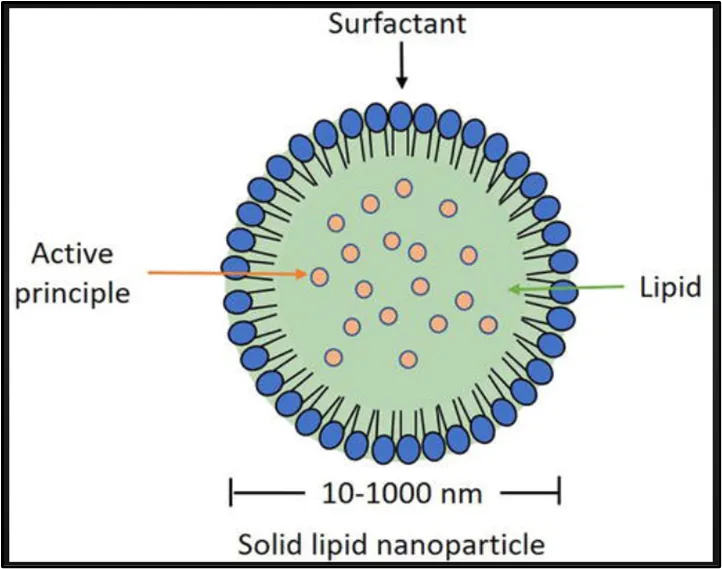
Figure No. 3: Structure Of Solid Lipid Nanoparticles
Aims Of Solid Lipid Nanoparticles: [65].
• To enhance the bioavailability of poorly soluble drugs.
• To provide controlled and sustained drug release.
• To protect drugs from chemical, enzymatic, and physical degradation.
• To improve drug stability during storage and in biological environments.
• To enable targeted drug delivery, reducing systemic side effects.
• To increase therapeutic efficacy while minimizing dose-related toxicity.
• To use biocompatible and biodegradable lipids for safer drug delivery.
Advantages Of Solid Lipid Nanoparticles: [66].
• Excellent biocompatibility.
• Improve stability of pharmaceuticals.
• Excellent reproducibility with a savvy high-weight homogenization technique as the readiness methodology.
• High and enhanced drug content.
• The achievability of consolidating both hydrophilic and hydrophobic medications.
Disadvantages Of Solid Lipid Nanoparticles: [67].
• Poor sedate stacking limit.
• Drug ejection after polymeric move amid capacity.
• Unpredictable gelation propensity.
• The low ability to stack hydrophilic medications because of apportioning impacts amid the generation procedure.
Importance Of Solid Lipid Nanoparticles: [68].
1.Enhanced Bioavailability
Solid lipid nanoparticles enhance the solubility and absorption of poorly water-soluble drugs by entrapping them within a lipid matrix, which ultimately leads to improved oral bioavailability.
2. Controlled And Sustained Drug Release
Due to their solid lipid core, solid lipid nanoparticles provide sustained and controlled drug release, which helps reduce dosing frequency and enhances patient compliance.
3. Protection Of Labile Drugs
The solid lipid matrix of solid lipid nanoparticles protects sensitive drug molecules such as antibiotics, peptides, and antioxidants from degradation caused by light exposure, ph variations, and enzymatic activity.
4. Biocompatibility And Safety
Solid lipid nanoparticles are formulated using physiological lipids, such as triglycerides and fatty acids, which are biocompatible, biodegradable and considered safe for long-term therapeutic use.
5. Reduced Toxicity Compared To Polymeric Nanocarriers
By avoiding the use of synthetic polymers, solid lipid nanoparticles reduce the risk of long-term accumulation and associated toxicity.
6. High Drug Loading Capacity
The solid lipid matrix efficiently encapsulates lipophilic drugs, making solid lipid nanoparticles particularly suitable for hydrophobic molecules with poor permeability, such as sparfloxacin.
7. Improved Targeting Ability
Surface modification strategies, such as pegylation and ligand attachment, enable solid lipid nanoparticles to achieve targeted drug delivery to specific tissues, including the brain, lungs, and tumour sites.
8. Enhanced Stability
Because of their solid-state core, solid lipid nanoparticles exhibit greater physical stability than emulsions and liposomes, thereby minimizing drug leakage and expulsion.
Types Of Solid Lipid Nanoparticles:
1. Homogeneous Matrix Type
A model of solid solutions, is categorized as a homogeneous matrix model due to the molecular dispersion of the active ingredient within the lipid core or its existence as amorphous groups. The model, as mentioned earlier, is derived through the utilization of high-pressure homogenization or cold homogenization methodologies conducted at temperatures surpassing the melting point of lipids.
The pharmaceutical agent is planned to be distributed at a molecular level, devoid of surfactants or solubility-enhancing agents, through a cold homogenization methodology [69].
2. Drug-Enriched Shell Model
Synthesized through a homogenization process that involves heat induction. This model asserts that the attainment of the lipid’s recrystallization temperature results in forming a solid lipid core. Upon the reduction of the dispersion temperature, the o/w nano emulsion experiences lipid precipitation, resulting in an elevation of drug concentration within the liquid lipid.
The drug tends to concentrate within the outer shell of the solid lipid nanoparticle that remains in a liquid state. The drug tends to concentrate within the outer shell of the solid lipid nanoparticle that remains in a liquid state [70].
3. Drug-Enriched Core Model
Drug precipitation takes place prior to the re-solidification of lipids. This phenomenon occurred when the drug concentration in the lipids approaching the point of solubility saturation, then a high drug concentration incorporated into the lipid. Upon cooling, the liquid lipid in the nano emulsion reaches a state of supersaturation concerning the drug. Therefore, lipid solidification takes place after precipitation. The subsequent drop in temperature causes the lipid to recrystallize near the core, which has been enriched with drug molecules and now resembles a membrane structure. This particular model is generally appropriate for drugs that require an extended-release profile within a specific time frame [71].
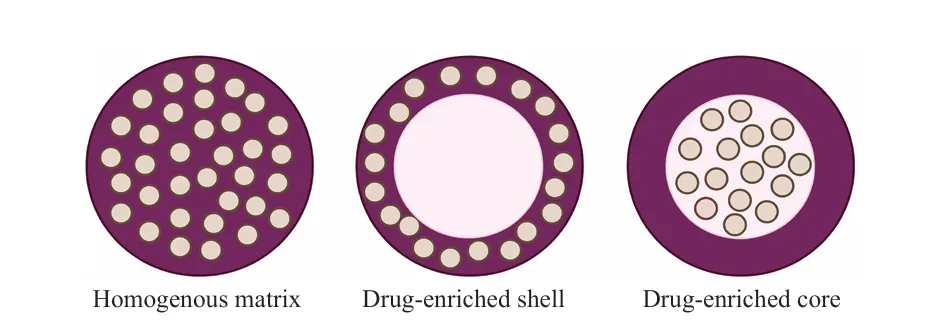
Figure No. 4: Drug Incorporation Model Of Sln: Homogenous Matrix Of Solid Solution (Left); Drug-Enriched Shell (Middle); And Drug-Enriched Core (Right).
Methods Of Preparation Of Solid Lipid Nanoparticles:
1. Hot Homogenization Method:
Solid lipid nanoparticles are prepared by the hot homogenization method by first melting the solid lipid slightly above its melting point and dissolving the drug in the molten lipid. Separately, an aqueous surfactant solution is prepared and heated to the same temperature as the lipid phase. The hot aqueous phase is then slowly added to the molten lipid with continuous stirring and subjected to hot homogenization to form a fine oil-in-water emulsion. Homogenization is continued for a few minutes to reduce the droplet size uniformly. The resulting hot emulsion is then allowed to cool to room temperature. During cooling, the lipid solidifies, leading to the formation of solid lipid nanoparticles [72].
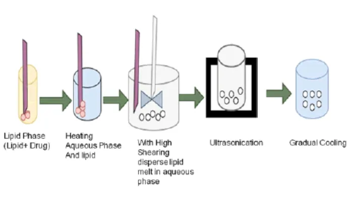
Figure No. 5: Hot Homogenization Method
2. Solvent Evaporation Method
Solid lipid nanoparticles are prepared by the solvent evaporation method by first dissolving the solid lipid and the drug in a suitable organic solvent. This organic phase is then slowly added to an aqueous surfactant solution with continuous stirring to form an oil-in-water emulsion. The emulsion is further stirred or homogenized to reduce the droplet size. The organic solvent is then removed by evaporation, usually under reduced pressure or continuous stirring. As the solvent gradually evaporates, the lipid separates out and solidifies. This leads to the formation of solid lipid nanoparticles uniformly dispersed in the aqueous medium [73].
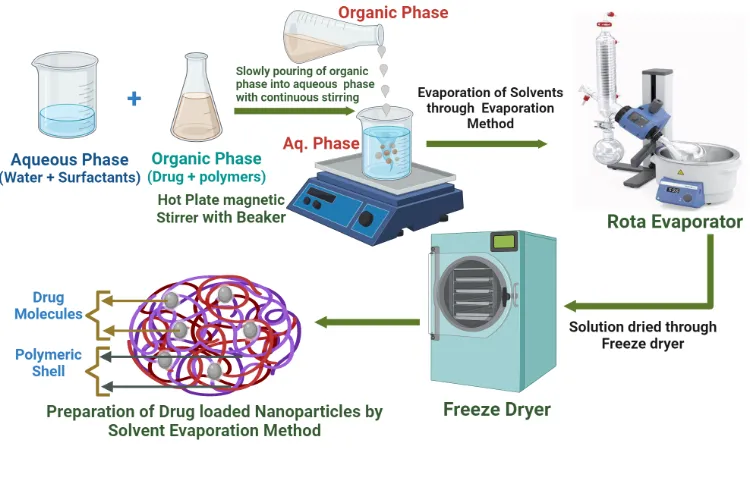
Figure No. 6: Solvent Evaporation Method
3. Micro Emulsion-Based Method
Solid lipid nanoparticles are prepared by the microemulsion-based method by first melting the solid lipid above its melting point and incorporating the drug into the molten lipid. A suitable surfactant and co-surfactant are then added and mixed until a clear and transparent system is obtained. Warm distilled water is added slowly with gentle stirring to form a hot oil-in-water microemulsion. This hot microemulsion is then rapidly dispersed into cold water under continuous stirring. The sudden drop in temperature causes the lipid to solidify. This leads to the formation of solid lipid nanoparticles uniformly dispersed in the aqueous medium [74].
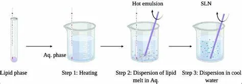
Figure No. 7: Micro Emulsion-Based Method
4. Solvent Emulsification-Diffusion Method
Solid lipid nanoparticles are prepared by the solvent emulsification–diffusion method by first dissolving the solid lipid and the drug in a partially water-miscible organic solvent. This organic phase is then slowly emulsified into an aqueous surfactant solution with continuous stirring to form an oil-in-water emulsion. After emulsification, additional water is added to the system to allow the organic solvent to diffuse into the aqueous phase. As the solvent diffuses out, the lipid becomes supersaturated. The lipid then separates and starts to precipitate. On complete diffusion of the solvent, the lipid solidifies to form solid lipid nanoparticles uniformly dispersed in the aqueous medium [75].
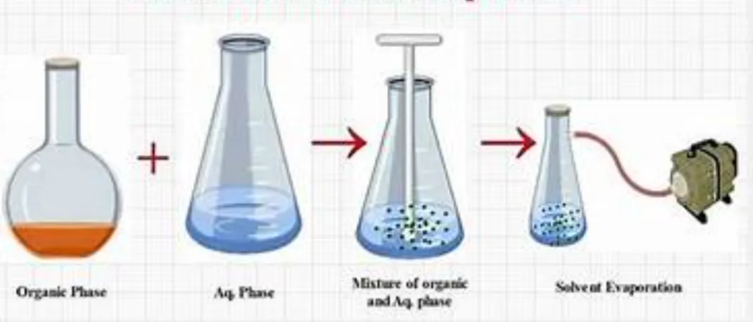
Figure No. 8: Solvent Emulsification-Diffusion Method
5. Ultra Sonication Method
Solid lipid nanoparticles are prepared by the ultrasonication method by first melting the solid lipid slightly above its melting point and dissolving the drug in it. A hot aqueous surfactant solution is prepared separately and added to the molten lipid with continuous stirring to form a coarse emulsion. This emulsion is then subjected to ultrasonication for a fixed period. The ultrasonic waves break the lipid droplets into very fine particles. After sonication, the dispersion is allowed to cool to room temperature. During cooling, the lipid solidifies, resulting in the formation of solid lipid nanoparticles [76].
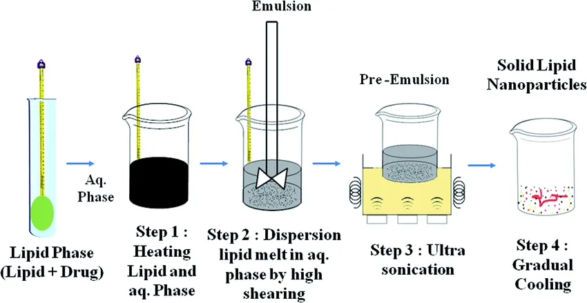
Figure No. 9: Ultra Sonication Method
6. Spray Drying Method
Solid lipid nanoparticles are prepared by the spray drying method by first making a dispersion of the lipid, drug, and surfactant in water or a suitable solvent. This dispersion is then sprayed through a nozzle into a stream of hot air, which quickly evaporates the solvent. As the droplets dry, the lipid solidifies, forming nanoparticles. The resulting solid lipid nanoparticles are collected as a fine, free-flowing powder. This method provides an easy way to obtain dry nanoparticles that are stable and convenient to store [77].
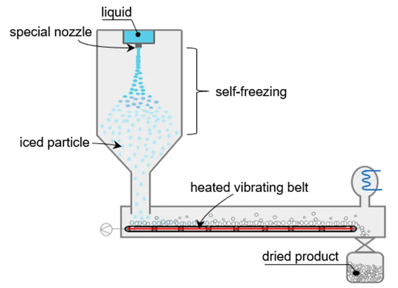
Figure No. 10: Spray Drying Method
Evaluation Of Solid Lipid Nanoparticles:
1. Physical Appearance
The physical appearance of solid lipid nanoparticles is evaluated by simple visual observation of the prepared solid lipid nanoparticles dispersion. A small quantity of the formulation is examined for its color, clarity, and overall uniformity. The dispersion should appear smooth and homogeneous without any visible particles or sedimentation. The presence of turbidity, aggregation, or phase separation is carefully noted. The formulation is also observed during storage to detect any changes in appearance. A stable Solid lipid nanoparticles formulation maintains a uniform and acceptable appearance over time [78].
2. Drug Content
The drug content of solid lipid nanoparticles is evaluated by taking a known amount of the Solid lipid nanoparticles formulation and dissolving it in a suitable solvent to break the lipid matrix. The mixture is then filtered or centrifuged to obtain a clear solution. The amount of drug present in the solution is measured using an appropriate method such as UV-visible spectrophotometry. The drug concentration is calculated from a previously prepared calibration curve. This test helps confirm that the drug is uniformly distributed in the formulation. Accurate drug content indicates good formulation quality [79].
3. Drug Entrapment Efficiency
The drug entrapment efficiency of solid lipid nanoparticles is evaluated by first separating the free drug from the nanoparticles. A measured amount of the Solid lipid nanoparticles dispersion is centrifuged at high speed to settle the nanoparticles. The clear supernatant, which contains the unentrapped drug, is carefully collected. The amount of free drug in the supernatant is then determined using UV–visible spectrophotometry. Entrapment efficiency is calculated by comparing the free drug with the total drug used in the formulation. This evaluation shows how efficiently the drug is incorporated into the lipid nanoparticles [80].
4. Particle Size
Particle size analysis, a small quantity of solid lipid nanoparticle dispersion is first diluted with distilled water to avoid particle aggregation. The diluted sample is then transferred into a clean cuvette and placed in the particle size analyser. Measurements are carried out at room temperature using dynamic light scattering. The average particle size and size distribution of the nanoparticles are then recorded [81].
5. Zeta Potential
The zeta potential of solid lipid nanoparticles is evaluated to understand their surface charge and stability. A small amount of the Solid lipid nanoparticles dispersion is first diluted with distilled water or a suitable buffer. The diluted sample is then placed in the zeta potential measuring cell. The measurement is carried out using a zeta potential analyser based on electrophoretic mobility. The obtained zeta potential value indicates the stability of the nanoparticles. Higher positive or negative values suggest better stability due to reduced particle aggregation [82].
6. In-Vitro Drug Release
The in-vitro drug release of solid lipid nanoparticles is evaluated using a diffusion method such as the dialysis bag technique. A measured amount of the solid lipid nanoparticles formulation is placed inside a dialysis membrane and properly sealed. The dialysis bag is immersed in a suitable release medium maintained at body temperature with continuous stirring. Samples are withdrawn at regular time intervals and replaced with fresh medium to maintain sink conditions. The withdrawn samples are analysed for drug content using uv spectrophotometry. The release profile obtained helps in understanding the drug release behaviour of the formulation [83].
Characterization Of Solid Lipid Nanoparticles:
1. FTIR (Fourier Transform Infrared Spectroscopy):
FTIR analysis of fexofenadine hydrochloride is performed using the KBR pellet method. A small quantity of the drug is gently mixed with dry, IR-grade potassium bromide and compressed to form a clear, thin pellet. This pellet is then placed in the FTIR sample holder and the spectrum is recorded in the range of 4000-400 cm⁻¹. The recorded spectrum is used to identify characteristic functional groups and to assess the purity and compatibility of the drug [84].
2. Transmission Electron Microscopy:
TEM analysis, a small amount of the solid lipid nanoparticle dispersion is first diluted with distilled water. A drop of this diluted sample is carefully placed onto a carbon-coated copper grid and allowed to stand for a short time so the particles can settle. Excess liquid is gently removed using filter paper, and the grid may be negatively stained to improve contrast. After drying at room temperature, the grid is observed under a transmission electron microscope to study the size and shape of the nanoparticles [85].
3. Scanning Electron Microscopy:
SEM analysis, a small quantity of solid lipid nanoparticle dispersion is placed on a clean metal stub and allowed to dry at room temperature. Once dried, the sample is coated with a thin layer of gold to improve conductivity. The stub is then placed inside the SEM chamber and scanned under suitable operating conditions. The obtained images are used to observe the surface morphology and shape of the solid lipid nanoparticles [86].
4. Differential Scanning Calorimetry:
DSC analysis, a small quantity of solid lipid nanoparticles is accurately weighed and sealed in an aluminium pan, with an empty pan used as a reference. The sample is placed in the DSC instrument and heated at a controlled rate under a nitrogen atmosphere. The temperature is scanned over an appropriate range covering the melting points of the lipid and drug. The obtained thermogram is used to study the thermal behaviour and crystallinity of the solid lipid nanoparticles [87].
Limitations Of Solid Lipid Nanoparticles:
The main limitations of solid lipid nanoparticles include their limited drug loading capacity, particularly for water-soluble drugs, because of the highly ordered lipid structure. During storage, drug expulsion may occur due to changes in the crystalline form of the lipid. Solid lipid nanoparticles can also exhibit an initial burst release, which may result in uncontrolled drug delivery. In addition, particle aggregation or growth over time can affect their physical stability, and large-scale production is often challenging and expensive [88].
Applications Of Solid Lipid Nanoparticles:
1. Per Oral Administration
Per oral administration forms of Solid lipid nanoparticles may include aqueous dispersions or Solid lipid nanoparticles loaded traditional dosage forms, e.g. Tablets, pellets or capsules. The microclimate of the stomach favors particle aggregation due to the acidity and high ionic strength. It can be expected, that food will have a large impact on Solid lipid nanoparticles performance.
The plasma levels and body distribution were determined after administration of ca-SLN suspension versus a ca solution (ca-sol). Two plasma peaks were observed after administration of ca-SLN. The first peak was attributed to the presence of free drug; the second peak can be attributed to controlled release or potential gut uptake of Solid lipid nanoparticles. These two peaks were also found in the total ca concentration-time profiles of all measured organs [89].
2. Parenteral Administration
SLN have been administered intravenously to animals. Pharmacokinetic studies of doxorubicin incorporated into Solid lipid nanoparticles showed higher blood levels in comparison to a commercial drug solution after iv injection in rats. Concerning the body distribution, Solid lipid nanoparticles were found to cause higher drug concentrations in lung, spleen and brain, while the solution led to a distribution more into liver and kidneys. Parenteral application is a very wide field for Solid lipid nanoparticles [90].
3. Transdermal Application
The smallest particle sizes are observed for Solid lipid nanoparticles dispersions with low lipid content (up to 5%). Both the low concentration of the dispersed lipid and the low viscosity are disadvantageous for dermal ad-ministration. In most cases, the incorporation of the Solid lipid nanoparticles dispersion in an ointment or gel is necessary in order to achieve a formulation which can be administered to the skin. The incorporation step implies a further reduction of the lipid content. An increase of the solid lipid content of the Solid lipid nanoparticles dispersion results in semisolid, gel-like systems, which might be acceptable for direct application on the skin [91].
4. Topical Application
Regarding the regularity aspect, topical application is relatively unproblematic. The major advantages for topical products are the protective properties of Solid lipid nanoparticles for chemically labile drugs against degradation and the occlusion effect due to film formation on the skin. Especially in the area of cosmetics there are many compounds such as retinol or vitamin c which cannot be incorporated because of the lack of chemical stability [92].
5. Ophthalmic Administration
Many investigations have been made to use nanoparticles for prolonged release of drugs to the eye. The basic problem of ophthalmologic formulation is the fast removal from the eye, which implies clearance of the applied drug through the nose.
it could be shown for nanoparticles that an increased adhesiveness is available leading to higher drug levels at desired site of action. However, the basic problem was that the nanoparticles are of limited toxicological acceptance. It was shown by gasco that Solid lipid nanoparticles have a prolonged retention time at the eye. The lipids of Solid lipid nanoparticles are easy to metabolize and open a new way for ophthalmological drug delivery without impairing vision [93].
6. Pulmonary Administration
A very interesting application appears to be the pulmonary administration of Solid lipid nanoparticles. Solid lipid nanoparticles powders cannot be administered to the lung because the particle size is too small and they will be exhaled. A very simple approach is the aerosolization of aqueous Solid lipid nanoparticles dispersions. The important point is that the Solid lipid nanoparticles should not aggregate during the aerosolization.
The aerosol droplets were collected by collision of aerosol with a glass wall of a beaker. This basically demonstrates that Solid lipid nanoparticles are suitable for lung delivery [94].
FUTURE PROSPECTIVE
Solid lipid nanoparticles hold great promise as advanced drug delivery systems due to their biocompatibility and ability to enhance bioavailability of poorly soluble drugs. Future research is expected to focus on surface-modified and targeted Solid lipid nanoparticles to improve site-specific delivery and therapeutic efficacy. Integration of Solid lipid nanoparticles with personalized medicine and biomarker-based therapies may further optimize treatment outcomes. Advances in large-scale manufacturing and formulation stability will support clinical translation and commercialization. Additionally, combining Solid lipid nanoparticles with biological and novel therapeutic agents may expand their applications in chronic and inflammatory diseases.
CONCLUSION
Solid lipid nanoparticles represent a promising and versatile drug delivery system for improving the therapeutic performance of conventional drugs. Their ability to enhance bioavailability, provide controlled drug release, and protect labile drugs makes them highly suitable for modern pharmaceutical applications. The use of biocompatible and biodegradable lipids ensures improved safety and patient compliance. Despite certain limitations, advancements in formulation strategies have helped overcome major challenges associated with Solid lipid nanoparticles. Overall, solid lipid nanoparticles offer significant potential for targeted and effective drug delivery in the treatment of various diseases.
REFERENCES




Arti Kumari, S. Swetha Malika Devi, Beny Baby, S. Rajarajan, A Review on Solid Lipid Nanoparticles as Targeted Drug Delivery Systems for The Treatment of Ulcerative Colitis., Int. J. of Pharm. Sci., 2026, Vol 4, Issue 7, 6108-6129, https://doi.org/10.5281/zenodo.21721861
 10.5281/zenodo.21721861