View Article

Abstract

Solid lipid nanoparticles (SLNs) are a novel drug delivery system for the treatment of vulvovaginal candidiasis, a skin infection of fungal origin from Candida species. SLNs encapsulate antifungal agents, thereby increasing bioavailability and stability while reducing systemic side effects. Targeted drug delivery through the nanoscale carriers results in enhanced treatment efficacy and recurrence reduction. Controlled and sustained release of antifungal agents may improve lesion clearance and therapeutic outcomes. SLNs provide deeper penetration into skin, circumventing the constraint of traditional topical preparations. Patient compliance is enhanced by reduced frequency of applications necessary for traditional treatments. SLNs can be customised with a range of antifungal compounds, making them an effective and versatile drug delivery system. Yet, stability concerns, drug expulsion, and regulatory approval must be overcome for clinical acceptance. Complex formulation approaches like polymer coating and surfactant optimisation can help improve SLN performance. Further studies should optimise nanoparticle size, drug loading efficiency, and long-term stability. Clinical trials are necessa ry to confirm the safety, efficacy, and commercial viability of SLN-based antifungal therapies. This review highlights the promise of SLNs in transforming dermatological treatments for fungal infections.

Keywords

Solid lipid nanoparticles(SLNs), Vulvovaginal candidiasis, Topical formulation, In-vitro drug release, Drug delivery system

Introduction

× Popup Image

Vulvovaginal candidiasis (VVC) is a common genital infection that impacts one in four women at least once in their lifetime with nearly 50% experiencing recurrent infections[9]. Vaginal discomfort, abnormal discharge, and irritation are common clinical manifestations that can markedly affect women’s health and daily well-being. Candidal vulvovaginitis (CVV) is highly prevalent and frequently recurrent, and its diagnosis may be complicated by limited awareness and overlapping symptoms with other vaginal infections. Early and accurate diagnosis, along with appropriate therapeutic management and supportive lifestyle interventions, is essential to reduce disease recurrence. In addition, ongoing research focused on antifungal resistance and the development of novel and more effective treatment strategies is crucial to improve long-term clinical outcomes [2]

Vaginal drug delivery is a unique drug delivery method that can be utilised for long-term treatment with both local and systemic effects, depending on the drug's properties and the medical situation. potential for minimal adverse effects. Traditionally, these systems have been used to administer medications to treat vaginal infections and contraceptives[1]

The antifungal drugs that can be used to treat Candida albicans infection currently include polyenes, azoles, and echinocandins. Amphotericin B , which is mainly used as a polyene drug, should be used with caution due to its severe nephrotoxicity and hepatotoxicity although it has a good therapeutic effect. Azoles can be divided into two subcategories according to their chemical structure: Imidazole’s (clotrimazole, ketoconazole and miconazole) and triazoles (fluconazole, itraconazole, voriconazole and posaconazole). Local application of imidazole drugs is used to treat mucosal candidiasis, while triazole drugs are generally used to treat mucosal and systemic infections caused by Candida albicans[43] Antimycotic medications are applied topically or taken orally for one to seven days or more in order to treat VVC. To avoid the disadvantages of the oral route, topical medication distribution is advised. Compared to oral delivery, this method requires a lower dosage and offers the advantage of targeted drug delivery to a precisely desired region[4].

Solid lipid nanoparticles (SLNs) have gained increasing attention as an advanced drug delivery system because of their excellent biocompatibility and versatility in carrying both hydrophilic and lipophilic drugs. By remaining solid at physiological temperatures, SLNs protect encapsulated drugs from degradation and enable controlled, sustained release. Their nanoscale size and lipidic nature enhance interaction with the vaginal mucosa, improving adhesion and local drug penetration. Compared with conventional antifungal formulations, SLNs can prolong drug retention at the site of infection, maintain effective therapeutic concentrations for longer periods, and reduce the need for frequent dosing, thereby potentially lowering recurrence rates and improving patient compliance[44].

The aim of this review is to critically assess solid lipid nanop  articles (SLNs) as a novel and promising drug delivery approach for the treatment of vulvovaginal candidiasis, with emphasis on their formulation strategies, mechanistic advantages, and ability to enhance local drug retention, controlled release, and therapeutic efficacy while minimizing systemic side effects

The advantages of vaginal drug delivery given below

  • Achieve quick drug absorption and an early onset of action[20].
  • Inhibits metabolism during the initial pass.
  • Increased vascularization.
  • Possibility of self-medication.
  • Minimal negative effects on the system.
  • Non-invasive delivery.

Disadvantages

  • Localized irritation.
  • Cultural sensitivity.
  • Personal hygiene.
  • Gender specificity.
  • Sometimes the drug will leak out of the vagina.
.                    


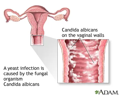
Fig no 1.  Diagrammatic representation of candidiasis[92].

History of vulvovaginal candidiasis.

  • Candida albicans is the main cause of vulvovaginal candidiasis (VVC), a common mucosal fungal infection of the female genital system[17]. In 1923, Berkhout formally classified the genus Candida, and Candida albicans was recognized as the most common etiological agent of vulvovaginal infections. By the early 20th century, VVC was recognised as a unique clinical entity, frequently related with pregnancy, diabetes, and poor genital hygiene[22].
  • During the 1950s and 1960s, greater use of broad-spectrum antibiotics led to an increase in VVC cases, highlighting the significance of vaginal microflora imbalance in disease progression. This era also saw the introduction of antifungal medications such as nystatin and azole derivatives, which dramatically improved the management.

Table no 1.  Pathogenesis of Vulvovaginal candidiasis[23]
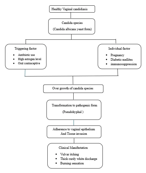
Symptoms of vaginal infection

  • Itching and irritation in the vagina and vulva.
  • Redness and swelling of the vulva.
  • Watery vaginal discharge.
  • Vaginal rash.
  • Vaginal pain and soreness.
  • Burning sensation during urination

Diagnosis

Diagnosis of candidal vaginitis may be supported by microscopic examination of vaginal discharge using a 10% potassium hydroxide (KOH) wet mount, which may reveal budding yeast cells, pseudo hyphae, In vulvovaginal candidiasis, the vaginal pH is usually less than 4.5, which may help differentiate it from other causes of vaginitis [1].

Topical treatment for vulvovaginal candidiasis.

Topical  treatment of vulvovaginal candidiasis  is mostly based on azole antifungal drugs, which are indicated as first-line therapy for simple infections. The CDC Sexually Transmitted Infections Treatment Guidelines (2021) state that intravaginal azoles such as clotrimazole, miconazole, terconazole, butoconazole, econazole, and tioconazole are highly efficacious and have comparable cure rates[30]. The use of topical medications like miconazole, terconazole, and boric acid for recurrent vulvovaginal candidiasis is also supported by expert consensus maintenance dosage schedules are used to prevent relapse[29,17].

Solid lipid nanoparticles (SLNs), or lipospheres, are pharmaceutical nanocarriers that are made to deliver drugs in a controlled way.[12,13] Solid lipid nanoparticles are colloidal dispersions that are biocompatible and well-tolerated in vivo. They range in size from 100 to 500 nm.[3]

The internal core structure of solid lipid nanoparticles enables them to concentrate lipophilic substances. The advantages of nanoparticles, which are extremely small particles with nanosize ranges, are related to their biopharmaceutical properties after being administered in vivo and the controlled release of loaded components at the intended site of action for improved therapeutic action.[14].
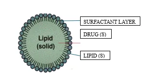
 

Figure no.2 Solid lipid nanoparticle

Advantages of Solid lipid Nanoparticle For Topical Application.[24]

  • Solid lipids are used to reduce drug mobility and allow for controlled and extended release.
  • The lipid matrix reduces the danger of toxicity because it is made up of physiological and biodegradable lipids.
  • Solid lipid nanoparticles improves stability. 
  • Solid lipid nanoparticles also improve the shelf life.
  • Solid lipid nanoparticles help to improve therapeutic effect at site of action.
  • Solid lipid nanoparticles are used because they reduce the irritation compared to other delivery system.

Disadvantages[26]

  • Lipids can undergo polymorphic transitions and form supercooled melts, which affect stability.
  • Solid lipid nanoparticles have the crystalline structure which limits the available for encapsulating drugs.
  • Sometimes degradation occurs in products of lipid and also present a safety risk based on formulation.

Drug incorporation models.

There are three drug incorporation models which describe drug release from SLN[91].

A) Homogenous matrix model.

B) Drug enriched shell with a lipid core.

 C) Drug enriched core with lipid shell.
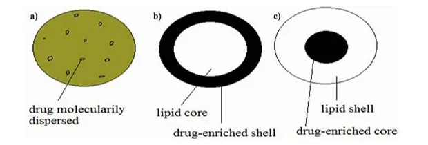
 

Fig no 3. Drug incorporation models[1,93].

Homogenous matrix model.

Drug is dispersed molecularly in the lipid matrix when solid lipid nanoparticles are prepared by cold homogenization method.

Drug enriched shell with a lipid core.

A solid lipid core is formed on the basis of recrystallization temperature of lipid.

 Drug enriched core with lipid shell.

Cooling the nano-emulsion, which may leads the super saturation of drug which is dissolved in lipid melt it may lead recrystallization of lipid

Role of solid lipid nanoparticle to treat vulvovaginal candidiasis[16,27,28]

  • SLNs encapsulate the drug and enhance the bioavailability of antifungal agents.
  • Many antifungal agents used in VVC  are poorly water-soluble. SLNs enhance their solubility and local bioavailability in the vaginal environment.
  • SLNs localize drug action at the vaginal site, minimizing systemic absorption and reducing side effects such as burning and irritation.
  • SLNs provide prolonged release of antifungal drugs, maintaining effective concentrations for longer periods and reducing dosing frequency.
  • SLNs lipid composition and nano size encourage adhesion to the vaginal mucosa, increasing drug retention and reducing leakage that comes with conventional creams and pessaries.
  • The solid lipid matrix protects antifungal drugs from chemical and enzymatic degradation in the vaginal environment.

Therapeutic Advantages of SLNs.

  1. Introduction to SLNs in Treatment:

Solid lipid nanoparticles (SLNs) are submicron colloidal carriers composed of physiological lipids that solidify at body temperature and room temperature. The limitations of conventional dosage forms, such as poor solubility, instability, and low drug bioavailability, are intended to be overcome by these enhanced drug delivery systems. SLNs increase treatment efficacy while maintaining safety and biocompatibility by encasing therapeutic chemicals in a solid lipid matrix[20].                                                                                                   

  1. Enhancement of Bioavailability and Stability:

SLNs significantly improve drug bioavailability by enhancing solubility and protecting drugs from chemical and enzymatic degradation. The solid lipid core stabilizes both hydrophilic and lipophilic drugs, leading to improved absorption and prolonged systemic circulation. This results in better therapeutic efficacy at lower doses, making treatment more effective and safer[21]

  1. Higher rates of clinical and mycological cure:

Patients treated with SLN-based antifungal formulations have higher rates of both clinical cure and mycological cure . Such double effectiveness not only provides quicker relief from symptoms but also reduces the possibility of relapse[22].

  1. Minimal side effects:

SLN-based formulations are capable of delivering drugs locally with minimum systemic absorption and, thus, associated adverse effects. Patients are also less prone to irritation, redness, or sensitivity than in the case of traditional topical antifungal treatments[23].

  1. Reduced Recurrence Rates:

The deep penetration along with sustained action of SLNs prevents fungal infections from coming back again. This is very useful in the case of vulvovaginal candidiasis as this condition often tends to recur[24,25].

  1. Versatile Drug Delivery System:

SLNs can be used to encapsulate a wide range of hydrophilic and lipophilic antifungal drugs, making it a versatile and adaptable delivery system. They may also be used in combination with other active ingredients, such as anti-inflammatory agents, to enhance therapeutic outcomes further[26].

Mechanistic Advantages of SLNs in Treating VVC

 1.Improved drug solubility and biopharmaceutical performance[38].

Solid lipid nanoparticles (SLNs) enhance the apparent solubility and overall biopharmaceutical performance of poorly water-soluble antifungal drugs by entrapping them within a solid lipid matrix. In this system, the drug is either molecularly dispersed or solubilized in the lipid core, which promotes uniform drug distribution at the site of application. Moreover, the nanoscale size of SLNs provides a high surface area, facilitating improved drug dissolution and closer interaction with the vaginal epithelium, ultimately leading to better local drug availability and therapeutic effectiveness.

 2.Mucoadhesion & prolonged retention at mucosal sites[39].

Solid lipid nanoparticles (SLNs) possess inherent mucoadhesive properties that help them remain in close contact with mucosal tissues such as the vaginal epithelium. Their lipid-based composition and nanoscale size enable effective interaction with the mucin layer, minimizing rapid removal by vaginal fluids. As a result, the formulation stays localized at the site of infection for a longer period.

3. Enhanced penetration and localization[40].

Solid lipid nanoparticles (SLNs) improve the ability of antifungal drugs to penetrate and remain within the vaginal tissue because of their very small size and lipid-based nature. These nanoparticles can closely interact with the vaginal mucus and epithelial cell membranes, allowing the drug to reach deeper tissue layers where Candida organisms often survive. The strong affinity of the lipid matrix for biological membranes helps concentrate the drug at the site of infection, leading to higher local drug levels with minimal systemic absorption.

 4. Sustained & controlled drug release[41].

Solid lipid nanoparticles provide sustained and controlled release of antifungal   drugs by retaining them within a solid lipid matrix, which enables gradual drug release at the vaginal mucosa. This prolonged drug availability maintains effective antifungal concentrations, enhances therapeutic efficacy, and reduces dosing frequency, making SLNs particularly beneficial for the treatment of vulvovaginal candidiasis.

5. Reduced dosing frequency & improved compliance[42].

 Solid lipid nanoparticles (SLNs) maintain therapeutic antifungal drug levels at the vaginal mucosa for extended periods through sustained and controlled drug release. This prolonged local drug availability reduces the need for frequent dosing, improving patient convenience and treatment adherence. Reduced dosing frequency also lowers the risk of local irritation and enhances overall compliance, which is particularly important in the management of recurrent vulvovaginal candidiasis.

SLN-Loaded Antifungal Drugs for vulvovaginal candidiasis.

Clotrimazole

Clotrimazole-loaded solid lipid nanoparticles (SLNs) have been investigated as a promising vaginal drug delivery system to improve the therapeutic efficacy of clotrimazole in vulvovaginal candidiasis. Clotrimazole exerts its antifungal activity by inhibiting the fungal cytochrome P450–dependent enzyme lanosterol 14-α-demethylase, thereby disrupting ergosterol biosynthesis, an essential component of the fungal cell membrane. Encapsulation of clotrimazole within solid lipid nanoparticles improves its physicochemical stability, overcomes poor aqueous solubility, and facilitates sustained drug release at the vaginal mucosa. SLNs enhance intimate contact with the vaginal epithelium, promoting prolonged residence time and improved penetration into infected tissues and fungal biofilms[28,89,90].

Miconazole

Solid lipid nanoparticles (SLNs) loaded with miconazole have shown significant potential as an effective vaginal delivery system for treating vulvovaginal candidiasis. Its primary mechanism of action involves inhibition of the fungal cytochrome P450–dependent enzyme lanosterol 14-α-demethylase, which is crucial for ergosterol biosynthesis. Miconazole acts through multiple antifungal mechanisms. Miconazole efficiently penetrates the stratum corneum of the skin and remains detectable for more than four days following topical application. Systemic exposure is minimal, with less than 1% absorbed into the bloodstream, while vaginal administration results in absorption of no more than 1.3%, indicating predominantly local activity.[94,33].

Ketoconazole

Ketoconazole-loaded solid lipid nanoparticles (SLNs) have been extensively studied to overcome the limitations of conventional ketoconazole formulations, including poor aqueous solubility, low bioavailability, and local irritation. Loading ketoconazole into SLNs enhances its solubility, allows sustained and controlled drug release, and improves penetration into the vaginal epithelium with minimal systemic absorption. Its primary mechanism involves inhibition of the fungal cytochrome P450–dependent enzyme lanosterol 14-α-demethylase, which disrupts ergosterol biosynthesis[32].

Econazole

Econazole-loaded solid lipid nanoparticles improve drug solubility, enhance formulation stability, and allow a sustained release of the drug. The lipid-based carrier interacts closely with the vaginal mucosa, helping the formulation remain at the site of infection for a longer duration and improving drug penetration into the vaginal epithelium[45,46]. This localized delivery enhances antifungal effectiveness while limiting systemic exposure. Econazole acts by inhibiting the cytochrome P450–dependent enzyme lanosterol 14-α-demethylase, which disrupts ergosterol synthesis, compromises fungal cell membrane integrity, and ultimately leads to fungal cell death[47].

Limitations that can be improved by nanoparticle applications.[31,28]

Antimycotic resistance refers to the persistence of fungal infection despite appropriate antifungal treatment. Antifungal-induced cellular stress promotes adaptive responses in fungi, including activation of stress signalling pathways and cellular defence mechanisms, ultimately leading to resistance to antimycotic agents.

Antimycotic resistance in dermatophytes is primarily mediated by the overexpression of efflux pump systems, which actively expel antifungal drugs and other toxic compounds from fungal cells. Multidrug resistance mainly results from increased expression of ATP-binding cassette (ABC) transporters, which transport structurally diverse molecules across cell membranes using ATP hydrolysis, thereby reducing intracellular drug accumulation.

Method of preparation of solid lipid nanoparticles

Table no 2.Method of preparation  SLN[34,35,36]
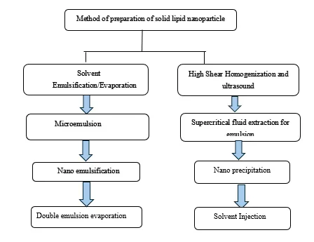


Preparation techniques for solid lipid nanoparticles[37].

The performance of SLNs depends on the method of preparation which may influence the particle size, drug loading capacity, drug release, drug stability etc.

  1. 1.High-pressure homogenization

   a. Hot homogenization

   b. Cold homogenization

2. Ultra-sonication or high-speed homogenization

   a. Probe ultrasonication

   b. Bath ultrasonication

3. Supercritical fluid method

4. Micro emulsion-based method

5. Spray drying method

6. Double emulsion method

7. Precipitation technique

8. Film-ultrasound dispersion

 1.High-pressure homogenization.

  1. Hot homogenization: The lipid is processed above its melting point to facilitate emulsification. A high-shear homogenizer is used to form a pre-emulsion by mixing the drug-loaded molten lipid with the aqueous emulsifier phase, which is then subjected to high-pressure homogenization at elevated temperature. Reduced lipid viscosity under these conditions promotes the formation of smaller, more uniform particles and improves the overall quality of the SLN dispersion. High-pressure homogenization may accelerate degradation of both the drug and the lipid carrier due to the elevated processing conditions. Typically, three to five homogenization cycles are sufficient to obtain high-quality formulations. During processing, the sample temperature increases by approximately 10 °C at 500 bar, and pressures in the range of 500–1500 bar are commonly employed. At excessively high homogenization intensities, the increased kinetic energy of particles can promote coalescence, resulting  in an increase in particle size[48,49].
  2. Cold homogenization: Cold homogenization is comparable to pressure-assisted milling, as it involves homogenization of solid lipids under controlled temperature conditions. This method was developed to overcome limitations of hot homogenization, such as drug loss into the aqueous phase and lipid polymorphic transitions caused by complex crystallization. Although the drug is initially dispersed in the molten lipid similar to hot homogenization, the lipid is subsequently cooled, solidified, and homogenized at low temperatures, reducing thermal stress and formulation instability. In cold homogenization, the drug-loaded lipid is rapidly solidified and ground into a fine powder, then mixed with a cooled emulsifier solution and high-pressure homogenized. This method produces slightly larger but more uniformly sized particles compared to hot homogenization[50].
  1. Ultra-sonication

This method uses ultrasonic waves to create high shear forces that break lipid           particles into smaller sizes. It can be performed with either a probe or bath sonicator. While it effectively reduces particle size, it may lead to metal contamination and physical instability[51].

3. Supercritical fluid method

This is a relatively new technique, in which supercritical carbon dioxide acts as a solvent. The lipid is dissolved in supercritical CO₂, and upon rapid expansion, it precipitates to form nanoparticles. This is a solvent free method that can give dry powders rather than suspensions[52,53

 4. Spray drying method

Spray drying is a widely used pharmaceutical technique for converting liquid formulations into dry powders. Converting lipid nanoparticle aqueous dispersions into dry form improves their stability and allows long-term storage . Spray drying is a single-step process in which a liquid feed, usually a solution or suspension, is atomized and rapidly dried by contact with hot gas, leading to fast solvent evaporation and particle formation. The dried particles are separated from the drying air using cyclone separators or electrostatic precipitators[54].

5. Double emulsion method

A new solvent emulsification–evaporation technique was employed to prepare hydrophilic drug-loaded solid lipid nanoparticles. In this method, the drug was stabilized within the inner aqueous phase of a w/o/w double emulsion, which helped prevent its migration into the outer aqueous phase during solvent evaporation[48].

Evaluation of SLNs

Physical evaluation


The SLN formulations were off-white with semi-solid texture. The pH was between 7.0 and 8.0, which is appropriate for dermal use. Rheological investigations indicated non-Newtonian flow behaviour, which suggests shear-thinning characteristics conducive to topical use. The viscosity values decreased with spindle speed increase, which verifies their suitability for use in diverse formulations. These characteristics ensure stability and easy application of the formulation[55].

Particle Size and Polydispersity Index (PDI)

 The particle size and size distribution of solid lipid nanoparticles are commonly evaluated using techniques such as photon correlation spectroscopy, laser diffraction, transmission electron microscopy, scanning electron microscopy, scanning tunnelling microscopy, atomic force microscopy, and freeze-fracture electron microscopy. The uniformity of the particle population is expressed by the polydispersity index (PDI), which reflects the distribution of particle sizes within the formulation[36].

Zeta potential

The zeta potential, a formulation characteristic of SLNs, indicates colloidal stability. The zeta meter measures the zeta potential. Zeta potential describes the storage stability of colloidal dispersions. High zeta potential causes electric charges to repel particles, preventing them from aggregating. To stabilise the colloidal dispersion, the zeta potential should be greater than ±30mV[56].

Differential Scanning Calorimetry(DSC) and Scanning Electron Microscopy(SEM)

DSC analysis revealed no significant interactions between excipients and the medication, assuring the stability of the active ingredient. SEM investigations revealed that SLNs had smooth, spherical surfaces, which increased their stability and potential for use. The results are consistent with the structural integrity and biocompatibility of the formulation with the skin[57,58].

Drug content and In vitro release studies

Drug content analysis revealed remarkable uniformity among formulations, with values greater than 94%. In vitro release experiments demonstrated sustained drug release behaviour over 24 hours, which is ideal for long-term therapeutic activity. SLNs released the medicine at a slower and more controlled rate than the commercial version, showing its potential to increase patient compliance[55].

Stability studies

Short-term stability testing at 40°C ± 2°C and 75% ± 5% RH for four weeks showed no significant changes in drug content, release pattern, or physical appearance. The findings confirm the formulation's stability under accelerated settings, ensuring the product's effectiveness throughout time[58,36].

Characteristics of Solid lipid nanoparticles[60,61].

  • SLNs have particle sizes in the nano range, which enhances permeation into biological membranes and increases surface area for better drug delivery.
  • The lipid matrix of SLNs can encapsulate and protect lipophilic drugs like clotrimazole, increasing drug loading and preventing premature degradation.
  • SLNs provide controlled release of the drug over time due to their solid lipid core, reducing dosing frequency and maintaining therapeutic levels.
  • The solid lipid core improves physical stability of the formulation, helping prevent drug leakage and degradation during storage.
  • SLNs are made of biodegradable, physiological lipids, reducing toxicity and making them suitable for vaginal or topical use.
  •  Due to their small size and lipidic nature, SLNs can improve drug solubility and absorption at the target site, enhancing therapeutic efficacy.

Application For Solid lipid nanoparticles to treat vulvovaginal candidiasis[62].

Solid lipid nanoparticles are emerging as a promising approach for treating vulvovaginal candidiasis because they can enhance the effectiveness of antifungal drugs while addressing the limitations of traditional formulations. By enclosing poorly water‑soluble antifungals within a lipid matrix, SLNs improve drug solubility and ensure that more of the drug reaches the vaginal tissue where it is needed. When incorporated into gels or mucoadhesive systems, they provide controlled, sustained release, maintaining effective drug levels for longer periods and reducing the need for frequent applications. Their tiny size and lipid-based nature also allow SLNs to stick to and penetrate the vaginal mucosa more efficiently than conventional creams or gels, increasing the drug’s residence time at the infection site. Solid lipid nanoparticles help improve drug delivery, support sustained antifungal action, and may reduce issues such as treatment failure, recurrence, or drug resistance.

Discussion

Solid lipid nanoparticles offer an innovative approach for treating Vulvovaginal Candidiasis. SLNs improve drug stability, solubility, and vaginal penetration, allowing antifungal agents to reach candida more effectively while minimizing systemic side effects. Their sustained release maintains therapeutic levels, reduces dosing frequency, and enhances patient adherence, which is particularly valuable for recurrent or chronic infections. Optimizing lipid composition, surfactants, and particle size can further improve drug loading, retention, and release profiles. Advanced strategies, including mucoadhesive or stimuli-responsive SLNs, hold promise for more personalized therapy. SLNs provide a safe, effective, and efficient alternative, offering better treatment outcomes and reduced recurrence compared to conventional formulations.

CONCLUSION

In conclusion, solid lipid nanoparticles (SLNs) are an important breakthrough in the topical therapy of vulvovaginal candidiasis. With their capacity for enhanced vaginal mucosal penetration and delivery of antifungal drugs to the site of infection, coupled with controlled and prolonged drug release, they are an effective treatment method. SLNs not only increase overall bioavailability and stability of encapsulated drugs but also provide a more patient-friendly option by decreasing the application frequency and lowering systemic side effects. Beyond already present drawbacks in the form of formulation stability and additional clinical studies needed, future research focused on optimising SLN systems promises to address better outcomes for patients in treating vulvovaginal candidiasis and other antifungal diseases

REFERENCES

  1. Facchinatto WM, Galante J, Mesquita L, Silva DS, Dos Santos DM, Moraes TB, Campana-Filho SP, Colnago LA, Sarmento B, das Neves J. Clotrimazole-loaded N-(2-hydroxy)-propyl-3-trimethylammonium, O-palmitoyl chitosan nanoparticles for topical treatment of vulvovaginal candidiasis. Acta biomaterialia. 2021 Apr 15;125:312-21
  2. Kumar A, Rajput HS, Agarwal S. Formulation and evaluation of nanogel-based fluconazole for the treatment of candidal vulvovaginitis.
  3. Priyanka PA, Sri Rekha M, Devi AS. Review on formulation and evaluation of solid lipid nanoparticles for vaginal application. Int J Pharm Pharm Sci. 2022 Jan;14(1):1-8.
  4. Gao Y, Cao Q, Xiao Y, Wu Y, Ding L, Huang H, Li Y, Yang J, Meng L. The progress and future of the treatment of Candida albicans infections based on nanotechnology. Journal of Nanobiotechnology. 2024 Sep 16;22(1):568.
  5. Aher S, Pitale MB. Mucoadhesive Nanostructured Lipid Carriers Based Gel to Treat Vulvovaginal Candidiasis. Systematic Reviews in Pharmacy. 2024 Jun 1;15(6).
  6. Madkhali OA. Perspectives and prospective on solid lipid nanoparticles as drug delivery systems. Molecules. 2022 Feb 24;27(5):1543.
  7. Okada H, Yamazaki I, Ogawa Y, Hirai S, Yashiki T, Mima H. Vaginal absorption of a potent luteinizing hormone-releasing hormone analog (leuprolide) in rats I: absorption by various routes and absorption enhancement. Journal of Pharmaceutical Sciences. 1982 Dec 1;71(12):1367-71.
  8. Lima Memorial Health System. Vulvovaginal candidiasis [Internet]. Available from: https://www.limamemorial.org/healthlibrary/HIE%20Multimedia-TextOnly/1/001511. [Last accessed 2021 May 28]
  9. Sobel JD. Vulvovaginal candidosis. The Lancet. 2007 Jun 9;369(9577):1961-71.
  10. Achkar JM, Fries BC. Candida infections of the genitourinary tract. Clinical microbiology reviews. 2010 Apr;23(2):253-73.
  11. Lopes JP, Lionakis MS. Pathogenesis and virulence of Candida albicans. Virulence. 2022 Dec 31;13(1):89-121.
  12. Phillips NA, Bachmann G, Haefner H, Martens M, Stockdale C. Topical treatment of recurrent vulvovaginal candidiasis: an expert consensus. Women's Health Reports. 2022 Jan 1;3(1):38-42.
  13. Sobel JD, Faro S, Force RW, Foxman B, Ledger WJ, Nyirjesy PR, Reed BD, Summers PR. Vulvovaginal candidiasis: epidemiologic, diagnostic, and therapeutic considerations. American journal of obstetrics and gynecology. 1998 Feb 1;178(2):203-11.
  14. Paliwal R, Rai S, Vaidya B, Khatri K, Goyal AK, Mishra N, Mehta A, Vyas SP. Effect of lipid core material on characteristics of solid lipid nanoparticles designed for oral lymphatic delivery. Nanomedicine: Nanotechnology, Biology and Medicine. 2009 Jun 1;5(2):184-91.
  15. Cortesi R, Esposito E, Luca G, Nastruzzi C. Production of lipospheres as carriers for bioactive compounds. Biomaterials. 2002 Jun 1;23(11):2283-94.
  16. Erla NC, Chennu MMP Rao. Review on formulation and evaluation methods of in situ solid lipid nanoparticles gel dosage forms
  17. Battaglia L, Serpe L, Muntoni E, Zara G, Trotta M, Gallarate M. Methotrexate-loaded SLNs prepared by coacervation technique: in vitro cytotoxicity and in vivo pharmacokinetics and biodistribution. Nanomedicine. 2011 Nov 1;6(9):1561-73.
  18. Mehnert W, Mäder K. Solid lipid nanoparticles: production, characterization and applications. Advanced drug delivery reviews. 2012 Dec 1;64:83-101.
  19. Phillips NA, Bachmann G, Haefner H, Martens M, Stockdale C. Topical Treatment of Recurrent Vulvovaginal Candidiasis : An Expert Consensus. 2022;3:38–42.
  20.  Uner M, Yener G. Importance of solid lipid nanoparticles (SLN) in various administration routes and future perspectives. Int J Nanomedicine. 2007;2(3):289-300. PMID 18019829
  21. Müller RH, Mäder K, Gohla S. Solid lipid nanoparticles (SLN) for controlled drug delivery. Eur J Pharm Biopharm. 2000;50(1):161–177
  22. Müller RH, Keck CM. Twenty years of drug nanocrystals: where are we, and where do we go?. European journal of pharmaceutics and biopharmaceutics: official journal of Arbeitsgemeinschaft fur Pharmazeutische Verfahrenstechnik eV. 2011 Sep 28;80(1):1-3.
  23. Westesen K, Bunjes H, Koch MH. Physicochemical characterization of lipid nanoparticles and evaluation of their drug loading capacity and sustained release potential. Journal of controlled release. 1997 Oct 13;48(2-3):223-36.
  24. Mehnert W, Mäder K. Solid lipid nanoparticles: production, characterization and applications. Adv Drug Deliv Rev. 2001;47(2–3):165–196
  25. Young-Tak K, Ramalingham P, Sang-Woo Y. A Liphophilic Curcumin-Loaded Solid Lipid Nanoparticle Surface-Modified With Chitosan. KR101871328B1; 2018
  26. Wang H, Wang H, Yang W, Yu M, Sun S, Xie B. Improved oral bioavailability and liver targeting of sorafenib solid lipid nanoparticles in rats. AAPS PharmSciTech. 2018;19(2):761–768. doi:10.1208/s12249-017-0901-3
  27. German-Cortés J, Vilar-Hernández M, Rafael D, Abasolo I, Andrade F. Solid lipid nanoparticles: multitasking nano-carriers for cancer treatment. Pharmaceutics. 2023;15(3):831. doi:10.3390/pharmaceutics15030831
  28. Almawash S. Solid lipid nanoparticles, an effective carrier for classical antifungal drugs. Saudi Pharm J. 2023;31(7):1167–1180. doi:10.1016/j.jsps.2023.05.011.
  29. Scioli Montoto S, Muraca G, Ruiz ME. Solid lipid nanoparticles for drug delivery: pharmacological and biopharmaceutical aspects. Frontiers in molecular biosciences. 2020 Oct 30;7:587997.
  30. Lin CH, Chen CH, Lin ZC, Fang JY. Recent advances in oral delivery of drugs and bioactive natural products using solid lipid nanoparticles as the carriers. Journal of food and drug analysis. 2017 Apr 1;25(2):219-34.
  31. Cassano R, Ferrarelli T, Mauro MV, Cavalcanti P, Picci N, Trombino S. Preparation, characterization and in vitro activities evaluation of solid lipid nanoparticles based on PEG-40 stearate for antifungal drugs vaginal delivery. Drug delivery. 2016 Mar 23;23(3):1037-46.
  32. Scioli Montoto S, Muraca G, Ruiz ME. Solid lipid nanoparticles for drug delivery: pharmacological and biopharmaceutical aspects. Frontiers in molecular biosciences. 2020 Oct 30;7:587997.
  33. Sreeharsha N, Prasanthi S, Rao GS, Gajula LR, Biradar N, Goudanavar P, Naveen NR, Shiroorkar PN, Meravanige G, Telsang M, Asif AH. Formulation optimization of chitosan surface coated solid lipid nanoparticles of griseofulvin: A Box-Behnken design and in vivo pharmacokinetic study. European Journal of Pharmaceutical Sciences. 2025 Jan 1;204:106951.
  34. German-Cortés J, Vilar-Hernández M, Rafael D, Abasolo I, Andrade F. Solid lipid nanoparticles as multifunctional carriers for antifungal therapy. Pharmaceutics. 2022;14(11):2458. doi:10.3390/pharmaceutics14112458
  35. Rautemaa-Richardson R, Alastruey-Izquierdo A. Antifungal resistance mechanisms and novel drug delivery strategies. Journal of Fungi. 2022;8(8):822. doi:10.3390/jof8080822
  36. Regidor, P. A., Thamkhantho, M., Chayachinda, C., & Palacios, S. (2023). Miconazole for the treatment of vulvovaginal candidiasis. In vitro, in vivo and clinical results. Review of the literature. Journal of Obstetrics and Gynaecology, 43(1). https://doi.org/10.1080/01443615.2023.2195001
  37. Ivanov M, Ćirić A, Stojković D. Emerging antifungal targets and strategies. International Journal of Molecular Sciences. 2022 Mar 2;23(5):2756.
  38. Almawash S. Solid lipid nanoparticles, an effective carrier for classical antifungal drugs. Saudi Pharmaceutical Journal. 2023 Jul 1;31(7):1167-80.
  39. Odds FC, Brown AJ, Gow NA. Antifungal agents: mechanisms of action. Trends in microbiology. 2003 Jun 1;11(6):272-9.
  40. Furfaro E, Signori A, Di Grazia C, Dominietto A, Raiola AM, Aquino S, Ghiggi C, Ghiso A, Ungaro R, Angelucci E, Viscoli C. Serial monitoring of isavuconazole blood levels during prolonged antifungal therapy. Journal of Antimicrobial Chemotherapy. 2019 Aug 1;74(8):2341-6.
  41. Parhi R, Suresh P. Preparation and characterization of solid lipid nanoparticles-a review. Current drug discovery technologies. 2012 Mar 1;9(1):2-16.
  42. Lippacher A, Müller RH, Mäder K. Investigation on the viscoelastic properties of lipid based colloidal drug carriers. International journal of pharmaceutics. 2000 Mar 10;196(2):227-30.
  43. Gonzalez-Mira E, Egea MA, Garcia ML, Souto EB. Design and ocular tolerance of flurbiprofen loaded ultrasound-engineered NLC. Colloids and Surfaces B: Biointerfaces. 2010 Dec 1;81(2):412-21.
  44. Müller RH, Radtke M, Wissing SA. Solid lipid nanoparticles (SLN) and nanostructured lipid carriers (NLC) in cosmetic and dermatological preparations. Advanced drug delivery reviews. 2002 Nov 1;54:S131-55.
  45. Souto EB, Müller RH. Lipid nanoparticles: effect on bioavailability and pharmacokinetic changes. Drug delivery. 2009 Nov 19:115-41.
  46. Odds FC, Brown AJ, Gow NA. Antifungal agents: mechanisms of action. Trends in microbiology. 2003 Jun 1;11(6):272-9.
  47. Verma S, Makkar D. Solid lipid nanoparticles: a comprehensive review. J Chem Pharm Res. 2016;8(8):102–114
  48. Avula PR, Vani GS. A review on solid lipid nanoparticles: preparation techniques, characterization, and future challenges. Int J Pharm Sci. 2025;3(7):3752–3763. doi:10.5281/zenodo.16526995
  49. Kumar H, Rajpoot DA, Sharma S, Kumar A. Solid Lipid Nanoparticles: A Strategy to Improve Oral Delivery of the Biopharmaceutics classification system (BCS) Class II Drugs. International Journal of Pharmaceutical & Biological Archives. 2018;9(4):204-15.
  50. López KL, Ravasio A, González-Aramundiz JV, Zacconi FC. Solid lipid nanoparticles (SLN) and nanostructured lipid carriers (NLC) prepared by microwave and ultrasound-assisted synthesis: promising green strategies for the nanoworld. Pharmaceutics. 2023 Apr 25;15(5):1333.
  51. Kim BD, Na K, Choi HK. Preparation and characterization of solid lipid nanoparticles (SLN) made of cacao butter and curdlan. European journal of pharmaceutical sciences. 2005 Feb 1;24(2-3):199-205.
  52. Jovanović N, Bouchard A, Hofland GW, Witkamp GJ, Crommelin DJ, Jiskoot W. Stabilization of proteins in dry powder formulations using supercritical fluid technology. Pharmaceutical research. 2004 Nov;21(11):1955-69.
  53. Chattopadhyay P, Shekunov BY, Yim D, Cipolla D, Boyd B, Farr S. Production of solid lipid nanoparticle suspensions using supercritical fluid extraction of emulsions (SFEE) for pulmonary delivery using the AERx system. Advanced drug delivery reviews. 2007 Jul 10;59(6):444-53.
  54. Khairnar SV, Pagare P, Thakre A, Nambiar AR, Junnuthula V, Abraham MC, Kolimi P, Nyavanandi D, Dyawanapelly S. Review on the scale-up methods for the preparation of solid lipid nanoparticles. Pharmaceutics. 2022 Sep 6;14(9):1886.
  55. Satapathy S, Patro CS. Solid lipid nanoparticles for efficient oral delivery of tyrosine kinase inhibitors: a nano targeted cancer drug delivery. Advanced pharmaceutical bulletin. 2021 Jul 3;12(2):298.
  56. Yoon G, Park JW, Yoon IS. Solid lipid nanoparticles (SLNs) and nanostructured lipid carriers (NLCs): recent advances in drug delivery. Journal of Pharmaceutical Investigation. 2013 Oct;43(5):353-62.
  57. Pinto MF, Moura CC, Nunes C, Segundo MA, Lima SA, Reis S. A new topical formulation for psoriasis: development of methotrexate-loaded nanostructured lipid carriers. International journal of pharmaceutics. 2014 Dec 30;477(1-2):519-26.
  58. Kohli AK, Alpar HO. Potential use of nanoparticles for transcutaneous vaccine delivery: effect of particle size and charge. International journal of pharmaceutics. 2004 May 4;275(1-2):13-7.
  59. Queiroz MD, Muehlmann LA. Characteristics and preparation of solid lipid nanoparticles and nanostructured lipid carriers. Journal of Nanotheranostics. 2024 Nov 25;5(4):188-211.
  60. Cassano R, Ferrarelli T, Mauro MV, Cavalcanti P, Picci N, Trombino S. Preparation, characterization and in vitro activities evaluation of solid lipid nanoparticles based on PEG-40 stearate for antifungal drugs vaginal delivery. Drug delivery. 2016 Mar 23;23(3):1037-46

Reference

  1. Facchinatto WM, Galante J, Mesquita L, Silva DS, Dos Santos DM, Moraes TB, Campana-Filho SP, Colnago LA, Sarmento B, das Neves J. Clotrimazole-loaded N-(2-hydroxy)-propyl-3-trimethylammonium, O-palmitoyl chitosan nanoparticles for topical treatment of vulvovaginal candidiasis. Acta biomaterialia. 2021 Apr 15;125:312-21
  2. Kumar A, Rajput HS, Agarwal S. Formulation and evaluation of nanogel-based fluconazole for the treatment of candidal vulvovaginitis.
  3. Priyanka PA, Sri Rekha M, Devi AS. Review on formulation and evaluation of solid lipid nanoparticles for vaginal application. Int J Pharm Pharm Sci. 2022 Jan;14(1):1-8.
  4. Gao Y, Cao Q, Xiao Y, Wu Y, Ding L, Huang H, Li Y, Yang J, Meng L. The progress and future of the treatment of Candida albicans infections based on nanotechnology. Journal of Nanobiotechnology. 2024 Sep 16;22(1):568.
  5. Aher S, Pitale MB. Mucoadhesive Nanostructured Lipid Carriers Based Gel to Treat Vulvovaginal Candidiasis. Systematic Reviews in Pharmacy. 2024 Jun 1;15(6).
  6. Madkhali OA. Perspectives and prospective on solid lipid nanoparticles as drug delivery systems. Molecules. 2022 Feb 24;27(5):1543.
  7. Okada H, Yamazaki I, Ogawa Y, Hirai S, Yashiki T, Mima H. Vaginal absorption of a potent luteinizing hormone-releasing hormone analog (leuprolide) in rats I: absorption by various routes and absorption enhancement. Journal of Pharmaceutical Sciences. 1982 Dec 1;71(12):1367-71.
  8. Lima Memorial Health System. Vulvovaginal candidiasis [Internet]. Available from: https://www.limamemorial.org/healthlibrary/HIE%20Multimedia-TextOnly/1/001511. [Last accessed 2021 May 28]
  9. Sobel JD. Vulvovaginal candidosis. The Lancet. 2007 Jun 9;369(9577):1961-71.
  10. Achkar JM, Fries BC. Candida infections of the genitourinary tract. Clinical microbiology reviews. 2010 Apr;23(2):253-73.
  11. Lopes JP, Lionakis MS. Pathogenesis and virulence of Candida albicans. Virulence. 2022 Dec 31;13(1):89-121.
  12. Phillips NA, Bachmann G, Haefner H, Martens M, Stockdale C. Topical treatment of recurrent vulvovaginal candidiasis: an expert consensus. Women's Health Reports. 2022 Jan 1;3(1):38-42.
  13. Sobel JD, Faro S, Force RW, Foxman B, Ledger WJ, Nyirjesy PR, Reed BD, Summers PR. Vulvovaginal candidiasis: epidemiologic, diagnostic, and therapeutic considerations. American journal of obstetrics and gynecology. 1998 Feb 1;178(2):203-11.
  14. Paliwal R, Rai S, Vaidya B, Khatri K, Goyal AK, Mishra N, Mehta A, Vyas SP. Effect of lipid core material on characteristics of solid lipid nanoparticles designed for oral lymphatic delivery. Nanomedicine: Nanotechnology, Biology and Medicine. 2009 Jun 1;5(2):184-91.
  15. Cortesi R, Esposito E, Luca G, Nastruzzi C. Production of lipospheres as carriers for bioactive compounds. Biomaterials. 2002 Jun 1;23(11):2283-94.
  16. Erla NC, Chennu MMP Rao. Review on formulation and evaluation methods of in situ solid lipid nanoparticles gel dosage forms
  17. Battaglia L, Serpe L, Muntoni E, Zara G, Trotta M, Gallarate M. Methotrexate-loaded SLNs prepared by coacervation technique: in vitro cytotoxicity and in vivo pharmacokinetics and biodistribution. Nanomedicine. 2011 Nov 1;6(9):1561-73.
  18. Mehnert W, Mäder K. Solid lipid nanoparticles: production, characterization and applications. Advanced drug delivery reviews. 2012 Dec 1;64:83-101.
  19. Phillips NA, Bachmann G, Haefner H, Martens M, Stockdale C. Topical Treatment of Recurrent Vulvovaginal Candidiasis?: An Expert Consensus. 2022;3:38–42.
  20.  Uner M, Yener G. Importance of solid lipid nanoparticles (SLN) in various administration routes and future perspectives. Int J Nanomedicine. 2007;2(3):289-300. PMID 18019829
  21. Müller RH, Mäder K, Gohla S. Solid lipid nanoparticles (SLN) for controlled drug delivery. Eur J Pharm Biopharm. 2000;50(1):161–177
  22. Müller RH, Keck CM. Twenty years of drug nanocrystals: where are we, and where do we go?. European journal of pharmaceutics and biopharmaceutics: official journal of Arbeitsgemeinschaft fur Pharmazeutische Verfahrenstechnik eV. 2011 Sep 28;80(1):1-3.
  23. Westesen K, Bunjes H, Koch MH. Physicochemical characterization of lipid nanoparticles and evaluation of their drug loading capacity and sustained release potential. Journal of controlled release. 1997 Oct 13;48(2-3):223-36.
  24. Mehnert W, Mäder K. Solid lipid nanoparticles: production, characterization and applications. Adv Drug Deliv Rev. 2001;47(2–3):165–196
  25. Young-Tak K, Ramalingham P, Sang-Woo Y. A Liphophilic Curcumin-Loaded Solid Lipid Nanoparticle Surface-Modified With Chitosan. KR101871328B1; 2018
  26. Wang H, Wang H, Yang W, Yu M, Sun S, Xie B. Improved oral bioavailability and liver targeting of sorafenib solid lipid nanoparticles in rats. AAPS PharmSciTech. 2018;19(2):761–768. doi:10.1208/s12249-017-0901-3
  27. German-Cortés J, Vilar-Hernández M, Rafael D, Abasolo I, Andrade F. Solid lipid nanoparticles: multitasking nano-carriers for cancer treatment. Pharmaceutics. 2023;15(3):831. doi:10.3390/pharmaceutics15030831
  28. Almawash S. Solid lipid nanoparticles, an effective carrier for classical antifungal drugs. Saudi Pharm J. 2023;31(7):1167–1180. doi:10.1016/j.jsps.2023.05.011.
  29. Scioli Montoto S, Muraca G, Ruiz ME. Solid lipid nanoparticles for drug delivery: pharmacological and biopharmaceutical aspects. Frontiers in molecular biosciences. 2020 Oct 30;7:587997.
  30. Lin CH, Chen CH, Lin ZC, Fang JY. Recent advances in oral delivery of drugs and bioactive natural products using solid lipid nanoparticles as the carriers. Journal of food and drug analysis. 2017 Apr 1;25(2):219-34.
  31. Cassano R, Ferrarelli T, Mauro MV, Cavalcanti P, Picci N, Trombino S. Preparation, characterization and in vitro activities evaluation of solid lipid nanoparticles based on PEG-40 stearate for antifungal drugs vaginal delivery. Drug delivery. 2016 Mar 23;23(3):1037-46.
  32. Scioli Montoto S, Muraca G, Ruiz ME. Solid lipid nanoparticles for drug delivery: pharmacological and biopharmaceutical aspects. Frontiers in molecular biosciences. 2020 Oct 30;7:587997.
  33. Sreeharsha N, Prasanthi S, Rao GS, Gajula LR, Biradar N, Goudanavar P, Naveen NR, Shiroorkar PN, Meravanige G, Telsang M, Asif AH. Formulation optimization of chitosan surface coated solid lipid nanoparticles of griseofulvin: A Box-Behnken design and in vivo pharmacokinetic study. European Journal of Pharmaceutical Sciences. 2025 Jan 1;204:106951.
  34. German-Cortés J, Vilar-Hernández M, Rafael D, Abasolo I, Andrade F. Solid lipid nanoparticles as multifunctional carriers for antifungal therapy. Pharmaceutics. 2022;14(11):2458. doi:10.3390/pharmaceutics14112458
  35. Rautemaa-Richardson R, Alastruey-Izquierdo A. Antifungal resistance mechanisms and novel drug delivery strategies. Journal of Fungi. 2022;8(8):822. doi:10.3390/jof8080822
  36. Regidor, P. A., Thamkhantho, M., Chayachinda, C., & Palacios, S. (2023). Miconazole for the treatment of vulvovaginal candidiasis. In vitro, in vivo and clinical results. Review of the literature. Journal of Obstetrics and Gynaecology, 43(1). https://doi.org/10.1080/01443615.2023.2195001
  37. Ivanov M, ?iri? A, Stojkovi? D. Emerging antifungal targets and strategies. International Journal of Molecular Sciences. 2022 Mar 2;23(5):2756.
  38. Almawash S. Solid lipid nanoparticles, an effective carrier for classical antifungal drugs. Saudi Pharmaceutical Journal. 2023 Jul 1;31(7):1167-80.
  39. Odds FC, Brown AJ, Gow NA. Antifungal agents: mechanisms of action. Trends in microbiology. 2003 Jun 1;11(6):272-9.
  40. Furfaro E, Signori A, Di Grazia C, Dominietto A, Raiola AM, Aquino S, Ghiggi C, Ghiso A, Ungaro R, Angelucci E, Viscoli C. Serial monitoring of isavuconazole blood levels during prolonged antifungal therapy. Journal of Antimicrobial Chemotherapy. 2019 Aug 1;74(8):2341-6.
  41. Parhi R, Suresh P. Preparation and characterization of solid lipid nanoparticles-a review. Current drug discovery technologies. 2012 Mar 1;9(1):2-16.
  42. Lippacher A, Müller RH, Mäder K. Investigation on the viscoelastic properties of lipid based colloidal drug carriers. International journal of pharmaceutics. 2000 Mar 10;196(2):227-30.
  43. Gonzalez-Mira E, Egea MA, Garcia ML, Souto EB. Design and ocular tolerance of flurbiprofen loaded ultrasound-engineered NLC. Colloids and Surfaces B: Biointerfaces. 2010 Dec 1;81(2):412-21.
  44. Müller RH, Radtke M, Wissing SA. Solid lipid nanoparticles (SLN) and nanostructured lipid carriers (NLC) in cosmetic and dermatological preparations. Advanced drug delivery reviews. 2002 Nov 1;54:S131-55.
  45. Souto EB, Müller RH. Lipid nanoparticles: effect on bioavailability and pharmacokinetic changes. Drug delivery. 2009 Nov 19:115-41.
  46. Odds FC, Brown AJ, Gow NA. Antifungal agents: mechanisms of action. Trends in microbiology. 2003 Jun 1;11(6):272-9.
  47. Verma S, Makkar D. Solid lipid nanoparticles: a comprehensive review. J Chem Pharm Res. 2016;8(8):102–114
  48. Avula PR, Vani GS. A review on solid lipid nanoparticles: preparation techniques, characterization, and future challenges. Int J Pharm Sci. 2025;3(7):3752–3763. doi:10.5281/zenodo.16526995
  49. Kumar H, Rajpoot DA, Sharma S, Kumar A. Solid Lipid Nanoparticles: A Strategy to Improve Oral Delivery of the Biopharmaceutics classification system (BCS) Class II Drugs. International Journal of Pharmaceutical & Biological Archives. 2018;9(4):204-15.
  50. López KL, Ravasio A, González-Aramundiz JV, Zacconi FC. Solid lipid nanoparticles (SLN) and nanostructured lipid carriers (NLC) prepared by microwave and ultrasound-assisted synthesis: promising green strategies for the nanoworld. Pharmaceutics. 2023 Apr 25;15(5):1333.
  51. Kim BD, Na K, Choi HK. Preparation and characterization of solid lipid nanoparticles (SLN) made of cacao butter and curdlan. European journal of pharmaceutical sciences. 2005 Feb 1;24(2-3):199-205.
  52. Jovanovi? N, Bouchard A, Hofland GW, Witkamp GJ, Crommelin DJ, Jiskoot W. Stabilization of proteins in dry powder formulations using supercritical fluid technology. Pharmaceutical research. 2004 Nov;21(11):1955-69.
  53. Chattopadhyay P, Shekunov BY, Yim D, Cipolla D, Boyd B, Farr S. Production of solid lipid nanoparticle suspensions using supercritical fluid extraction of emulsions (SFEE) for pulmonary delivery using the AERx system. Advanced drug delivery reviews. 2007 Jul 10;59(6):444-53.
  54. Khairnar SV, Pagare P, Thakre A, Nambiar AR, Junnuthula V, Abraham MC, Kolimi P, Nyavanandi D, Dyawanapelly S. Review on the scale-up methods for the preparation of solid lipid nanoparticles. Pharmaceutics. 2022 Sep 6;14(9):1886.
  55. Satapathy S, Patro CS. Solid lipid nanoparticles for efficient oral delivery of tyrosine kinase inhibitors: a nano targeted cancer drug delivery. Advanced pharmaceutical bulletin. 2021 Jul 3;12(2):298.
  56. Yoon G, Park JW, Yoon IS. Solid lipid nanoparticles (SLNs) and nanostructured lipid carriers (NLCs): recent advances in drug delivery. Journal of Pharmaceutical Investigation. 2013 Oct;43(5):353-62.
  57. Pinto MF, Moura CC, Nunes C, Segundo MA, Lima SA, Reis S. A new topical formulation for psoriasis: development of methotrexate-loaded nanostructured lipid carriers. International journal of pharmaceutics. 2014 Dec 30;477(1-2):519-26.
  58. Kohli AK, Alpar HO. Potential use of nanoparticles for transcutaneous vaccine delivery: effect of particle size and charge. International journal of pharmaceutics. 2004 May 4;275(1-2):13-7.
  59. Queiroz MD, Muehlmann LA. Characteristics and preparation of solid lipid nanoparticles and nanostructured lipid carriers. Journal of Nanotheranostics. 2024 Nov 25;5(4):188-211.
  60. Cassano R, Ferrarelli T, Mauro MV, Cavalcanti P, Picci N, Trombino S. Preparation, characterization and in vitro activities evaluation of solid lipid nanoparticles based on PEG-40 stearate for antifungal drugs vaginal delivery. Drug delivery. 2016 Mar 23;23(3):1037-46

Photo
Harish Gowda S.M.
Corresponding author

M Pharm Pharmaceutics, Karnataka College Of Pharmacy Banglore

Photo
Swetha Malika Devi
Co-author

Professor, Karnataka College Of Pharmacy Banglore

Photo
Dr. Beny Baby
Co-author

HOD, Karnataka College Of Pharmacy Banglore

Photo
Punith Gowda L.
Co-author

M Pharm Pharmaceutics, Karnataka College Of Pharmacy Banglore

Photo
Sumanth N.
Co-author

M Pharm Pharmaceutics, Karnataka College Of Pharmacy Banglore

Harish Gowda S.M., Swetha Malika Devi, Dr. Beny Baby, Punith Gowda L., Sumanth N., Systemic Review: Novel Approach For Solid Lipid Nanoparticles To Treat Vulvovaginal Candidiasis, Int. J. of Pharm. Sci., 2026, Vol 4, Issue 9, 4136-4152. https://doi.org/ 10.5281/zenodo.23058415

More related articles
Simultaneous Estimation of Finasteride and Tamsulo...
Nithyashree H P, Dr. Shravan L Nargund, Dr. Shachindra L Nargund,...
Distribution of Common CYP2C19 Alleles in the Tela...
Swarnalatha Daram, Venkata Subramanian, Sugumar Lakshmanan, Harir...
Development and In Vitro Evaluation of a Herbal Ointment Containing Hydroethanol...
Yograj, Vineet Kapoor, Nishant Sharma, Dr. Chinu Kumari, Dr. Abhishek Soni, Dr. Bhupendra Tomar...
A Comprehensive Review of Annona glabra: Phyto-chemistry, Pharmacological Activi...
Abhilasha K B, Sagar Gowda K P, Dr Suresha B S, Maheshwari L...
Related Articles
Antimicrobial Activity of Crude Extract of Swertia chirata and TLC Analysis...
Shazia Mansoor, Jagrati Ravat, Mohammad Nehal, Sakshi Mishra...
Intranasal Delivery of Atypical Antipsychotics via Cubosome-Loaded in Situ Gels ...
Kartik More, Nishant Mali, Harshada Kadam, Avinash Hosmani, Shivani Dadas...
Design and Characterization of Smart Thermo-Sensitive Hydrogel by Using Pluronic...
Aviraj Phadtare, Vasant Lokhande, Kailas Karande, Vishal Yadav...
Simultaneous Estimation of Finasteride and Tamsulosin Hydrochloride in Pharmaceu...
Nithyashree H P, Dr. Shravan L Nargund, Dr. Shachindra L Nargund, Kavana M, Dr. S. Vijaya Bhaskar...
More related articles
Simultaneous Estimation of Finasteride and Tamsulosin Hydrochloride in Pharmaceu...
Nithyashree H P, Dr. Shravan L Nargund, Dr. Shachindra L Nargund, Kavana M, Dr. S. Vijaya Bhaskar...
Distribution of Common CYP2C19 Alleles in the Telangana Population of South Indi...
Swarnalatha Daram, Venkata Subramanian, Sugumar Lakshmanan, Hariram Pampana...
Simultaneous Estimation of Finasteride and Tamsulosin Hydrochloride in Pharmaceu...
Nithyashree H P, Dr. Shravan L Nargund, Dr. Shachindra L Nargund, Kavana M, Dr. S. Vijaya Bhaskar...
Distribution of Common CYP2C19 Alleles in the Telangana Population of South Indi...
Swarnalatha Daram, Venkata Subramanian, Sugumar Lakshmanan, Hariram Pampana...