

We use cookies to ensure our website works properly and to personalise your experience. Cookies policy
Shivlingeshwar College of Pharmacy, Almala, Latur, Maharashtra, India
Breast cancer is one of the leading causes of cancer-related mortality among women worldwide, necessitating the development of more effective therapeutic approaches. The present study was undertaken to perform the analytical characterization and in vitro cytotoxic evaluation of a Vincristine–Tannic Acid combination against MCF-7 human breast cancer cells. The analytical characterization included pH determination, Fourier Transform Infrared (FTIR) spectroscopy, UV-Visible spectroscopy, and Reverse Phase High-Performance Liquid Chromatography (RP-HPLC). The pH of the prepared combination indicated its suitability for analytical studies. FTIR analysis confirmed the compatibility of Vincristine and Tannic Acid without significant chemical interaction. UV-Visible spectroscopy showed characteristic absorption maxima (?max) at 212 nm and 267 nm, confirming the presence of UV-active chromophores. RP-HPLC analysis demonstrated satisfactory chromatographic separation with acceptable retention time, peak symmetry, theoretical plates, and resolution, indicating the reliability of the developed analytical method. The in vitro cytotoxic activity of the Vincristine–Tannic Acid combination was evaluated by the MTT assay using MCF-7 human breast cancer cells, while L929 mouse fibroblast cells were used to assess cytotoxicity toward normal cells. The combination exhibited concentration-dependent cytotoxic activity with an IC?? value of 59.50 µg/mL against MCF-7 cells. In contrast, the IC?? value for L929 cells was greater than 100 µg/mL, indicating comparatively lower toxicity toward normal cells. These findings suggest that the Vincristine–Tannic Acid combination possesses promising in vitro anticancer activity against breast cancer cells while exhibiting better safety toward normal cells. Further in vivo and preclinical investigations are required to validate its therapeutic potential.
Breast Cancer
Breast cancer is one of the most frequently diagnosed cancers among women worldwide and represents a major global health burden. It is characterized by the uncontrolled growth and proliferation of abnormal cells within the breast tissue, particularly in the milk ducts and lobules. These abnormal cells can invade surrounding tissues and may spread to distant organs through the lymphatic and circulatory systems, a process known as metastasis. Breast cancer is a heterogeneous disease with diverse molecular and histopathological characteristics, resulting in varying clinical outcomes and therapeutic responses [1-2].
According to the World Health Organization (WHO), breast cancer was responsible for approximately 670,000 deaths worldwide in 2022, with nearly 2.3 million new cases reported globally. It remains the most commonly diagnosed cancer among women and is a leading cause of cancer-related mortality worldwide (WHO, 2026). Breast cancer can occur at any age after puberty; however, its incidence increases significantly with advancing age [3-5].
Several risk factors contribute to the development of breast cancer, including genetic predisposition, family history, mutations in BRCA1 and BRCA2 genes, obesity, alcohol consumption, hormonal imbalance, radiation exposure, and reproductive factors. Nevertheless, a large proportion of breast cancer cases occur in women without any identifiable risk factors, indicating the multifactorial nature of the disease [6-7].
Breast cancer is broadly classified into non-invasive and invasive types. Non-invasive cancers remain confined to the ducts or lobules, whereas invasive cancers penetrate surrounding tissues and possess metastatic potential. Among these, invasive ductal carcinoma (IDC) is the most prevalent subtype. Molecularly, breast cancer is categorized into Luminal A, Luminal B, HER2-positive, and Triple-Negative Breast Cancer (TNBC), each exhibiting distinct biological behavior and treatment responses [8-9].
Early diagnosis through mammography, clinical breast examination, and advanced imaging techniques has significantly improved patient survival rates. Current treatment modalities include surgery, chemotherapy, radiotherapy, hormone therapy, targeted therapy, and immunotherapy. Despite considerable advances in treatment strategies, challenges such as drug resistance, recurrence, systemic toxicity, and metastasis continue to limit therapeutic success [10-11].
The MCF-7 human breast adenocarcinoma cell line is widely employed in anticancer research due to its estrogen receptor-positive characteristics and its ability to mimic hormone-responsive breast cancer. Therefore, MCF-7 cells serve as an important in-vitro model for evaluating the cytotoxic and antiproliferative effects of novel anticancer agents and drug combinations [12-13].
In the present study, the anticancer potential of a Vincristine–Polyphenol combination was evaluated against MCF-7 breast cancer cells using the MTT assay, while simultaneous analytical estimation was performed by HPLC. Such combination approaches may offer enhanced therapeutic efficacy with reduced toxicity compared to conventional chemotherapy alone [13-14].
Vincristine: Introduction, Source and Historical Background
Vincristine is a naturally occurring vinca alkaloid obtained from the plant Catharanthus roseus (L.) G. Don, commonly known as Madagascar periwinkle. It belongs to a group of indole-indoline alkaloids that have demonstrated significant anticancer properties. Vincristine was first isolated during investigations aimed at identifying antidiabetic compounds from Catharanthus roseus. However, researchers observed a marked reduction in white blood cell counts, which subsequently led to its development as an anticancer agent [15-16]
The discovery of vincristine revolutionized cancer chemotherapy and established plant-derived alkaloids as an important class of anticancer drugs. Since its approval for clinical use in the 1960s, vincristine has become an essential component of combination chemotherapy regimens used for the treatment of hematological malignancies and solid tumours [17-19].
Botanical Source of Vincristine
Scientific Classification
The plant contains more than 130 alkaloids, among which vincristine and vinblastine are considered the most pharmacologically important. These alkaloids are produced through complex biosynthetic pathways involving tryptophan and terpenoid precursors [20-21].
Chemistry of Vincristine
Vincristine is a dimeric indole alkaloid formed by coupling of catharanthine and vindoline molecules. The molecular formula of vincristine sulphate is:C46H56N4O10·H2SO4[22].
The molecular weight is approximately 923.04 g/mol for the sulphate salt. Vincristine appears as a white to slightly yellow crystalline powder and is freely soluble in water when present as the sulphate salt [23].
The molecule possesses complex stereochemistry and several functional groups responsible for its biological activity. Minor structural differences between vincristine and vinblastine produce significant differences in pharmacological and toxicological properties [24].
Structure of Vincristine:
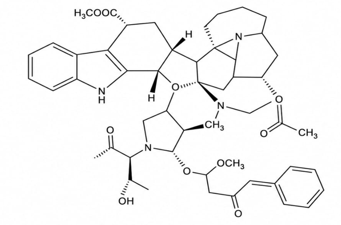
Fig. No. 1.1 Structure of vincristine
Pharmacological Classification
Vincristine belongs to:
It is classified as a cell cycle-specific anticancer drug because it acts primarily during the M phase of the cell cycle [25].
Mechanism of Action of Vincristine
The anticancer activity of vincristine results from its ability to bind specifically to β-tubulin molecules within microtubules. Microtubules are essential structural components required for chromosome segregation during mitosis [26-27].
Vincristine binds to tubulin dimers and prevents their polymerization into functional microtubules. As a result, mitotic spindle formation is inhibited, leading to metaphase arrest and eventual apoptotic cell death [28].
Cancer cells exposed to vincristine exhibit:
The process can be represented as:
Vincristine → Tubulin Binding → Microtubule Depolymerization → Mitotic Arrest → Apoptosis → Cancer Cell Death
Apart from disrupting mitosis, vincristine also influences several intracellular signaling pathways involved in cell survival and programmed cell death [29-30].
Pharmacokinetics of Vincristine
Absorption
Vincristine is not administered orally because of poor gastrointestinal absorption and extensive first-pass metabolism. Therefore, it is administered intravenously [31].
Distribution
Following intravenous administration, vincristine rapidly distributes into tissues. It exhibits extensive tissue binding and has a large volume of distribution.
High concentrations are detected in:
Penetration into the central nervous system is limited because of the blood-brain barrier [30].
Metabolism
Vincristine undergoes hepatic metabolism primarily through cytochrome P450 enzymes, especially CYP3A4 and CYP3A5 [31].
Elimination
The drug is eliminated mainly through biliary excretion, while only a small percentage is excreted through urine. The terminal elimination half-life ranges from 19 to 155 hours depending on patient-specific factors [32].
Therapeutic Applications of Vincristine
Vincristine is widely used in the treatment of several cancers.
Hematological Malignancies
Solid Tumours
Vincristine is generally administered as part of multidrug chemotherapy protocols because combination therapy produces superior therapeutic outcomes compared with monotherapy [33-34].
Vincristine in Combination Chemotherapy
Combination chemotherapy is a standard strategy in modern oncology. Vincristine forms an integral component of several established treatment regimens.
Examples include:
CHOP Regimen
Used for non-Hodgkin lymphoma.
MOPP Regimen
Used for Hodgkin lymphoma.
Hyper-CVAD Regimen
Includes vincristine for treatment of acute lymphoblastic leukemia [35].
Combination therapy improves efficacy by targeting multiple cellular pathways simultaneously and reducing the probability of drug resistance.
Adverse Effects and Toxicity of Vincristine
Although vincristine is highly effective, its clinical use is limited by toxicity.
Neurotoxicity
Peripheral neuropathy is the most common dose-limiting adverse effect.
Symptoms include:
Neurotoxicity occurs because microtubules are essential for axonal transport in neurons.
Gastrointestinal Toxicity
Hematological Effects
Compared with many anticancer drugs, vincristine causes relatively less bone marrow suppression.
Other Toxicities
Dose reduction is frequently required in patients with severe toxicity [36-37].
Multidrug Resistance Associated with Vincristine
One of the major challenges associated with vincristine therapy is the development of multidrug resistance (MDR).
Cancer cells acquire resistance through:
These mechanisms reduce intracellular drug concentration and limit therapeutic efficacy [38-39].
Need for Novel Vincristine-Based Combination Therapy
Despite its clinical success, vincristine therapy is often associated with toxicity and resistance. Therefore, researchers are exploring combination approaches involving natural bioactive compounds capable of enhancing therapeutic efficacy while reducing adverse effects.
Polyphenolic compounds are particularly attractive because they exhibit:
Among these compounds, tannic acid has emerged as a promising candidate for combination therapy. Several studies suggest that tannic acid can modulate oxidative stress, inhibit tumour cell proliferation, suppress angiogenesis, and enhance the activity of conventional chemotherapeutic agents.
Therefore, combining vincristine with tannic acid may provide a synergistic anticancer effect through complementary mechanisms of action, potentially improving therapeutic outcomes and overcoming drug resistance.
Tannic Acid:
Tannic acid is a naturally occurring hydrolysable polyphenolic compound widely distributed in various plants, fruits, nuts, seeds, bark, leaves, and beverages such as tea and wine. It belongs to the tannin family and has attracted considerable scientific interest because of its diverse biological and pharmacological activities. Traditionally, tannic acid has been used in medicine for its astringent, antimicrobial, antioxidant, and anti-inflammatory properties. In recent years, increasing evidence has demonstrated its potential role in cancer prevention and treatment [40].
Natural polyphenols have emerged as promising therapeutic agents due to their ability to modulate multiple molecular pathways involved in carcinogenesis. Among these compounds, tannic acid has shown significant anticancer activity against a wide range of cancer cell lines including breast cancer, lung cancer, liver cancer, colon cancer, prostate cancer, ovarian cancer, and leukemia [41].
The ability of tannic acid to interfere with cancer cell proliferation, induce apoptosis, suppress angiogenesis, and regulate oxidative stress pathways makes it an attractive candidate for combination chemotherapy strategies [42].
Natural Sources of Tannic Acid
Tannic acid is widely distributed throughout the plant kingdom. It is particularly abundant in:
Fruits
Nuts
Plant Materials
Beverages
Among these sources, gall nuts contain exceptionally high concentrations of tannic acid and are commonly used for industrial extraction [43-44].
Chemical Structure and Properties of Tannic Acid
Tannic acid is a hydrolysable tannin composed of a central glucose molecule esterified with multiple gallic acid residues.
Structure:
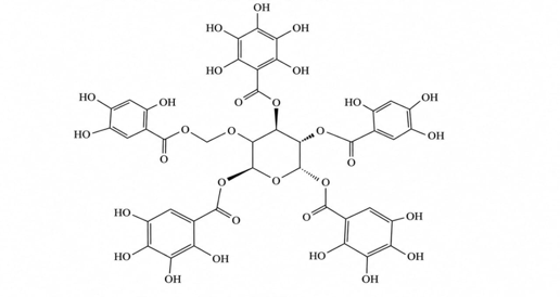
Fig. no. 1.2 structure of Tannic acid
Molecular Formula
C76H52O46
Molecular Weight
Approximately 1701.2 g/mol
The molecule contains numerous hydroxyl groups that contribute to its strong antioxidant and metal-chelating properties. Under acidic or enzymatic conditions, tannic acid undergoes hydrolysis to yield glucose and gallic acid molecules [45].
Physicochemical characteristics include:
These characteristics are responsible for many of its biological effects [46].
Pharmacological Activities of Tannic Acid
Tannic acid exhibits a broad spectrum of pharmacological activities.
Antioxidant Activity
Tannic acid is considered one of the most potent naturally occurring antioxidants. It scavenges reactive oxygen species (ROS), inhibits lipid peroxidation, and protects cellular macromolecules from oxidative damage [47].
Anti-inflammatory Activity
Tannic acid suppresses inflammatory mediators such as:
This anti-inflammatory effect may contribute significantly to its anticancer activity because chronic inflammation promotes tumour initiation and progression [48].
Antimicrobial Activity
Several studies have reported antibacterial, antiviral, and antifungal activities of tannic acid. The compound can disrupt microbial cell membranes and inhibit essential enzymatic processes [49].
Antidiabetic Activity
Tannic acid improves glucose metabolism and exhibits inhibitory effects on digestive enzymes such as α-amylase and α-glucosidase [50].
Cardioprotective Activity
By reducing oxidative stress and inflammation, tannic acid may contribute to cardiovascular protection [51].
Role of Oxidative Stress in Cancer
Oxidative stress plays a crucial role in cancer development.
Reactive oxygen species (ROS) contribute to:
Persistent oxidative stress promotes activation of oncogenic signaling pathways and facilitates malignant transformation [52].
Cancer cells generally exhibit elevated ROS levels compared with normal cells. Therefore, agents capable of modulating oxidative stress may serve as effective anticancer therapeutics.
Tannic acid exerts its antioxidant activity by:
These mechanisms contribute significantly to its cancer preventive and therapeutic effects [53].
Anticancer Activity of Tannic Acid
Recent investigations have demonstrated remarkable anticancer properties of tannic acid.
Studies indicate that tannic acid can:
Importantly, tannic acid affects multiple molecular targets simultaneously, reducing the likelihood of resistance development [54].
Mechanisms of Anticancer Action of Tannic Acid
Induction of Apoptosis
Apoptosis is a highly regulated process of programmed cell death essential for eliminating damaged or malignant cells.
Tannic acid induces apoptosis through:
Consequently, cancer cells undergo irreversible apoptotic death [55].
Cell Cycle Arrest
Uncontrolled cell cycle progression is a hallmark of cancer.
Tannic acid has been reported to arrest cancer cells at:
depending on cancer type and experimental conditions.
Cell cycle arrest suppresses proliferation and enhances susceptibility to apoptosis [56].
Inhibition of Angiogenesis
Tumour growth depends on angiogenesis, the formation of new blood vessels supplying oxygen and nutrients.
Tannic acid inhibits angiogenesis by suppressing:
As a result, tumour expansion becomes restricted [57].
Suppression of Metastasis
Metastasis remains the primary cause of cancer-related mortality.
Tannic acid inhibits metastasis through:
These effects reduce the spread of malignant cells to distant organs [58].
Molecular Targets of Tannic Acid
Numerous signaling pathways are regulated by tannic acid.
Important molecular targets include:
NF-κB Pathway
NF-κB promotes survival, inflammation, and proliferation in cancer cells.
Tannic acid inhibits NF-κB activation and reduces expression of inflammatory mediators.
PI3K/Akt/mTOR Pathway
This pathway controls cell growth and survival.
Tannic acid suppresses PI3K/Akt signalling, resulting in growth inhibition and apoptosis.
MAPK Pathway
Modulation of MAPK signalling contributes to antiproliferative effects observed in several cancer models.
p53 Pathway
Tannic acid enhances p53-mediated apoptosis and cell cycle arrest in various tumour cells [59-60].
In Vitro Anticancer Studies of Tannic Acid
Numerous in vitro investigations have confirmed the anticancer potential of tannic acid.
Breast Cancer
Tannic acid significantly inhibited proliferation of MCF-7 and MDA-MB-231 breast cancer cells through apoptosis induction and cell cycle arrest [59].
Lung Cancer
Studies demonstrated inhibition of A549 lung cancer cells via oxidative stress modulation and mitochondrial apoptosis pathways [60].
Colon Cancer
Tannic acid suppressed growth of HT-29 and HCT-116 colon cancer cells by regulating apoptotic proteins [61].
Liver Cancer
HepG2 liver cancer cells exhibited reduced viability following treatment with tannic acid .
Leukemia
Tannic acid induced apoptosis and inhibited proliferation in leukaemia cell models, suggesting potential utility in haematological malignancies [62].
Tannic Acid as a Chemosensitizing Agent
One of the most promising applications of tannic acid is its ability to enhance the activity of conventional chemotherapeutic agents.
Research indicates that tannic acid can:
These properties make tannic acid a valuable candidate for combination therapy with anticancer drugs such as vincristine [63].
Rationale for Combining Tannic Acid with Vincristine
Vincristine exerts its anticancer activity through disruption of microtubule dynamics, whereas tannic acid acts through multiple complementary mechanisms including antioxidant regulation, apoptosis induction, inhibition of survival pathways, and suppression of drug resistance.
The combination may therefore provide:
This scientific rationale forms the basis for evaluating the synergistic anticancer activity of vincristine and tannic acid using HPLC and in vitro experimental models [64].
In Vitro Cytotoxicity Study by MTT Assay
Introduction
The cytotoxic potential of vincristine, tannic acid, and their combination was evaluated using the MTT assay. The MTT assay is one of the most widely used colorimetric methods for assessing cell viability, proliferation, and cytotoxicity in cultured cells. The assay is based on the ability of metabolically active cells to reduce the yellow tetrazolium salt, MTT [3-(4,5-dimethylthiazol-2-yl)-2,5-diphenyl tetrazolium bromide], into insoluble purple formazan crystals by mitochondrial succinate dehydrogenase enzymes. The amount of formazan formed is directly proportional to the number of viable cells present in the culture [65].
The MTT assay is commonly employed in anticancer drug screening because it is simple, sensitive, reproducible, and cost-effective. Reduction in cell viability after treatment with test compounds indicates cytotoxic activity against cancer cells [66].
In the present study, the MTT assay was employed to evaluate the cytotoxic activity of the Vincristine–Tannic Acid combination against MCF-7 breast cancer cells and L929 normal fibroblast cells.
Principle
Viable cells possess active mitochondrial enzymes capable of reducing MTT into purple-colored formazan crystals. Dead or damaged cells lose this metabolic activity and therefore cannot convert MTT into formazan.
The intensity of purple colour produced is measured spectrophotometrically at 570 nm. A decrease in absorbance compared with untreated control cells reflects reduced cell viability and increased cytotoxicity [67].
MATERIALS AND METHODS
Materials, Vincristine, Tannic Acid, MCF-7 cells, L929 cells, MTT reagent, DMSO, DMEM, FBS
Preparation of Drug Solution:
Vincristine and tannic acid stock solutions were prepared using analytical grade reagents. Vincristine (0.1 mg) was accurately weighed and dissolved in 10 mL of distilled water to obtain a stock solution of 10 µg/ml. Similarly, tannic acid (1 mg) was accurately weighed and dissolved in 10 mL of distilled water to prepare a stock solution of 100 µg/ml. Both solutions were freshly prepared before the experiment and mixed in the required proportions to obtain the Vincristine–Tannic Acid combination. The prepared combination was used for analytical characterization by pH determination, FTIR spectroscopy, UV–Visible spectroscopy, RP-HPLC analysis, and in vitro cytotoxic evaluation using the MTT assay.
pH Determination:
The pH of vincristine, tannic acid, and the Vincristine–Tannic Acid combination was determined using a calibrated digital pH meter at room temperature. Prior to measurement, the pH meter was calibrated using standard buffer solutions (pH 4.0, 7.0, and 9.2). Approximately 10 mL of each sample solution was transferred into a clean beaker, and the electrode was immersed completely in the solution. The pH value was recorded after obtaining a stable reading. Each measurement was performed in triplicate, and the average pH value was reported.
Fourier Transform Infrared (FTIR) Spectroscopic Analysis:
The compatibility of vincristine and tannic acid was evaluated using Fourier Transform Infrared (FTIR) spectroscopy. The FTIR spectra of vincristine, tannic acid, and their combination were recorded over the spectral range of 4000–400 cm⁻¹ using an FTIR spectrophotometer. The characteristic absorption peaks of the individual compounds and the combination were compared to identify the presence of functional groups and to assess any possible chemical interaction. The obtained spectra were analysed for peak position, intensity, and functional group identification.
UV–Visible Spectroscopic Analysis:
The UV–Visible spectroscopic analysis of vincristine, tannic acid, and the Vincristine–Tannic Acid combination was carried out using a UV–Visible spectrophotometer. The prepared solutions were scanned over the wavelength range of 200–400 nm using the appropriate solvent as blank. The absorption maxima (λmax) of the samples were recorded, and the obtained spectra were analysed to confirm the presence of characteristic chromophoric groups and to support the analytical characterization of the prepared combination.
RP-HPLC Analysis:
RP-HPLC analysis was performed to evaluate the chromatographic characteristics of the Vincristine–Tannic Acid combination. The analysis was carried out using a reverse-phase C18 column under optimized chromatographic conditions. The mobile phase consisted of methanol and water in the optimized ratio, which was filtered and degassed before use. The samples were injected into the HPLC system, and detection was carried out at the selected wavelength. Chromatographic parameters including retention time, peak area, tailing factor, theoretical plates, and resolution were recorded to assess the analytical performance of the developed method.
In Vitro Cytotoxic Evaluation by MTT Assay:
The in vitro cytotoxic activity of the Vincristine–Tannic Acid combination was evaluated against MCF-7 human breast cancer cells using the MTT assay. MCF-7 cells were cultured in Dulbecco's Modified Eagle Medium (DMEM) supplemented with 10% fetal bovine serum (FBS) and 1% antibiotic solution under standard cell culture conditions (37°C, 5% CO₂). Cells were seeded into 96-well microplates and incubated for 24 hours to allow cell attachment.
After incubation, the culture medium was replaced with fresh medium containing different concentrations of the Vincristine–Tannic Acid combination. The treated cells were further incubated for 24 hours. Following treatment, MTT reagent was added to each well and incubated for an additional 3–4 hours to allow the formation of purple formazan crystals by metabolically active cells. The medium was carefully removed, and the formazan crystals were dissolved using dimethyl sulfoxide (DMSO). The absorbance was measured at 570 nm using a microplate reader. Cell viability (%) and growth inhibition (%) were calculated, and the IC₅₀ value of the combination was determined from the dose–response curve.
The cytotoxicity of the Vincristine–Tannic Acid combination toward normal cells was also evaluated using L929 mouse fibroblast cells under similar experimental conditions to assess its safety profile.
RESULT AND DISCUSSION
PH Measurement
Table 1. measurement of PH range
|
Sr. No. |
Sample |
pH |
|
1 |
Tannic Acid |
3.5 |
|
2 |
Vincristine |
4.6 |
|
3 |
Vincristine + Tannic acid |
5.0 |
“Tannic acid showed acidic pH of 3.5, while vincristine exhibited slightly acidic pH of 4.6. The pH of the combined Sample was found to be 5.0, indicating mildly acidic nature of the Sample.”
Fourier Transform Infrared Spectroscopy (FTIR)
FTIR analysis was performed using an Agilent Technologies FTIR spectrophotometer. The instrument was operated in the wavenumber range of 4000–650 cm⁻¹ with a resolution of 8 cm⁻¹. A total of 32 sample scans and 32 background scans were recorded before analysis. The system status was found to be good during the experiment.
For analysis, the sample containing Vincristine and Tannic Acid combination was prepared and placed on the ATR/sample holder properly. Initially, background scanning was performed to remove atmospheric interference. After background correction, the sample scan was carried out and the spectrum was recorded.
The obtained FTIR spectrum was analyzed for characteristic absorption peaks corresponding to different functional groups. The presence of peaks corresponding to O–H/N–H, C–H, C=O, aromatic C=C, and C–O stretching confirmed and no specific interaction of sample. FTIR
Spectrum and their interpretation data are represented in the following table 3. Mix sample.
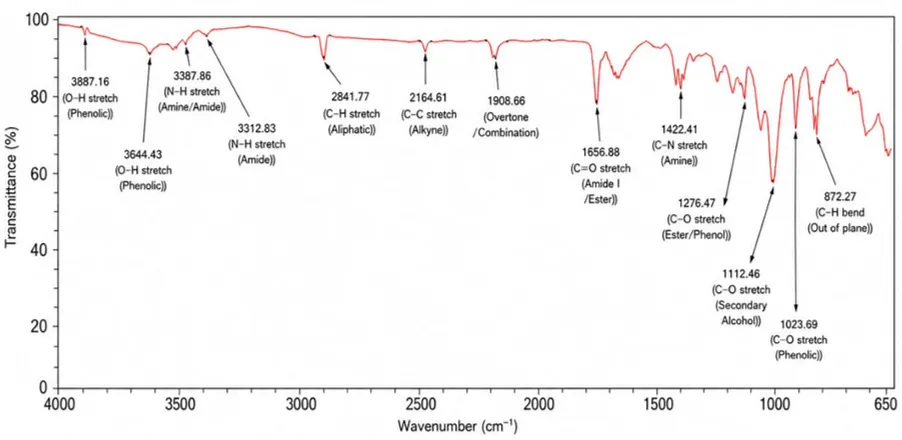
Fig.3. FTIR of Mix Sample
Table 2. Interpretation data of Mix Sample
|
Sr. No. |
Peak (cm-1) |
Functional Group |
|
1 |
3887.16 |
O-H Stretching (phenolic) |
|
2 |
3644.43 |
O-H Stretching (phenolic) |
|
3 |
3387.86 |
N-H Stretching (Amine/ Amide) |
|
4 |
3312.83 |
N-H Stretching ( Amide) |
|
5 |
2926.43 |
C-H C (Aluphatic) |
|
6 |
2164.61 |
C≡C Stretching |
|
7 |
1908.66 |
Combination Bond |
|
8 |
1656.88 |
C=O N-H Stretching (Amide Ester) |
|
9 |
1422.41 |
C-N Stretching (Phenol/ester) |
|
10 |
1276.47 |
C-O Stretching (Phenol/Ester) |
|
11 |
1112.46 |
C-O Stretching |
|
12 |
1023.69 |
C-O Stretching (Phenolic) |
|
13 |
872.27 |
Aromatic C-H Bending |
The FTIR spectrum of the Vincristine–Tannic Acid physical mixture exhibited characteristic absorption peaks corresponding to the functional groups present in both compounds. The broad absorption bands observed at 3887.16 and 3644.43 cm⁻¹ were attributed to phenolic O–H stretching vibrations of tannic acid. Peaks at 3387.86 and 3312.83 cm⁻¹ corresponded to N–H stretching vibrations of amine and amide groups present in vincristine. The absorption band at 2926.43 cm⁻¹ indicated aliphatic C–H stretching. The peak at 1656.88 cm⁻¹ was assigned to C=O stretching vibration of amide/ester groups, while the band at 1422.41 cm⁻¹ represented C–N stretching vibration. The characteristic peaks observed at 1276.47, 1112.46 and 1023.69 cm⁻¹ corresponded to C–O stretching vibrations of ester, phenolic and alcohol groups. The peak at 872.27 cm⁻¹ was attributed to aromatic C–H bending vibration. The presence of these characteristic peaks confirmed the existence of both vincristine and tannic acid in the mixture and indicated the absence of any significant chemical interaction between them.
UV-Visible spectrophotometric Analysis
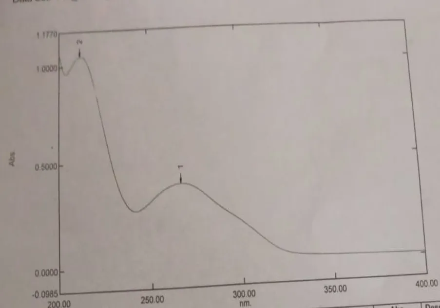
Fig.4. UV-Visible spectrophotometric Analysis
The UV-visible spectrum exhibited two characteristic absorption peaks at 212 nm and 267 nm. The highest absorbance (1.0484) was observed at 212 nm, indicating the principal absorption maximum (λ max) of the sample. A second absorption peak with an absorbance of 0.3902 was observed at 267 nm. These absorption maxima indicate the presence of chromophoric groups capable of electronic transitions within the UV region.
Table:3. UV-Visible spectral Data
|
Peak No. |
Wavelength(nm) |
Absorbance |
|
1 |
212 |
1.0484 |
|
2 |
267 |
0.3902 |
The UV-visible spectrum demonstrated two distinct absorption peaks at 212 nm and 267 nm. The peak observed at 212 nm showed the highest absorbance and was considered as the principal λ max of the sample. This absorption may be attributed to π→π* electronic transitions associated with aromatic or conjugated functional groups present in the compound.
The second absorption peak at 267 nm may correspond to n→π* transitions or extended conjugation within the molecular structure. The presence of these characteristic absorption peaks confirms the UV-active nature of the sample. The spectrum obtained was smooth, well-defined, and free from significant interference, indicating the purity of the prepared solution and the suitability of the analytical method.
The observed λmax values can be utilized for the further quantitative estimation and analytical characterization of the sample by UV-visible spectroscopy.
High performance Liquid Chromatography (HPLC)
HPLC Analysis of Vincristine
The HPLC analysis of Vincristine was carried out using RP-HPLC method with C18 column and UV detection at 254 nm. The chromatogram showed a distinct and sharp peak of Vincristine at a retention time of approximately 4.00 min, confirming the presence of the drug in the sample.
The obtained peak was symmetrical with acceptable chromatographic parameters, indicating good separation efficiency and suitability of the analytical method. No significant interfering peaks were observed near the retention time of Vincristine, which demonstrated the specificity of the method.
The HPLC study confirmed the purity and stability of Vincristine under the selected chromatographic conditions. Therefore, the developed RP-HPLC method was found to be simple, reliable, and suitable for routine analysis of Vincristine is shown in fig 5.
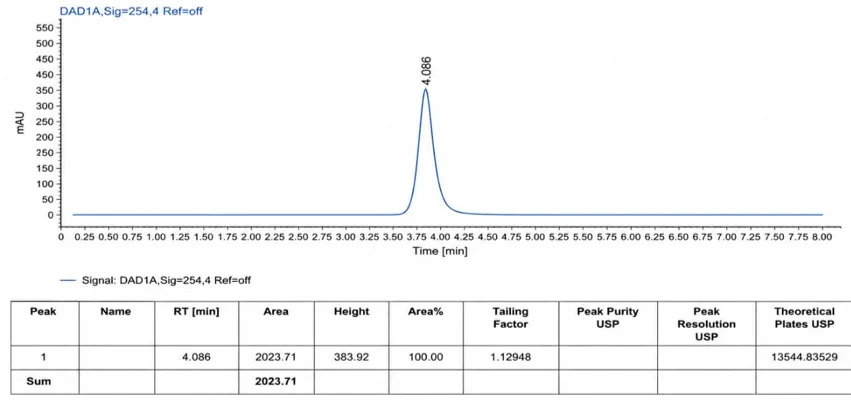
Fig.5. HPLC Analysis of Vincristine
HPLC Analysis of Tannic Acid
The HPLC analysis of Tannic Acid was carried out using RP-HPLC method with UV detection at 254 nm. The chromatogram showed a sharp and prominent peak at approximately 2.712 min retention time, confirming the presence of Tannic Acid in the sample.
The obtained chromatographic peak was symmetrical with acceptable peak area and tailing factor values, indicating satisfactory analytical performance of the method. A minor peak was also observed at 3.118 min, which may be due to impurities or slight peak splitting during analysis.
The chromatogram demonstrated good separation efficiency and confirmed the identity of Tannic Acid under the selected chromatographic conditions. Therefore, the developed RP-HPLC method was found to be suitable for the analysis of Tannic Acid is shown in figure 6.
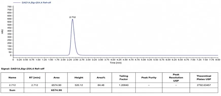
Fig.6. HPLC Analysis of Tannic Acid
HPLC Analysis of Combination
The HPLC chromatogram of the combined formulation of Vincristine and Tannic Acid showed distinct and well-resolved peaks at retention times 2.706 min, 3.086 min, and 4.079 min. The major peak was observed at 2.706 min with an area percentage of 73.54%, indicating the predominant presence of the compound in the sample.
The chromatographic peaks were sharp and symmetrical with acceptable tailing factor values. Peak resolution and theoretical plate values indicated satisfactory separation efficiency of the method. No significant interfering or impurity peaks were observed in the chromatogram.
The HPLC chromatogram confirmed satisfactory purity and compatibility of Vincristine and Tannic Acid combination with absence of major impurity peaks.
Therefore, the developed HPLC method was found to be suitable for the identification and analysis of Vincristine and Tannic Acid combination is shown figure 7.
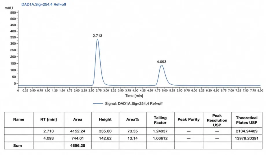
Fig.7.HPLC Analysis of Combination
Table 4. Interpretation Data of Combination Compound
|
Parameter |
Vincristine |
Tannic Acid |
|
Retention Time (RT) |
4.093 |
2.713 |
|
Area |
744.013 |
4152.237 |
|
Tailing |
1.066 |
1.249 |
|
USP Theoretical Plats |
13978.20391 |
2134.944 |
|
Resolution |
- |
3.80541 |
HPLC analysis was performed to evaluate the chromatographic behavior of Vincristine and Tannic Acid. The obtained chromatographic parameters are summarized in the table.
Vincristine showed a retention time (RT) of 4.093 min, whereas Tannic Acid exhibited a retention time of 2.713 min, indicating that Tannic Acid eluted earlier than Vincristine under the selected chromatographic conditions.
The peak area of Tannic Acid (4152.237) was significantly higher than that of Vincristine (744.013), suggesting a comparatively higher detector response or concentration of Tannic Acid.
The tailing factor values for Vincristine (1.066) and Tannic Acid (1.249) were close to 1, indicating good peak symmetry and acceptable chromatographic performance.
The USP theoretical plates for Vincristine (13,978.20) were considerably higher than those for Tannic Acid (2,134.94), demonstrating better column efficiency and sharper peak formation for Vincristine.
A resolution value of 3.805 was observed between the two compounds, which is greater than the USP recommended value of 2.0, indicating excellent separation and no significant overlap between the peaks of Vincristine and Tannic Acid.
Cytotoxic Activity of Tannic Acid–Vincristine Combination Against MCF-7 Cell Line.
Table no.5. Effects Of 5-Flurouracilagainst MCF-7 (Human Mammary gland Breast Adenocarcinoma Cell line) MTT Assay
|
Sr. No. |
Concentration (µg/ml) |
Absorbance (O D) |
Cell Viability (%) |
IC50 (µg/ml) |
|||
|
1 |
2 |
3 |
Mean±SD |
||||
|
1 |
Control |
2.107 |
2.114 |
2.11 |
2.110±0.002 |
|
|
|
2 |
Standard-5FU 100 |
0.563 |
0.561 |
0.569 |
0.564±0.004 |
26.74143 |
10.56 µg/ml |
|
50 |
0.744 |
0.561 |
0.746 |
0.745±0.001 |
35.30248 |
||
|
25 |
0.851 |
0.745 |
0.856 |
0.854±0.002 |
40.46754 |
||
|
12.5 |
0.995 |
0.855 |
0.994 |
0.993±0.002 |
47.06997 |
||
|
6.25 |
1,253 |
1.256 |
1.253 |
1.254±0.001 |
59.42189 |
||
The MTT assay results demonstrated that 5-Fluorouracil exhibited potent cytotoxic activity against MCF-7 breast cancer cells. A concentration-dependent reduction in cell viability was observed, indicating that higher concentrations of the drug effectively inhibited cell growth and proliferation. The lowest cell viability (26.74%) was observed at 100 µg/ml, whereas the highest viability (59.42%) was recorded at 6.25 µg/ml. This trend suggests that the anticancer activity of 5-FU increases with concentration. The IC₅₀ value of 10.56 µg/ml indicates that a relatively low concentration of the drug is sufficient to inhibit 50% of the cancer cell population.
These findings are consistent with the established mechanism of 5-Fluorouracil, which interferes with DNA synthesis and induces cell death in rapidly dividing cancer cells. Therefore, the obtained results confirm the effectiveness of 5-FU as a standard anticancer drug against MCF-7 breast cancer cells and validate its use as a positive control in cytotoxicity studies.
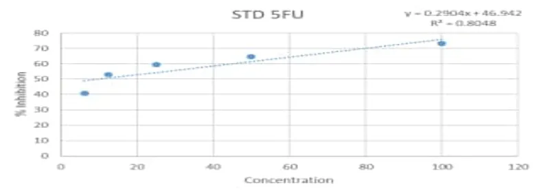
Fig. 8. STD 5FU
Figure shows the concentration-dependent cytotoxic effect of standard 5-Fluorouracil on MCF-7 cells. An increase in drug concentration resulted in a gradual decrease in cell viability, demonstrating enhanced anticancer activity. The calculated IC₅₀ value of 10.56 µg/ml confirms the potent cytotoxic potential of 5-FU against MCF-7 breast cancer cells.
Table No.6. Effects of Samples MCF-7 (Human Mammary gland Breast Adenocarcinoma Cell line) MTT Assay
|
Sr. No. |
Concentration (µg/ml) |
Absorbance (O D) |
Cell Viability (%) |
IC50 (µg/ml) |
|||
|
1 |
2 |
3 |
Mean±SD |
||||
|
1 |
Controld |
2.107 |
2.114 |
2.11 |
2.110±0.002 |
|
|
|
2 |
Standard-5FU 100 |
0.856 |
0.852 |
0.851 |
0.853±0.002646 |
40.42654 |
59.50 µg/ml |
|
50 |
0.992 |
0.995 |
0.996 |
0.994±002082 |
47.1248 |
||
|
25 |
1.236 |
1.235 |
1.239 |
1.236±002082 |
58.60979 |
||
|
12.5 |
1.389 |
1.387 |
1.385 |
1.387±0.002 |
65.7346 |
||
|
6.25 |
1.489 |
1.485 |
1.489 |
1.487±0.002309 |
70.50553 |
||
The cytotoxic activity of the sample combination against MCF-7 human breast adenocarcinoma cells was evaluated using the MTT assay. The results demonstrated a concentration-dependent decrease in cell viability with increasing concentration of the sample combination.
The percentage cell viability was found to be 70.51%, 65.73%, 58.61%, 47.12%, and 40.43% at concentrations of 6.25, 12.5, 25, 50, and 100 µg/ml, respectively. The lowest cell viability was observed at 100 µg/ml, indicating maximum cytotoxic activity, whereas the highest cell viability was observed at 6.25 µg/ml.
The IC₅₀ value of the sample combination was calculated as 59.50 µg/ml, suggesting moderate cytotoxic potential against MCF-7 breast cancer cells. The reduction in absorbance values with increasing concentration further confirmed inhibition of cell proliferation and decreased metabolic activity of viable cells.
The graph exhibited a concentration-dependent increase in percentage inhibition, demonstrating that the cytotoxic effect of the sample combination increased with concentration. These findings indicate that the sample combination possesses significant anticancer activity and effectively suppresses the growth of MCF-7 cells.
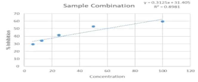
Fig. 9. Sample Combination MCF-7 cell
Figure shows the concentration-dependent cytotoxic effect of the sample combination on MCF-7 cells. An increase in concentration resulted in increased percentage inhibition and reduced cell viability. The maximum inhibition was observed at 100 µg/ml, while the minimum inhibition was recorded at 6.25 µg/ml. The IC₅₀ value of 59.50 µg/ml indicates moderate cytotoxic activity of the sample combination against MCF-7 breast cancer cells.
Cytotoxic Activity of Tannic Acid–Vincristine Combination Against L929 Cell line:
Table No.7. Cytotoxic Effect of Tannic Acid–Vincristine Combination on L929 Cell Line Determined by MTT Assay.
|
Sr. No. |
Concentration (µg/ml) |
Absorbance (O D) |
Cell Viability (%) |
|||
|
1 |
2 |
3 |
Average |
|||
|
1 |
Control |
2.107 |
2.114 |
2.11 |
2.110±0.003512 |
|
|
2 |
Sample Combination 100 |
1.102 |
1.103 |
1.105 |
1.103±0.001528 |
52.29068 |
|
50 |
1.353 |
1.256 |
1.259 |
1.256±0.003 |
59.52607 |
|
|
25 |
1.3256 |
1.329 |
1.327 |
1.327±0.001709 |
62.90047 |
|
|
12.5 |
1.415 |
1.412 |
1.416 |
1.414±0.002082 |
67.03002 |
|
|
6.25 |
1.513 |
1.514 |
1.512 |
1.513±0.001 |
71.70616 |
|
The results presented in Table 1 indicate that the Tannic Acid–Vincristine combination exhibited a concentration-dependent reduction in the viability of L929 cells. The control group showed an average absorbance of 2.110 ± 0.003, representing 100% viable cells. Treatment with the sample at concentrations of 6.25, 12.5, 25, 50, and 100 µg/mL resulted in cell viabilities of 71.71%, 67.03%, 62.90%, 59.53%, and 52.29%, respectively. The lowest cell viability was observed at 100 µg/mL, indicating the highest cytotoxic activity of the sample.
The decrease in absorbance values and corresponding cell viability percentages with increasing sample concentration indicates that the Tannic Acid–Vincristine combination exerted a dose-dependent cytotoxic effect on L929 cells. As the concentration increased, fewer metabolically active cells were present, leading to reduced MTT reduction and lower formazan formation. These findings suggest that the combination effectively inhibited cellular growth and viability, with maximum inhibition observed at the highest tested concentration. The results demonstrate moderate cytotoxic potential of the formulation against L929 cells.
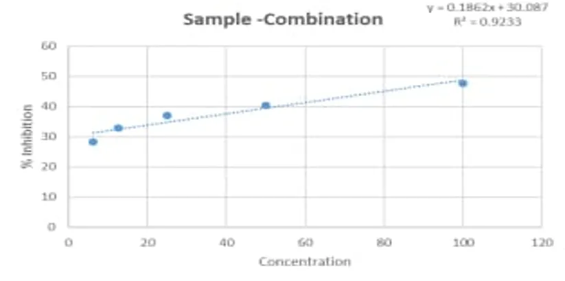
Fig 10. Concentration-Dependent Effect of Tannic Acid–Vincristine Combination on L929 Cell Viability.
Figure 10 illustrates the relationship between sample concentration and percentage cell viability. A gradual decline in cell viability was observed as the concentration of the Tannic Acid–Vincristine combination increased from 6.25 µg/ml to 100 µg/ml. Cell viability decreased from 71.71% at the lowest concentration to 52.29% at the highest concentration, indicating a concentration-dependent cytotoxic response
The graphical representation clearly demonstrates a negative correlation between sample concentration and cell viability. The continuous downward trend confirms that increasing concentrations of the Tannic Acid–Vincristine combination enhanced its cytotoxic activity. The reduction in viability may be attributed to inhibition of mitochondrial metabolic activity and cell proliferation. Since cell viability remained slightly above 50% even at 100 µg/mL, the IC₅₀ value is estimated to be greater than 100 µg/mL. Overall, the graph confirms the dose-dependent cytotoxic effect of the combination and supports the observations obtained from the tabulated data
CONCLUSION
The present study successfully performed the analytical characterization and in vitro cytotoxic evaluation of the Vincristine–Tannic Acid combination against MCF-7 human breast cancer cells. The analytical investigations, including pH determination, FTIR spectroscopy, UV–Visible spectroscopy, and RP-HPLC analysis, confirmed the compatibility of vincristine and tannic acid and demonstrated the suitability of the developed analytical method. The MTT assay revealed concentration-dependent cytotoxic activity of the combination against MCF-7 cells, with an IC₅₀ value of 59.50 µg/ml. Furthermore, the combination exhibited comparatively lower toxicity toward L929 normal fibroblast cells (IC₅₀ >100 µg/ml), indicating a favorable safety profile in vitro. Overall, the findings suggest that the Vincristine–Tannic Acid combination possesses promising in vitro anticancer potential and may serve as a candidate for further preclinical investigations.
REFERENCES


Wagalgave Priyanka, Dr. Dharashive Vishweshwar, Analytical Characterization and In Vitro Cytotoxic Evaluation of a Vincristine–Tannic Acid Combination Against MCF-7 Human Breast Cancer Cells, Int. J. of Pharm. Sci., 2026, Vol 4, Issue 7, 2850-2873. https://doi.org/10.5281/zenodo.21363025
 10.5281/zenodo.21363025